Activated Kras and Ink4a/Arf deficiency cooperate to produce metastatic pancreatic ductal adenocarcinoma - PubMed (original) (raw)
. 2003 Dec 15;17(24):3112-26.
doi: 10.1101/gad.1158703. Epub 2003 Dec 17.
Affiliations
- PMID: 14681207
- PMCID: PMC305262
- DOI: 10.1101/gad.1158703
Activated Kras and Ink4a/Arf deficiency cooperate to produce metastatic pancreatic ductal adenocarcinoma
Andrew J Aguirre et al. Genes Dev. 2003.
Abstract
Pancreatic ductal adenocarcinoma ranks among the most lethal of human malignancies. Here, we assess the cooperative interactions of two signature mutations in mice engineered to sustain pancreas-specific Cre-mediated activation of a mutant Kras allele (KrasG12D) and deletion of a conditional Ink4a/Arf tumor suppressor allele. The phenotypic impact of KrasG12D alone was limited primarily to the development of focal premalignant ductal lesions, termed pancreatic intraepithelial neoplasias (PanINs), whereas the sole inactivation of Ink4a/Arf failed to produce any neoplastic lesions in the pancreas. In combination, KrasG12D expression and Ink4a/Arf deficiency resulted in an earlier appearance of PanIN lesions and these neoplasms progressed rapidly to highly invasive and metastatic cancers, resulting in death in all cases by 11 weeks. The evolution of these tumors bears striking resemblance to the human disease, possessing a proliferative stromal component and ductal lesions with a propensity to advance to a poorly differentiated state. These findings in the mouse provide experimental support for the widely accepted model of human pancreatic adenocarcinoma in which activated KRAS serves to initiate PanIN lesions, and the INK4A/ARF tumor suppressors function to constrain the malignant conversion of these PanIN lesions into lethal ductal adenocarcinoma. This faithful mouse model may permit the systematic analysis of genetic lesions implicated in the human disease and serve as a platform for the identification of early disease markers and for the efficient testing of novel therapies.
Figures
Figure 1.
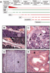
Preinvasive ductal lesions arising in Pdx1-Cre; LSL-KrasG12D mice. (A) Genetic progression model of human pancreatic adenocarcinoma. The cellular phenotypes of the increasing grades of ductal neoplastic lesions are indicated. Previous studies have cataloged the presence of genetic alterations at specific disease stages, as depicted in the temporal sequence. The thickness of the line corresponds to the frequency of a lesion. Loss-of-function events are depicted in red, whereas gain-of-function lesions are shown in green. (B, top) Hematoxylin and eosin (H&E) stain showing a normal islet (arrow) and duct (arrowheads) in the background of normal acinar tissue (asterisks) in a 12-week-old Pdx1-Cre; LSL-KrasG12D mouse. An adjacent blood vessel (BV) is also indicated. (Bottom) Higher-power view of the single-layer cuboidal ductal epithelium. (C) PanIN-1 lesions detected in a 9-week-old Pdx1-Cre; LSL-KrasG12D mouse (H&E staining). Note PanIN lesions with mucinous columnar epithelium (arrows) and papillary architecture (dashed box). (D) Focus of ductal proliferation (dashed box) with prominent stromal response (asterisk) in a 12-week-old Pdx1-Cre; LSL-KrasG12D mouse (H&E staining). (E) Extensive PanIN lesions with a classical picture of intimately associated fibrotic stroma (asterisk) in the pancreas of a Pdx1-Cre; LSL-KrasG12D mouse at 26 weeks of age (H&E staining).
Figure 2.
Ink4a/Arf deficiency promotes progression to invasive pancreatic adenocarcinoma. (A) Complete excision of the Ink4a/Arf locus in the pancreas with Pdx1-Cre. PstI Southern blot on pancreas (P) or spleen (S) DNA from Ink4a/Arflox/+ mice that harbor or lack the Pdx1-Cre transgene. The wild-type or lox allele migrates at 9.0 kb, and the recombined _Ink4a/Arf_-null allele corresponds to the 4.6-kb band. (B) Kaplan-Meier pancreatic tumor-free survival curve for Pdx1-Cre; LSL-KrasG12D; Ink4a/Arflox/lox mice (denoted Ink/Arf L/L; n = 26 mice) and control cohorts (denoted Ctrl; all combinations of Pdx1-Cre, LSL-KrasG12D, and Ink4a/Arflox/lox alleles, n = 186 mice). Clinically, mice presented in a moribund state and were killed for autopsy. (C) Gross photograph of a pancreatic adenocarcinoma obstructing the common bile duct and causing dilation of the gall bladder (asterisk). Note that jaundice is readily apparent in the abdominal skin (J). T indicates tumor; D, duodenum; and L, liver. Bar, 0.6 cm. Dashed circle denotes the tumor. (D) Well-differentiated ductal adenocarcinoma observed in a Pdx1-Cre; LSL-KrasG12D; Ink4a/Arflox/lox animal at 7.9 weeks of age. Glandular tumor cells (arrowheads) are surrounded by abundant stroma (asterisk). (E) Poorly differentiated adenocarcinoma arising in the same mouse as that from D. Irregular, ill-formed formed glands (arrowheads) are present with mixture of highly mitotic, atypical tumor cells. (F) Region of tumor with sarcomatoid features from the same mouse as in D and E. (_G_-I) Regions of well-differentiated (G), poorly differentiated (H), and sarcomatoid (I) tumor stained for the ductal marker, cytokeratin-19. (J) PAS+D stain for apical mucins (maroon) in well-differentiated tumor cells. (K) Trichrome stain for collagen (blue) reveals the fibrotic nature of the tumor stroma.
Figure 3.
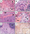
Murine pancreatic tumors invade and metastasize. (A) Duodenal invasion by pancreatic ductal adenocarcinoma. Tumor (T), muscle wall (M, arrowheads), and intestinal epithelium (IE) are indicated. (B) High-magnification photomicrograph of a lymph node metastasis (T, arrowhead). LN denotes normal lymph node architecture. (C) Tumor cells (T) invading the stomach wall (M). Adjacent gastric epithelium is indicated (GE). (D) High-power micrograph of metastatic tumor cells (arrowheads) within a portal tract in the liver. Hepatocytes (H), portal vein (PV), and a reactive bile duct (B) are indicated. (E) Pancreatic tumor cells (T) invading the spleen. White pulp of the spleen is indicated (WP). (F) Immunohistochemical stain for cytokeratin-19 on tumor cells in E invading the spleen. (Inset) Higher-power image of ck-19+ invading tumor cells.
Figure 4.
Early-stage pancreatic lesions in Pdx1-Cre; LSL-KrasG12D; Ink/Arflox/lox animals. (A) High-magnification view of a low-grade PanIN lesion (arrowhead) seen in a 5-week Pdx1-Cre; LSL-KrasG12D; Ink4a/Arflox/+ animal. (B) Low-grade preinvasive ductal lesion in a 3-week-old Pdx1-Cre; LSL-KrasG12D; Ink4a/Arflox/lox mouse. (C) High-grade preinvasive ductal lesion in a Pdx1-Cre; LSL-KrasG12D; Ink4a/Arflox/lox mouse at 4 weeks. (D) Early focus of pancreatic adenocarcinoma in a 4-week-old Pdx1-Cre; LSL-KrasG12D; Ink4a/Arflox/lox mouse. Note both the ductal and anaplastic components of this early cancer. (E) High-grade PanIN lesion in a 5-week Pdx1-Cre; LSL-KrasG12D; Ink4a/Arflox/lox mouse. Serial sectioning through the entire pancreas at 10-μm intervals failed to discover any foci of adenocarcinoma in this animal. (F) High-grade PanIN lesion (asterisk) surrounded by anaplastic tumor cells in a 5-week Pdx1-Cre; LSL-KrasG12D; Ink4a/Arflox/lox mouse.
Figure 5.
Molecular analysis of murine pancreatic adenocarcinomas. (A) Ras activation assay. Lysates from wild-type pancreas (lanes 1,2), Pdx1-Cre; LSL-KrasG12D pancreas (lanes 3,4), and the murine pancreatic adenocarcinomas (lanes 5,6) affinity precipitated with Raf RBD agarose (Upstate) and then subjected to immunoblot analysis with anti-Ras antibodies. (B) PCR analysis of the Ink4a/Arf locus in murine pancreatic adenocarcinoma cell lines. Multiplex PCR was performed on DNA from the pancreatic cancer cell lines (lanes _3_-16) with primers that amplify the _Ink4a/Arf_+ (lower band), Ink4a/Arf lox (middle band), and _Ink4a/Arf_- (upper band) alleles. DNA from Ink4a/Arf+/+ (+/+, lanes 1,18) and Ink4a/Arflox/lox (L/L, lanes 2,17) mice served as controls. All cell lines show only the _Ink4a/Arf_- allele. (C) Immunoblot analysis of the tumor lysates. Membranes were immunoblotted for p16Ink4a, p19Arf, Smad4, and α-tubulin (as a loading control). Lysates from primary mouse embryonic fibroblasts (MEF, lane 1) served as a positive control. (D) Immunoblot analysis of p53 expression. Primary MEFs (lane 1) and p53-/- MEFs (lane 2), were positive and negative controls, respectively. (E) Induction of p53 and p21 in pancreatic adenocarcinoma cells by ionizing irradiation. Mouse pancreatic cancer cell lines were either untreated (-) or gamma irradiated (+; lanes _1_-8). Lysates were immunoblotted for p53, p21, and α-tubulin. MEFs with a mutant p53 allele (p53*) were a control for p53 overexpression. Note that the tumors show modest expression of p53 compared with that of cells with mutant stabilized p53 and that ionizing radiation can effectively induce p53 and p21 in these tumor cells. (F) Amplification of the Kras gene and elevated Kras protein expression in a subset of pancreatic adenocarcinomas. The upper panel shows the relative Kras gene copy number as measured by quantitative real-time PCR. Wild-type specimens have a ratio of 1.0; -indicates not done. The middle and lower panels show Western blot analysis of the corresponding Kras levels and the tubulin (tub) loading control, respectively. Lane 1 is a control MEF specimen. Lanes _2_-15 are tumor cell line specimens. Note that lanes 10 and 14 show both high-level Kras gene amplification and protein overexpression. (G) The mutant Kras allele is amplified in tumors showing increased Kras gene copy number. RT-PCR/RFLP analysis was performed on pancreatic adenocarcinoma cell line RNA to evaluate the whether the wild-type and KrasG12D alleles are expressed based on the _KrasG12D_-specific HindIII site. PCR-amplified cDNA was untreated (-) or digested with HindIII (+). Lanes _1_-12 are tumor cell lines. Lanes 13 and 14 are control testes cDNA. All tumors express both alleles. Note that tumors 58 and 65 (lanes 2,4)—corresponding to lanes 10 and 14 in _F_—show an increased relative ratio of the lower KrasG12D allele, consistent with amplification and overexpression of this mutant allele.
Figure 6.
Expression of Egfr and Her2 in pancreatic adenocarcinomas. (A,B) Immunohistochemistry with anti-Egfr (A) or anti-Her2 (B) antibodies shows robust expression of these proteins in the glandular regions of the tumors. (C,D) Immunohistochemistry for Egfr and Her2 reveals very weak or absent expression in the poorly differentiated regions of these tumors. Note that C and D were photographed from adjacent regions of the slides depicted in A and B.
Similar articles
- Both p16(Ink4a) and the p19(Arf)-p53 pathway constrain progression of pancreatic adenocarcinoma in the mouse.
Bardeesy N, Aguirre AJ, Chu GC, Cheng KH, Lopez LV, Hezel AF, Feng B, Brennan C, Weissleder R, Mahmood U, Hanahan D, Redston MS, Chin L, Depinho RA. Bardeesy N, et al. Proc Natl Acad Sci U S A. 2006 Apr 11;103(15):5947-52. doi: 10.1073/pnas.0601273103. Epub 2006 Apr 3. Proc Natl Acad Sci U S A. 2006. PMID: 16585505 Free PMC article. - Obligate roles for p16(Ink4a) and p19(Arf)-p53 in the suppression of murine pancreatic neoplasia.
Bardeesy N, Morgan J, Sinha M, Signoretti S, Srivastava S, Loda M, Merlino G, DePinho RA. Bardeesy N, et al. Mol Cell Biol. 2002 Jan;22(2):635-43. doi: 10.1128/MCB.22.2.635-643.2002. Mol Cell Biol. 2002. PMID: 11756558 Free PMC article. - Smad4 is dispensable for normal pancreas development yet critical in progression and tumor biology of pancreas cancer.
Bardeesy N, Cheng KH, Berger JH, Chu GC, Pahler J, Olson P, Hezel AF, Horner J, Lauwers GY, Hanahan D, DePinho RA. Bardeesy N, et al. Genes Dev. 2006 Nov 15;20(22):3130-46. doi: 10.1101/gad.1478706. Genes Dev. 2006. PMID: 17114584 Free PMC article. - Mouse models of pancreatic cancer.
Herreros-Villanueva M, Hijona E, Cosme A, Bujanda L. Herreros-Villanueva M, et al. World J Gastroenterol. 2012 Mar 28;18(12):1286-94. doi: 10.3748/wjg.v18.i12.1286. World J Gastroenterol. 2012. PMID: 22493542 Free PMC article. Review. - Pancreatic intraepithelial neoplasia revisited and updated.
Sipos B, Frank S, Gress T, Hahn S, Klöppel G. Sipos B, et al. Pancreatology. 2009;9(1-2):45-54. doi: 10.1159/000178874. Epub 2008 Dec 12. Pancreatology. 2009. PMID: 19077454 Review.
Cited by
- Targeting a chemo-induced adaptive signaling circuit confers therapeutic vulnerabilities in pancreatic cancer.
Saito Y, Xiao Y, Yao J, Li Y, Liu W, Yuzhalin AE, Shyu YM, Li H, Yuan X, Li P, Zhang Q, Li Z, Wei Y, Yin X, Zhao J, Kariminia SM, Wu YC, Wang J, Yang J, Xia W, Sun Y, Jho EH, Chiao PJ, Hwang RF, Ying H, Wang H, Zhao Z, Maitra A, Hung MC, DePinho RA, Yu D. Saito Y, et al. Cell Discov. 2024 Oct 29;10(1):109. doi: 10.1038/s41421-024-00720-w. Cell Discov. 2024. PMID: 39468013 Free PMC article. - RUNX3 Controls a Metastatic Switch in Pancreatic Ductal Adenocarcinoma.
Whittle MC, Izeradjene K, Rani PG, Feng L, Carlson MA, DelGiorno KE, Wood LD, Goggins M, Hruban RH, Chang AE, Calses P, Thorsen SM, Hingorani SR. Whittle MC, et al. Cell. 2015 Jun 4;161(6):1345-60. doi: 10.1016/j.cell.2015.04.048. Epub 2015 May 21. Cell. 2015. PMID: 26004068 Free PMC article. - Mouse models of Kras activation in gastric cancer.
Won Y, Choi E. Won Y, et al. Exp Mol Med. 2022 Nov;54(11):1793-1798. doi: 10.1038/s12276-022-00882-1. Epub 2022 Nov 11. Exp Mol Med. 2022. PMID: 36369466 Free PMC article. Review. - Mouse models for studying angiogenesis and lymphangiogenesis in cancer.
Eklund L, Bry M, Alitalo K. Eklund L, et al. Mol Oncol. 2013 Apr;7(2):259-82. doi: 10.1016/j.molonc.2013.02.007. Epub 2013 Mar 5. Mol Oncol. 2013. PMID: 23522958 Free PMC article. Review. - Hyaluronan impairs vascular function and drug delivery in a mouse model of pancreatic cancer.
Jacobetz MA, Chan DS, Neesse A, Bapiro TE, Cook N, Frese KK, Feig C, Nakagawa T, Caldwell ME, Zecchini HI, Lolkema MP, Jiang P, Kultti A, Thompson CB, Maneval DC, Jodrell DI, Frost GI, Shepard HM, Skepper JN, Tuveson DA. Jacobetz MA, et al. Gut. 2013 Jan;62(1):112-20. doi: 10.1136/gutjnl-2012-302529. Epub 2012 Mar 30. Gut. 2013. PMID: 22466618 Free PMC article.
References
- Bachoo R.M., Maher, E.A., Ligon, K.L., Sharpless, N.E., Chan, S.S., You, M.J., Tang, Y., DeFrances, J., Stover, E., Weissleder, R., et al. 2002. Epidermal growth factor receptor and Ink4a/Arf: Convergent mechanisms governing terminal differentiation and transformation along the neural stem cell to astrocyte axis. Cancer Cell 1: 269-277. - PubMed
- Bardeesy N. and DePinho, R.A. 2002. Pancreatic cancer biology and genetics. Nat. Rev. Cancer 2: 897-909. - PubMed
- Bardeesy N., Sinha, M., Hezel, A.F., Signoretti, S., Hathaway, N.A., Sharpless, N.E., Loda, M., Carrasco, D.R., and DePinho, R.A. 2002b. Loss of the Lkb1 tumour suppressor provokes intestinal polyposis but resistance to transformation. Nature 419: 162-167. - PubMed
Publication types
MeSH terms
Substances
LinkOut - more resources
Full Text Sources
Other Literature Sources
Medical
Molecular Biology Databases
Miscellaneous


