Cortical synaptogenesis and motor map reorganization occur during late, but not early, phase of motor skill learning - PubMed (original) (raw)
Cortical synaptogenesis and motor map reorganization occur during late, but not early, phase of motor skill learning
Jeffrey A Kleim et al. J Neurosci. 2004.
Abstract
Extensive motor skill training induces reorganization of movement representations and synaptogenesis within adult motor cortex. Motor skill does not, however, develop uniformly across training sessions. It is characterized by an initial fast phase, followed by a later slow phase of learning. How cortical plasticity emerges during these phases is unknown. Here, we examine motor map topography and synapse number within rat motor cortex during the early and late phases of motor learning. Adult rats were placed in either a skilled or unskilled reaching condition (SRC and URC, respectively) for 3, 7, or 10 d. Intracortical microstimulation of layer V was used to determine the topography of forelimb movement representations within caudal forelimb area of motor cortex contralateral to the trained paw. Quantitative electron microscopy was used to measure the number of synapses per neuron within layer V. SRC animals showed significant increases in reaching accuracy after 3, 7, and 10 d of training. In comparison with URC animals, SRC animals had significantly larger distal forelimb representations after 10 d of training only. Furthermore, SRC animals had significantly more synapses per neuron than URC animals after 7 and 10 d of training. These results show that both motor map reorganization and synapse formation occur during the late phase of skill learning. Furthermore, synaptogenesis precedes map reorganization. We propose that motor map reorganization and synapse formation do not contribute to the initial acquisition of motor skills but represent the consolidation of motor skill that occurs during late stages of training.
Figures
Figure 1.
A, Performance of animals on the skilled reaching task after 3, 7, or10 d of training. Closed circles represent performance on the first day of training (Baseline), and open squares represent performance on the last day of training (Test). In comparison with baseline levels, all animals showed a significant increase in the percentage of successful reaches on the test day (*p < 0.05; Student's dependent t test). B, Performance on the last day of training for all three training durations. Reaching accuracy did not significantly differ between the 7 and 10 d animals, but both were significantly higher than the 3 d animals (*p < 0.05; Fisher's PLSD).
Figure 2.
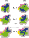
Representative motor maps from SRC and URC animals after 3, 7, and 10 d of training. SRC animals exhibited a significant increase in the proportion of the CFA occupied by distal movement (green) representations in comparison with URC animals after 10 d of training. URC animals had a significantly greater proportion of CFA occupied by proximal movement (blue) than SRC animals after 10 d of training. Vibrissae representations are shown in purple, head/neck representations are shown in yellow, and nonresponse sites are shown in gray. Bregma is indicated by a red line.
Figure 3.
Left, Total area of the CFA in SRC and URC animals in the three different training schedules. The mean percentage of the CFA ± SEM occupied by distal (middle) and proximal (right) forelimb movement representations. SRC animals had a significantly greater proportion of the CFA occupied by distal movement representations than the URC animals after 10 d of training (*p < 0.05; Fisher's PLSD). Conversely, URC animals had a significantly greater proportion of the CFA occupied by proximal movement representations than SRC animals after 10 d (*p < 0.05; PLSD).
Figure 4.
Neuron density (Nvneuron) (A), synapse density (Nvsynapse) (B), and number of synapses per neuron (Syn/Neuron) (C) within layer V of the CFA. SRC animals had a significantly lower density of neurons than URC animals after 7 and 10 d of training (*p < 0.05; Fisher's PLSD). No significant differences in synapse density were found between SRC and URC animals in any of the three training schedules. SRC animals had significantly more synapses per neuron than URC animals after 7 and 10 d of training (*p < 0.05; Fisher's PLSD).
Similar articles
- Functional reorganization of the rat motor cortex following motor skill learning.
Kleim JA, Barbay S, Nudo RJ. Kleim JA, et al. J Neurophysiol. 1998 Dec;80(6):3321-5. doi: 10.1152/jn.1998.80.6.3321. J Neurophysiol. 1998. PMID: 9862925 - Motor learning-dependent synaptogenesis is localized to functionally reorganized motor cortex.
Kleim JA, Barbay S, Cooper NR, Hogg TM, Reidel CN, Remple MS, Nudo RJ. Kleim JA, et al. Neurobiol Learn Mem. 2002 Jan;77(1):63-77. doi: 10.1006/nlme.2000.4004. Neurobiol Learn Mem. 2002. PMID: 11749086 - Sensitivity of cortical movement representations to motor experience: evidence that skill learning but not strength training induces cortical reorganization.
Remple MS, Bruneau RM, VandenBerg PM, Goertzen C, Kleim JA. Remple MS, et al. Behav Brain Res. 2001 Sep 14;123(2):133-41. doi: 10.1016/s0166-4328(01)00199-1. Behav Brain Res. 2001. PMID: 11399326 - In search of the motor engram: motor map plasticity as a mechanism for encoding motor experience.
Monfils MH, Plautz EJ, Kleim JA. Monfils MH, et al. Neuroscientist. 2005 Oct;11(5):471-83. doi: 10.1177/1073858405278015. Neuroscientist. 2005. PMID: 16151047 Review. - Motor training induces experience-specific patterns of plasticity across motor cortex and spinal cord.
Adkins DL, Boychuk J, Remple MS, Kleim JA. Adkins DL, et al. J Appl Physiol (1985). 2006 Dec;101(6):1776-82. doi: 10.1152/japplphysiol.00515.2006. Epub 2006 Sep 7. J Appl Physiol (1985). 2006. PMID: 16959909 Review.
Cited by
- Maintaining a Dynamic Brain: A Review of Empirical Findings Describing the Roles of Exercise, Learning, and Environmental Enrichment in Neuroplasticity from 2017-2023.
Milbocker KA, Smith IF, Klintsova AY. Milbocker KA, et al. Brain Plast. 2024 May 14;9(1-2):75-95. doi: 10.3233/BPL-230151. eCollection 2024. Brain Plast. 2024. PMID: 38993580 Free PMC article. Review. - Small, correlated changes in synaptic connectivity may facilitate rapid motor learning.
Feulner B, Perich MG, Chowdhury RH, Miller LE, Gallego JA, Clopath C. Feulner B, et al. Nat Commun. 2022 Sep 2;13(1):5163. doi: 10.1038/s41467-022-32646-w. Nat Commun. 2022. PMID: 36056006 Free PMC article. - Transspinal direct current stimulation immediately modifies motor cortex sensorimotor maps.
Song W, Truong DQ, Bikson M, Martin JH. Song W, et al. J Neurophysiol. 2015 Apr 1;113(7):2801-11. doi: 10.1152/jn.00784.2014. Epub 2015 Feb 11. J Neurophysiol. 2015. PMID: 25673738 Free PMC article. - Changes in corticospinal excitability during reach adaptation in force fields.
Orban de Xivry JJ, Ahmadi-Pajouh MA, Harran MD, Salimpour Y, Shadmehr R. Orban de Xivry JJ, et al. J Neurophysiol. 2013 Jan;109(1):124-36. doi: 10.1152/jn.00785.2012. Epub 2012 Oct 3. J Neurophysiol. 2013. PMID: 23034365 Free PMC article. - Cortical Motor Circuits after Piano Training in Adulthood: Neurophysiologic Evidence.
Houdayer E, Cursi M, Nuara A, Zanini S, Gatti R, Comi G, Leocani L. Houdayer E, et al. PLoS One. 2016 Jun 16;11(6):e0157526. doi: 10.1371/journal.pone.0157526. eCollection 2016. PLoS One. 2016. PMID: 27309353 Free PMC article.
References
- Anderson BJ, Li X, Alcantara AA, Isaacs KR, Black JE, Greenough WT (1994) Glial hypertrophy is associated with synaptogenesis following motor-skill learning, but not with angiogenesis following exercise. Glia 11: 73-80. - PubMed
- Classen J, Liepert J, Wise SP, Hallett M, Cohen LG (1998) Rapid plasticity of human cortical movement representation induced by practice. J Neurophysiol 79: 1117-1123. - PubMed
- Clements MP, Rose SP (1996) Time-dependent increase in release of arachidonic acid following passive avoidance training in the day-old chick. J Neurochem 67: 1317-1323. - PubMed
Publication types
MeSH terms
LinkOut - more resources
Full Text Sources
Miscellaneous


