HER-2 gene amplification can be acquired as breast cancer progresses - PubMed (original) (raw)
. 2004 Jun 22;101(25):9393-8.
doi: 10.1073/pnas.0402993101. Epub 2004 Jun 11.
Debasish Tripathy, Sanjay Shete, Raheela Ashfaq, Barbara Haley, Steve Perkins, Peter Beitsch, Amanullah Khan, David Euhus, Cynthia Osborne, Eugene Frenkel, Susan Hoover, Marilyn Leitch, Edward Clifford, Ellen Vitetta, Larry Morrison, Dorothee Herlyn, Leon W M M Terstappen, Timothy Fleming, Tanja Fehm, Thomas Tucker, Nancy Lane, Jianqiang Wang, Jonathan Uhr
Affiliations
- PMID: 15194824
- PMCID: PMC438987
- DOI: 10.1073/pnas.0402993101
HER-2 gene amplification can be acquired as breast cancer progresses
Songdong Meng et al. Proc Natl Acad Sci U S A. 2004.
Abstract
Amplification and overexpression of the HER-2 oncogene in breast cancer is felt to be stable over the course of disease and concordant between primary tumor and metastases. Therefore, patients with HER-2-negative primary tumors rarely will receive anti-Her-2 antibody (trastuzumab, Herceptin) therapy. A very sensitive blood test was used to capture circulating tumor cells (CTCs) and evaluate their HER-2 gene status by fluorescence in situ hybridization. The HER-2 status of the primary tumor and corresponding CTCs in 31 patients showed 97% agreement, with no false positives. In 10 patients with HER-2-positive tumors, the HER-2/chromosome enumerator probe 17 ratio in each tumor was about twice that of the corresponding CTCs (mean 6.64 +/- 2.72 vs. 2.8 +/- 0.6). Hence, the ratio of the CTCs is a reliable surrogate marker for the expected high ratio in the primary tumor. Her-2 protein expression of 10 CTCs was sufficient to make a definitive diagnosis of the HER-2 gene status of the whole population of CTCs in 19 patients with recurrent breast cancer. Nine of 24 breast cancer patients whose primary tumor was HER-2-negative each acquired HER-2 gene amplification in their CTCs during cancer progression, i.e., 37.5% (95% confidence interval of 18.8-59.4%). Four of the 9 patients were treated with Herceptin-containing therapy. One had a complete response and 2 had a partial response.
Figures
Fig. 1.
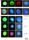
Fluorescent microscopic images of cytomorphology, immunophenotype, and FISH evaluation of CTCs of patients with metastatic breast cancer. (A) Decomposing the immunophenotype and displaying aneuploidy of a CTC from a metastatic breast patient. Single-band filters were used to block out the fluorescence of one or more fluorochromes used to stain for CTCs. Horizontally, the photos show the same cell stained for the combination of CK (green), mam (red), and nucleic acid (blue); CK only; mam only; and FISH analysis. For FISH, CEP 1 (SpectrumOrange), CEP 8 (SpectrumAqua), and CEP 17 (SpectrumGreen) were used (counts were 5, 2, and 5, respectively). The photos are taken in only one _Z_-plane, whereas the microscopist can focus on the entire range of _Z_-planes; therefore, spots that are not seen or overlap in the single _Z_-plane of the photo can be distinguished by microscopy. (B) Sequential genotyping of a CTC isolated from blood of a metastatic breast cancer patient. First photo, epithelial cell detected by an anti-CK-antibody (green) and nucleic acid (blue). Second photo, same cell hybridized with a locus-specific probe for c-myc (orange) and CEP 8 (aqua) (7, 3). Third photo, same cell hybridized with Her-2 (orange), CEP 10 (green), and CEP 17 (aqua) (6, 2, and 3). Fourth photo, same cell hybridized with CEP 1 (orange), CEP 8 (aqua), and CEP 17 (green) (6, 3, and 2). (C) Comparison of the intensity of Her-2 IF staining with HER-2 gene amplification of CTCs from metastatic breast cancer. Three rows representing Her-2 IF staining intensity of a single cell of 1+ (first row), 2+ (second row) and 3+ (third row) are shown. Photo 1 of each row shows staining for the combination of CK, Her-2, and nucleic acid. Photo 2 shows IF staining for Her-2 and nucleic acid. Photo 3 shows FISH results for HER-2 (SpectrumOrange) and CEP 17 (SpectrumGreen). Her-2 IF staining intensity (IFI) and HER-2/CEP 17 ratio (Ratio) for each cell are shown.
Similar articles
- Patterns of her-2/neu amplification and overexpression in primary and metastatic breast cancer.
Simon R, Nocito A, Hübscher T, Bucher C, Torhorst J, Schraml P, Bubendorf L, Mihatsch MM, Moch H, Wilber K, Schötzau A, Kononen J, Sauter G. Simon R, et al. J Natl Cancer Inst. 2001 Aug 1;93(15):1141-6. doi: 10.1093/jnci/93.15.1141. J Natl Cancer Inst. 2001. PMID: 11481385 - Predicting the efficacy of trastuzumab-based therapy in breast cancer: current standards and future strategies.
Singer CF, Köstler WJ, Hudelist G. Singer CF, et al. Biochim Biophys Acta. 2008 Dec;1786(2):105-13. doi: 10.1016/j.bbcan.2008.02.003. Epub 2008 Mar 4. Biochim Biophys Acta. 2008. PMID: 18375208 Review. - Discrepancies in clinical laboratory testing of eligibility for trastuzumab therapy: apparent immunohistochemical false-positives do not get the message.
Tubbs RR, Pettay JD, Roche PC, Stoler MH, Jenkins RB, Grogan TM. Tubbs RR, et al. J Clin Oncol. 2001 May 15;19(10):2714-21. doi: 10.1200/JCO.2001.19.10.2714. J Clin Oncol. 2001. PMID: 11352964 - HER2 testing in patients with breast cancer: poor correlation between weak positivity by immunohistochemistry and gene amplification by fluorescence in situ hybridization.
Perez EA, Roche PC, Jenkins RB, Reynolds CA, Halling KC, Ingle JN, Wold LE. Perez EA, et al. Mayo Clin Proc. 2002 Feb;77(2):148-54. doi: 10.4065/77.2.148. Mayo Clin Proc. 2002. PMID: 11838648 - The Her-2/neu gene and protein in breast cancer 2003: biomarker and target of therapy.
Ross JS, Fletcher JA, Linette GP, Stec J, Clark E, Ayers M, Symmans WF, Pusztai L, Bloom KJ. Ross JS, et al. Oncologist. 2003;8(4):307-25. doi: 10.1634/theoncologist.8-4-307. Oncologist. 2003. PMID: 12897328 Review.
Cited by
- Molecular profiling of individual tumor cells by hyperspectral microscopic imaging.
Uhr JW, Huebschman ML, Frenkel EP, Lane NL, Ashfaq R, Liu H, Rana DR, Cheng L, Lin AT, Hughes GA, Zhang XJ, Garner HR. Uhr JW, et al. Transl Res. 2012 May;159(5):366-75. doi: 10.1016/j.trsl.2011.08.003. Epub 2011 Sep 3. Transl Res. 2012. PMID: 22500509 Free PMC article. - Prognostic Impact of HER2 and ER Status of Circulating Tumor Cells in Metastatic Breast Cancer Patients with a HER2-Negative Primary Tumor.
Beije N, Onstenk W, Kraan J, Sieuwerts AM, Hamberg P, Dirix LY, Brouwer A, de Jongh FE, Jager A, Seynaeve CM, Van NM, Foekens JA, Martens JW, Sleijfer S. Beije N, et al. Neoplasia. 2016 Nov;18(11):647-653. doi: 10.1016/j.neo.2016.08.007. Epub 2016 Oct 17. Neoplasia. 2016. PMID: 27764697 Free PMC article. - Persistence of disseminated tumor cells after neoadjuvant treatment for locally advanced breast cancer predicts poor survival.
Mathiesen RR, Borgen E, Renolen A, Løkkevik E, Nesland JM, Anker G, Ostenstad B, Lundgren S, Risberg T, Mjaaland I, Kvalheim G, Lønning PE, Naume B. Mathiesen RR, et al. Breast Cancer Res. 2012 Aug 14;14(4):R117. doi: 10.1186/bcr3242. Breast Cancer Res. 2012. PMID: 22889108 Free PMC article. - Clinical application of circulating tumor cells in breast cancer: overview of the current interventional trials.
Bidard FC, Fehm T, Ignatiadis M, Smerage JB, Alix-Panabières C, Janni W, Messina C, Paoletti C, Müller V, Hayes DF, Piccart M, Pierga JY. Bidard FC, et al. Cancer Metastasis Rev. 2013 Jun;32(1-2):179-88. doi: 10.1007/s10555-012-9398-0. Cancer Metastasis Rev. 2013. PMID: 23129208 Free PMC article. Review. - Mutation analysis of BRAF and KIT in circulating melanoma cells at the single cell level.
Sakaizawa K, Goto Y, Kiniwa Y, Uchiyama A, Harada K, Shimada S, Saida T, Ferrone S, Takata M, Uhara H, Okuyama R. Sakaizawa K, et al. Br J Cancer. 2012 Feb 28;106(5):939-46. doi: 10.1038/bjc.2012.12. Epub 2012 Jan 26. Br J Cancer. 2012. PMID: 22281663 Free PMC article.
References
- Slamon, D. J., Godolphin, W., Jones, L. A., Holt, J. A., Wong, S. G., Keith, D. E., Levin, W. J., Stuart, S. G., Udove, J., Ullrich, A., et al. (1989) Science 244, 707-712. - PubMed
- Ross, J. S. & Gray, G. S. (2003) Clin. Leadersh. Manag. Rev. 17, 333-340. - PubMed
- Vogel, C., Cobleigh, M. A., Tripathy, D., Gutheil, J. C., Harris, L. N., Fehrenbacher, L., Slamon, D. J., Murphy, M., Novotny, W. F., Burchmore, M., et al. (2001) Eur. J. Cancer 37, 25-29. - PubMed
- Slamon, D. J., Leyland-Jones, B., Shak, S., Fuchs, H., Paton, V., Bajamonde, A., Fleming, T., Eiermann, W., Wolter, J., Pegram, M., et al. (2001) N. Engl. J. Med. 344, 783-792. - PubMed
- Fehm, T., Sagalowsky, A. I., Clifford, E., Beitsch, P. D., Saboorian, H., Euhus, D., Meng, S., Morrison, L., Tucker, T., Lane, N., et al. (2002) Clin. Cancer Res. 8, 2073-2084. - PubMed
Publication types
MeSH terms
Substances
LinkOut - more resources
Full Text Sources
Other Literature Sources
Medical
Research Materials
Miscellaneous
