Mice lacking the type I interferon receptor are resistant to Listeria monocytogenes - PubMed (original) (raw)
Comparative Study
. 2004 Aug 16;200(4):527-33.
doi: 10.1084/jem.20040976. Epub 2004 Aug 9.
Affiliations
- PMID: 15302899
- PMCID: PMC2211930
- DOI: 10.1084/jem.20040976
Comparative Study
Mice lacking the type I interferon receptor are resistant to Listeria monocytogenes
Victoria Auerbuch et al. J Exp Med. 2004.
Abstract
Listeria monocytogenes is a facultative intracellular pathogen that induces a cytosolic signaling cascade resulting in expression of interferon (IFN)-beta. Although type I IFNs are critical in viral defense, their role in immunity to bacterial pathogens is much less clear. In this study, we addressed the role of type I IFNs by examining the infection of L. monocytogenes in BALB/c mice lacking the type I IFN receptor (IFN-alpha/betaR-/-). During the first 24 h of infection in vivo, IFN-alpha/betaR-/- and wild-type mice were similar in terms of L. monocytogenes survival. In addition, the intracellular fate of L. monocytogenes in macrophages cultured from IFN-alpha/betaR-/- and wild-type mice was indistinguishable. However, by 72 h after inoculation in vivo, IFN-alpha/betaR-/- mice were approximately 1,000-fold more resistant to a high dose L. monocytogenes infection. Resistance was correlated with elevated levels of interleukin 12p70 in the blood and increased numbers of CD11b+ macrophages producing tumor necrosis factor alpha in the spleen of IFN-alpha/betaR-/- mice. The results of this study suggest that L. monocytogenes might be exploiting an innate antiviral response to promote its pathogenesis.
Figures
Figure 1.
L. monocytogenes infection of BALB/c and IFN-α/βR−/− mice. Naive BALB/c and IFN-α/βR−/− mice (three per group) were infected with 2 × 10⁴ wild-type L. monocytogenes and bacterial growth in the spleen (A) and liver (B) was followed over 3 d. (C) BALB/c and IFN-α/βR−/− mice were immunized with 2 × 10³ L. monocytogenes 3–4 wk before challenge with 4 × 10⁴ bacteria. Naive animals were infected with 4 × 10⁴ bacteria. Dashed line denotes limit of detection. CFUs from the spleen 48 h after inoculation are shown. Data represents two separate experiments.
Figure 2.
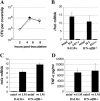
IFN-α/βR−/− BMDMs support normal L. monocytogenes growth. BALB/c and IFN-α/βR−/− BMDMs were (A) infected with wild-type L. monocytogenes and bacterial growth was monitored in the presence of 50 μg/ml gentamicin. Black diamonds, BALB/c; gray diamonds, IFN-α/βR−/−. BMDMs were infected with an MOI of 10:1 wild-type or 100:1 Δ_hly L. monocytogenes_, which does not enter the host cytosol and does not grow intracellularly, and ifnb (B) or tnfa (C) mRNA levels (normalized to β actin) and TNF-α supernatant protein levels (D) were quantified 6 h after inoculation. All experiments were performed at least twice.
Figure 3.
Elevated IL-12p70 and TNF-α, but not IFN-γ, in the absence of type I IFN signaling. (A) BALB/c and IFN-α/βR−/− mice were infected with 2.5 × 10⁴ wild-type L. monocytogenes, bled 24 h after inoculation, and serum was assayed for cytokine levels. Data represents the average from seven mice per group over two separate experiments ± SEM. Splenocytes from uninfected BALB/c and IFN-α/βR−/− mice (B) or from mice infected 48 h previously with 2 × 10⁴ L. monocytogenes (C) were cultured ± 4 × 10⁶ HKLM/ml for 18 h. Culture supernatants were assayed for TNF-α. Data represent the average from six mice per group over two separate experiments ± SEM.
Figure 4.
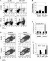
Increased number of CD11b+ TNF-α–producing cells in IFN-α/βR−/− mice. Splenocytes from uninfected (uninf) or _L. monocytogenes_–infected (inf) BALB/c and IFN-α/βR−/− mice were cultured ± 4 × 10⁶ HKLM/ml for 4 h. The absolute number of splenocytes was similar between BALB/c and IFN-α/βR−/− mice. Cells were stained for cell surface markers and intracellular TNF-α and Mac-3. (A–C) Cells were stained for CD11b, CD3e, and TNF-α, gated on CD3e− events, and expression of CD11b and TNF-α was analyzed. Data represents results from six mice per group over two separate experiments. (A) Representative dot plots from infected mouse splenocytes. Average percentage of total splenocytes ± SEM that are TNF-α–producing CD11b− (top left) and CD11b+ (top right) cells are shown. (B) Average percentage of total splenocytes that are CD11b+/CD3e−. (C) Average percentage of total splenocytes stimulated with 4 × 10⁶ HKLM/ml that are CD11b+/TNF-α+ or CD11b−/TNF-α+. (D and E) Cells stimulated with 4 × 10⁶ HKLM/ml were stained for CD11b, Mac-3, and TNF-α. (D) Representative dot plots from one of three mice per group. Values shown are average percentage of total splenocytes ± SEM residing within the R1 and R2 populations. (E) Cells within the R1 and R2 gates were analyzed for intracellular TNF-α staining. Average percentage of total splenocytes ± SEM that are CD11bint/Mac-3high/TNF-α+ (R1) or CD11bhigh/Mac-3int/high/TNF-α+ (R2). Statistical significance was determined by using the Mann-Whitney nonparametric test. *, P = 0.081; **, P = 0.005.
Similar articles
- Assessment of interleukin-12, gamma interferon, and tumor necrosis factor alpha secretion in sera from mice fed with dietary lipids during different stages of Listeria monocytogenes infection.
Puertollano MA, Cruz-Chamorro L, Puertollano E, Pérez-Toscano MT, Alvarez de Cienfuegos G, de Pablo MA. Puertollano MA, et al. Clin Diagn Lab Immunol. 2005 Sep;12(9):1098-103. doi: 10.1128/CDLI.12.9.1098-1103.2005. Clin Diagn Lab Immunol. 2005. PMID: 16148177 Free PMC article. - Type I IFN Does Not Promote Susceptibility to Foodborne Listeria monocytogenes.
Pitts MG, Myers-Morales T, D'Orazio SE. Pitts MG, et al. J Immunol. 2016 Apr 1;196(7):3109-16. doi: 10.4049/jimmunol.1502192. Epub 2016 Feb 19. J Immunol. 2016. PMID: 26895837 Free PMC article. - Type I interferon production enhances susceptibility to Listeria monocytogenes infection.
O'Connell RM, Saha SK, Vaidya SA, Bruhn KW, Miranda GA, Zarnegar B, Perry AK, Nguyen BO, Lane TF, Taniguchi T, Miller JF, Cheng G. O'Connell RM, et al. J Exp Med. 2004 Aug 16;200(4):437-45. doi: 10.1084/jem.20040712. Epub 2004 Aug 9. J Exp Med. 2004. PMID: 15302901 Free PMC article. - Innate immunity to a facultative intracellular bacterial pathogen.
Portnoy DA. Portnoy DA. Curr Opin Immunol. 1992 Feb;4(1):20-4. doi: 10.1016/0952-7915(92)90118-x. Curr Opin Immunol. 1992. PMID: 1596365 Review. - Confounding roles for type I interferons during bacterial and viral pathogenesis.
Carrero JA. Carrero JA. Int Immunol. 2013 Dec;25(12):663-9. doi: 10.1093/intimm/dxt050. Epub 2013 Oct 24. Int Immunol. 2013. PMID: 24158954 Free PMC article. Review.
Cited by
- Innate IFN-γ is essential for programmed death ligand-1-mediated T cell stimulation following Listeria monocytogenes infection.
Rowe JH, Ertelt JM, Way SS. Rowe JH, et al. J Immunol. 2012 Jul 15;189(2):876-84. doi: 10.4049/jimmunol.1103227. Epub 2012 Jun 18. J Immunol. 2012. PMID: 22711893 Free PMC article. - CD169+ macrophages orchestrate plasmacytoid dendritic cell arrest and retention for optimal priming in the bone marrow of malaria-infected mice.
Moore-Fried J, Paul M, Jing Z, Fooksman D, Lauvau G. Moore-Fried J, et al. Elife. 2022 Oct 24;11:e78873. doi: 10.7554/eLife.78873. Elife. 2022. PMID: 36278864 Free PMC article. - Age-dependent differences in systemic and cell-autonomous immunity to L. monocytogenes.
Sherrid AM, Kollmann TR. Sherrid AM, et al. Clin Dev Immunol. 2013;2013:917198. doi: 10.1155/2013/917198. Epub 2013 Apr 7. Clin Dev Immunol. 2013. PMID: 23653659 Free PMC article. Review. - Induction of PGRN by influenza virus inhibits the antiviral immune responses through downregulation of type I interferons signaling.
Wei F, Jiang Z, Sun H, Pu J, Sun Y, Wang M, Tong Q, Bi Y, Ma X, Gao GF, Liu J. Wei F, et al. PLoS Pathog. 2019 Oct 4;15(10):e1008062. doi: 10.1371/journal.ppat.1008062. eCollection 2019 Oct. PLoS Pathog. 2019. PMID: 31585000 Free PMC article. - Absent in melanoma 2 is required for innate immune recognition of Francisella tularensis.
Jones JW, Kayagaki N, Broz P, Henry T, Newton K, O'Rourke K, Chan S, Dong J, Qu Y, Roose-Girma M, Dixit VM, Monack DM. Jones JW, et al. Proc Natl Acad Sci U S A. 2010 May 25;107(21):9771-6. doi: 10.1073/pnas.1003738107. Epub 2010 May 10. Proc Natl Acad Sci U S A. 2010. PMID: 20457908 Free PMC article.
References
- Taki, S. 2002. Type I interferons and autoimmunity: lessons from the clinic and from IRF-2-deficient mice. Cytokine Growth Factor Rev. 13:379–391. - PubMed
- Toshchakov, V., B.W. Jones, A. Lentschat, A. Silva, P.Y. Perera, K. Thomas, M.J. Cody, S. Zhang, B.R. Williams, J. Major, et al. 2003. TLR2 and TLR4 agonists stimulate unique repertoires of host resistance genes in murine macrophages: interferon-beta-dependent signaling in TLR4-mediated responses. J. Endotoxin Res. 9:169–175. - PubMed
- van den Broek, M.F., U. Müller, S. Huang, R.M. Zinkernagel, and M. Aguet. 1995. Immune defence in mice lacking type I and/or type II interferon receptors. Immunol. Rev. 148:5–18. - PubMed
Publication types
MeSH terms
Substances
Grants and funding
- R01 AI027655/AI/NIAID NIH HHS/United States
- R37 AI029619/AI/NIAID NIH HHS/United States
- R01 AI27655/AI/NIAID NIH HHS/United States
- R01 AI29619/AI/NIAID NIH HHS/United States
LinkOut - more resources
Full Text Sources
Other Literature Sources
Medical
Molecular Biology Databases
Research Materials


