Expression of constitutively nuclear cyclin D1 in murine lymphocytes induces B-cell lymphoma - PubMed (original) (raw)
Expression of constitutively nuclear cyclin D1 in murine lymphocytes induces B-cell lymphoma
A B Gladden et al. Oncogene. 2006.
Abstract
Mantle cell lymphoma (MCL) is a B-cell lymphoma characterized by overexpression of cyclin D1 due to the t(11;14) chromosomal translocation. While expression of cyclin D1 correlates with MCL development, expression of wild-type (WT) cyclin D1 transgene in murine lymphocytes is unable to drive B-cell lymphoma. As cyclin D1 mutants that are refractory to nuclear export display heighten oncogenicity in vitro compared with WT D1, we generated mice expressing FLAG-D1/T286A, a constitutively nuclear mutant, under the control of the immunoglobulin enhancer, Emu. D1/T286A transgenic mice universally develop a mature B-cell lymphoma. Expression of D1/T286A in B lymphocytes results in S phase entry in resting lymphocytes and increased apoptosis in spleens of young premalignant mice. Lymphoma onset correlates with perturbations in p53/MDM2/p19Arf expression and with BcL-2 overexpression suggesting that alterations in one or both of these pathways may contribute to lymphoma development. Our results describe a cyclin D1-driven model of B-cell lymphomagenesis and provide evidence that nuclear-retention of cyclin D1 is oncogenic in vivo.
Figures
Figure 1. D1/T286A associates with CDK4 and p27 in transgenic splenocytes
a) Cyclin D1 direct Western of whole tissue protein lysates from either 6-week-old non-transgenic wild type (WT) or Eµ-D1/T286A (Eµ) mice. L.E. denotes longer exposure of the Western blot. b) Western analysis of protein lysates from NIH-3T3 cells (3T3), wild type spleens (WT), D1/T286A spleens (Eµ) or two human MCL cell lines performed with antibodies specific for β- Tubulin, human and mouse cyclin D1 (DCS6) or mouse D1 only (7213G). A non-specific background band recognized by the DCS6 antibody is noted by the (*). c) Western analysis with the indicated antibodies was performed on splenic extracts prepared from non-transgenic wild type (WT), D1/T286A (Eµ) mice, or NIH-3T3 fibroblasts. d) Protein lysates from (c) were precipitated using either a cyclin D1 antibody (D1), the M2 antibody (FLAG), or with normal rabbit serum (NRS). Proteins were separated by electrophoresis and probed with the indicated antibodies.
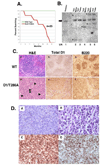
Figure 2. D1/T286A mice have a decreased lifespan due to the onset of B-cell lymphoma
a) Survival curve representing mean survival of either wild type (WT) (Green Line) or D1/T286A (Eµ) (Red Line) cohort over a 24-month period. b) Southern blot analysis of immunoglobulin heavy chain gene rearrangements in transgenic splenic tumors and available peripheral tumors from the identical mouse. Bold line represents the germline size of the IgH gene and (*) notes rearranged gene products. c) Representative histology of either wild type spleen (a, b, c) or a tumor burden D1/T286A spleen (d, e, f) stained with hematoxylin and eosin (a, d) with arrowheads denoting mitotic figures. Immunohistochemistry for either total cyclin D1 (b, e) or B220 (c, f). d) Higher magnification of D1/T286A tumor burden spleens stained with hematoxylin and eosin (A,B), for cyclin D1 (C) or B220 (D).
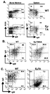
Figure 3. D1/T286A splenic B-cells display an altered surface immunoglobulin profile
a) Bone marrow cells from either non-transgenic wild type (WT) or D1/T286A (Eµ-TA) six-week old mice were analyzed by flow cytometry with antibodies specific for either B220 or surface IgM. Likewise total splenic lymphocytes from either wild type or Eµ-D1/T286A mice were stained with B220, surface IgD and surface IgM. B220 positive cells were gated on and surface IgM and IgD expression was analyzed. b) Splenic cells from six-week-old pre-malignant mice were stained with B220, surface IgM and IgD, B220 cells were gated on and surface immunoglobulin levels were analyzed. c) Cells from a tumor burden spleen (EµTA) or an age-matched mouse were stained with B220, surface IgM or IgD. B220 positive cells were gated on and surface immunoglobulin levels were analyzed by flow cytometry.
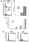
Figure 4. D1/T286A B-cells have a decreased dependence on mitogen stimulation
a) Purified splenic B-cells from six-week old wild type or D1/T286A mice were stained with propidium iodide and DNA content was determined by flow cytometry. b) Six-week-old wild type or D1/T286A mice were injected with BrdU 24 hours prior to sacrifice. BrdU incorporation was quantitated by ELISA from purified splenic B or thymic T-cells. c) Purified splenic B-cells from pre-malignant transgenic (Eµ) and age matched wild type (WT) mice were stimulated with the indicated concentrations of LPS for 48 hrs. Proliferation was determined by incorporation of 3H-Thymidine. d) Cells from a tumor burden spleen (Eµ) or an age-matched mouse were stained with propidium iodide and DNA content was determined by flow cytometry.
Figure 5. D1/T286A transgenic spleens display an increased proportion of apoptotic cells
Representative TUNEL analysis performed on a) non-transgenic spleens, b) age matched pre-malignant D1/T286A spleens, and c) tumors from malignant transgenic mice. d) Quantitation of TUNEL analysis.
Figure 6. The p53 pathway is targeted D1/T286A tumors
a) Direct western analysis was performed on protein lysates from either wild type spleen (WT) or D1/T286A (TA) tumors using antibodies specific for MDM2 (top panel) (*) denotes cross reacting band, p53 (middle panel) or p19ARF (bottom panel). b) Total protein lysates from wild type spleens or D1/T286A tumors were separated electrophoretically, transferred to nitrocellulose and probed with antibodies for either BcL-2 (top panel) or BcL-XL (bottom panel).
Similar articles
- A defect of the INK4-Cdk4 checkpoint and Myc collaborate in blastoid mantle cell lymphoma-like lymphoma formation in mice.
Vincent-Fabert C, Fiancette R, Rouaud P, Baudet C, Truffinet V, Magnone V, Guillaudeau A, Cogné M, Dubus P, Denizot Y. Vincent-Fabert C, et al. Am J Pathol. 2012 Apr;180(4):1688-701. doi: 10.1016/j.ajpath.2012.01.004. Epub 2012 Feb 9. Am J Pathol. 2012. PMID: 22326754 - Detection of cyclin D1 overexpression by real-time reverse-transcriptase-mediated quantitative polymerase chain reaction for the diagnosis of mantle cell lymphoma.
Suzuki R, Takemura K, Tsutsumi M, Nakamura S, Hamajima N, Seto M. Suzuki R, et al. Am J Pathol. 2001 Aug;159(2):425-9. doi: 10.1016/S0002-9440(10)61713-0. Am J Pathol. 2001. PMID: 11485900 Free PMC article. - Flipping the cyclin D1 switch in mantle cell lymphoma.
Hasanali Z, Sharma K, Epner E. Hasanali Z, et al. Best Pract Res Clin Haematol. 2012 Jun;25(2):143-52. doi: 10.1016/j.beha.2012.03.001. Epub 2012 May 4. Best Pract Res Clin Haematol. 2012. PMID: 22687450 Review. - Cyclin D1 expression in B-cell lymphomas.
Gladkikh A, Potashnikova D, Korneva E, Khudoleeva O, Vorobjev I. Gladkikh A, et al. Exp Hematol. 2010 Nov;38(11):1047-57. doi: 10.1016/j.exphem.2010.08.002. Epub 2010 Aug 18. Exp Hematol. 2010. PMID: 20727381 - Pathogenesis of mantle-cell lymphoma: all oncogenic roads lead to dysregulation of cell cycle and DNA damage response pathways.
Fernàndez V, Hartmann E, Ott G, Campo E, Rosenwald A. Fernàndez V, et al. J Clin Oncol. 2005 Sep 10;23(26):6364-9. doi: 10.1200/JCO.2005.05.019. J Clin Oncol. 2005. PMID: 16155021 Review.
Cited by
- Cyclin D1, cancer progression, and opportunities in cancer treatment.
Qie S, Diehl JA. Qie S, et al. J Mol Med (Berl). 2016 Dec;94(12):1313-1326. doi: 10.1007/s00109-016-1475-3. Epub 2016 Oct 2. J Mol Med (Berl). 2016. PMID: 27695879 Free PMC article. Review. - In the wrong place at the wrong time: does cyclin mislocalization drive oncogenic transformation?
Moore JD. Moore JD. Nat Rev Cancer. 2013 Mar;13(3):201-8. doi: 10.1038/nrc3468. Epub 2013 Feb 7. Nat Rev Cancer. 2013. PMID: 23388618 Review. - Development of a murine model for blastoid variant mantle-cell lymphoma.
Ford RJ, Shen L, Lin-Lee YC, Pham LV, Multani A, Zhou HJ, Tamayo AT, Zhang C, Hawthorn L, Cowell JK, Ambrus JL Jr. Ford RJ, et al. Blood. 2007 Jun 1;109(11):4899-906. doi: 10.1182/blood-2006-08-038497. Epub 2007 Feb 20. Blood. 2007. PMID: 17311992 Free PMC article. - The cyclin D1b splice variant: an old oncogene learns new tricks.
Knudsen KE. Knudsen KE. Cell Div. 2006 Jul 24;1:15. doi: 10.1186/1747-1028-1-15. Cell Div. 2006. PMID: 16863592 Free PMC article. - Mantle cell lymphoma cells express predominantly cyclin D1a isoform and are highly sensitive to selective inhibition of CDK4 kinase activity.
Marzec M, Kasprzycka M, Lai R, Gladden AB, Wlodarski P, Tomczak E, Nowell P, Deprimo SE, Sadis S, Eck S, Schuster SJ, Diehl JA, Wasik MA. Marzec M, et al. Blood. 2006 Sep 1;108(5):1744-50. doi: 10.1182/blood-2006-04-016634. Epub 2006 May 11. Blood. 2006. PMID: 16690963 Free PMC article.
References
- Adams JM, Harris AW, Pinkert CA, Corcoran LM, Alexander WS, Cory S, Palmiter RD, Brinster RL. Nature. 1985;318:533–538. - PubMed
- Akiyama N, Tsuruta H, Sasaki H, Sakamoto H, Hamaguchi M, Ohmura Y, Seto M, Ueda R, Hirai H, Yazaki Y, et al. Cancer Res. 1994;54:377–379. - PubMed
- Argatoff LH, Connors JM, Klasa RJ, Horsman DE, Gascoyne RD. Blood. 1997;89:2067–2078. - PubMed
Publication types
MeSH terms
Substances
Grants and funding
- R01 CA093237/CA/NCI NIH HHS/United States
- R01 CA093237-05/CA/NCI NIH HHS/United States
- CA93237/CA/NCI NIH HHS/United States
- R01 CA089194/CA/NCI NIH HHS/United States
- CA89194/CA/NCI NIH HHS/United States
LinkOut - more resources
Full Text Sources
Molecular Biology Databases
Research Materials
Miscellaneous


