Two internal ribosome entry sites mediate the translation of p53 isoforms - PubMed (original) (raw)
Two internal ribosome entry sites mediate the translation of p53 isoforms
Partho Sarothi Ray et al. EMBO Rep. 2006 Apr.
Abstract
The p53 tumour suppressor protein has a crucial role in cell-cycle arrest and apoptosis. Previous reports show that the p53 messenger RNA is translated to produce an amino-terminal-deleted isoform (DeltaN-p53) from an internal initiation codon, which acts as a dominant-negative inhibitor of full-length p53. Here, we show that two internal ribosome entry sites (IRESs) mediate the translation of both full-length and DeltaN-p53 isoforms. The IRES directing the translation of full-length p53 is in the 5'-untranslated region of the mRNA, whereas the IRES mediating the translation of DeltaN-p53 extends into the protein-coding region. The two IRESs show distinct cell-cycle phase-dependent activity, with the IRES for full-length p53 being active at the G2-M transition and the IRES for DeltaN-p53 showing highest activity at the G1-S transition. These results indicate a novel translational control of p53 gene expression and activity.
Figures
Figure 1
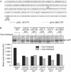
p53 5′-untranslated regions mediate cap-independent translation. (A) Nucleotide sequence of p53 5′-untranslated region (5′UTR) and part of the protein-coding region. The 134 nt canonical 5′UTR is underlined by a solid line, whereas the 251 nt 5′UTR for ΔN-p53 is underlined by a dotted line. (B) In vitro translation of capped and uncapped monocistronic RNAs in the presence or absence of exogenously added cap analogue. The band intensities were quantified and are represented graphically.
Figure 2
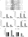
p53 5′-untranslated region sequences mediate internal ribosome entry site-dependent translation. (A) Schematic representation of bicistronic plasmids used in transient transfections. (B(i)) Transfection of bicistronic plasmids pRnullF, pRp53(+39)F and pRp53(−1)F containing a 264 nt unrelated sequence, p53 5′UTR (+39) and (−1) sequences, respectively, in the intercistronic space into HeLa cells. (ii) Transfection of bicistronic plasmids containing p53 5′UTR (+39) and (−1) downstream of the ΔEMCV sequence. The control bicistronic plasmid pRΔEF contained only the ΔEMCV sequence. Transfection efficiencies were normalized by co-transfecting with a β-galactosidase plasmid. The luciferase activities for Fluc and Rluc are shown separately as fold increase compared with that from control, taken as 1. (C(i,ii)) Transfection of the same set of bicistronic plasmids into H1299 cells. The data are represented as in (B). (D) Transfection of bicistronic plasmids containing p53 5′UTR (+39) or (−1) sequences in the intercistronic place and the ΔEMCV sequence upstream of Rluc into HeLa cells. The Fluc and Rluc activities from the ΔEMCV-containing plasmids are shown as fold increase or decrease with respect to the corresponding controls, taken as 100.
Figure 3
p53 bicistronic plasmid does not show splicing or cryptic promoter activity. (A) Reverse transcription–PCR analysis, using two sets of primers P1/P2 and P3/P4, of RNA extracted from pRp53(+39)F bicistronic plasmid-transfected (T) and untransfected (UT) HeLa cells. Lane 5 shows reverse transcriptase-negative control, whereas lanes 6 and 7 show PCR products amplified from the bicistronic DNA. (B) Northern blot of total RNA extracted from HeLa cells transfected with pRp53(+39)F (lane 1), pRp53(−1)F (lane 2), pR(coxsackievirusB3-IRES)F (lane 3) DNAs and _in vitro_-synthesized Fluc RNA (lane 4) using a 32P-labelled riboprobe corresponding to Fluc. (C) Transfection of bicistronic plasmids pBS-Rp53(+39)F and pBSRnullF lacking eukaryotic promoters into HeLa cells in the absence and presence of infection by VTF7-3. The luciferase activity values are indicated above the respective bars. (D) Transfection of HeLa cells with capped bicistronic RNAs containing the p53(+39) and (−1) 5′UTRs. Fluc/Rluc ratios are shown as fold increase compared with that from a control bicistronic RNA lacking p53 sequences.
Figure 4
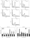
Cell cycle-dependent p53 internal ribosome entry site activity in G2/M synchronized cells. (A) Flow cytometric analysis of HeLa cells collected at different time points after being synchronized at G2/M phase by nocodazole treatment. The percentage of cells in S phase at each time point is indicated. (B) Luciferase assay of cells transfected with p53(+39) and (−1) bicistronic constructs and synchronized at G2/M phase at various time points after release. Fluc and Rluc activities at each time point are expressed as fold of the activity obtained from non-synchronized, transfected cells taken as control. The data mean±s.d. from three independent experiments.
Similar articles
- The translation initiation factor DAP5 promotes IRES-driven translation of p53 mRNA.
Weingarten-Gabbay S, Khan D, Liberman N, Yoffe Y, Bialik S, Das S, Oren M, Kimchi A. Weingarten-Gabbay S, et al. Oncogene. 2014 Jan 30;33(5):611-8. doi: 10.1038/onc.2012.626. Epub 2013 Jan 14. Oncogene. 2014. PMID: 23318444 - IRES mediated translational regulation of p53 isoforms.
Sharathchandra A, Katoch A, Das S. Sharathchandra A, et al. Wiley Interdiscip Rev RNA. 2014 Jan-Feb;5(1):131-9. doi: 10.1002/wrna.1202. Epub 2013 Nov 7. Wiley Interdiscip Rev RNA. 2014. PMID: 24343861 Review. - Effect of a natural mutation in the 5' untranslated region on the translational control of p53 mRNA.
Khan D, Sharathchandra A, Ponnuswamy A, Grover R, Das S. Khan D, et al. Oncogene. 2013 Aug 29;32(35):4148-59. doi: 10.1038/onc.2012.422. Epub 2012 Oct 1. Oncogene. 2013. PMID: 23027126 - Polypyrimidine tract binding protein regulates IRES-mediated translation of p53 isoforms.
Grover R, Ray PS, Das S. Grover R, et al. Cell Cycle. 2008 Jul 15;7(14):2189-98. doi: 10.4161/cc.7.14.6271. Epub 2008 May 11. Cell Cycle. 2008. PMID: 18635961 - p53 and little brother p53/47: linking IRES activities with protein functions.
Grover R, Candeias MM, Fåhraeus R, Das S. Grover R, et al. Oncogene. 2009 Jul 30;28(30):2766-72. doi: 10.1038/onc.2009.138. Epub 2009 Jun 1. Oncogene. 2009. PMID: 19483723 Review.
Cited by
- 5'-UTR recruitment of the translation initiation factor eIF4GI or DAP5 drives cap-independent translation of a subset of human mRNAs.
Haizel SA, Bhardwaj U, Gonzalez RL Jr, Mitra S, Goss DJ. Haizel SA, et al. J Biol Chem. 2020 Aug 14;295(33):11693-11706. doi: 10.1074/jbc.RA120.013678. Epub 2020 Jun 22. J Biol Chem. 2020. PMID: 32571876 Free PMC article. - The amyloid precursor protein (APP) intracellular domain regulates translation of p44, a short isoform of p53, through an IRES-dependent mechanism.
Li M, Pehar M, Liu Y, Bhattacharyya A, Zhang SC, O'Riordan KJ, Burger C, D'Adamio L, Puglielli L. Li M, et al. Neurobiol Aging. 2015 Oct;36(10):2725-36. doi: 10.1016/j.neurobiolaging.2015.06.021. Epub 2015 Jun 21. Neurobiol Aging. 2015. PMID: 26174856 Free PMC article. - RNA binding protein/RNA element interactions and the control of translation.
Pichon X, Wilson LA, Stoneley M, Bastide A, King HA, Somers J, Willis AE. Pichon X, et al. Curr Protein Pept Sci. 2012 Jun;13(4):294-304. doi: 10.2174/138920312801619475. Curr Protein Pept Sci. 2012. PMID: 22708490 Free PMC article. Review. - Control of Translation at the Initiation Phase During Glucose Starvation in Yeast.
Janapala Y, Preiss T, Shirokikh NE. Janapala Y, et al. Int J Mol Sci. 2019 Aug 19;20(16):4043. doi: 10.3390/ijms20164043. Int J Mol Sci. 2019. PMID: 31430885 Free PMC article. Review. - Long non-coding RNA UCA1 promotes breast tumor growth by suppression of p27 (Kip1).
Huang J, Zhou N, Watabe K, Lu Z, Wu F, Xu M, Mo YY. Huang J, et al. Cell Death Dis. 2014 Jan 23;5(1):e1008. doi: 10.1038/cddis.2013.541. Cell Death Dis. 2014. PMID: 24457952 Free PMC article.
References
- Barraille P, Chinestra P, Bayard F, Faye JC (1999) Alternative initiation of translation accounts for a 67/45 kDa dimorphism of the human estrogen receptor ERα. Biochem Biophys Res Commun 257: 84–88 - PubMed
- Bunz F, Dutriaux A, Lengauer C, Waldman T, Zhou S, Brown JP, Sedivy JM, Kinzler KW, Vogelstein B (1998) Requirement for p53 and p21 to sustain G2 arrest after DNA damage. Science 282: 1497–1501 - PubMed
- Cornelis S, Bruynooghe Y, Denecker G, Van Huffel S, Tinton S, Beyaert R (2000) Identification and characterization of a novel cell cycle-regulated internal ribosome entry site. Mol Cell 5: 597–605 - PubMed
- Courtois S, Verhaegh G, North S, Luciani MG, Lassus P, Hibner U, Oren M, Hainaut P (2002) N-p53, a natural isoform of p53 lacking the first transactivation domain, counteracts growth suppression by wild-type p53. Oncogene 21: 6722–6728 - PubMed
Publication types
MeSH terms
Substances
LinkOut - more resources
Full Text Sources
Other Literature Sources
Research Materials
Miscellaneous


