Herbal remedy magnolol suppresses IL-6-induced STAT3 activation and gene expression in endothelial cells - PubMed (original) (raw)
Herbal remedy magnolol suppresses IL-6-induced STAT3 activation and gene expression in endothelial cells
Shih-Chung Chen et al. Br J Pharmacol. 2006 May.
Abstract
Magnolol (Mag), an active constituent isolated from the Chinese herb Hou p'u (Magnolia officinalis) has long been used to suppress inflammatory processes. Chronic inflammation is well known to be involved in vascular injuries such as atherosclerosis in which interleukin (IL)-6 may participate. Signal transducer and activator of transcription protein 3 (STAT3), a transcription factor involved in inflammation and the cell cycle, is activated by IL-6. In this study, we evaluated whether Mag can serve as an anti-inflammatory agent during endothelial injuries. The effects of Mag on IL-6-induced STAT3 activation and downstream target gene induction in endothelial cells (ECs) were examined. Pretreatment of ECs with Mag dose dependently inhibited IL-6-induced Tyr705 and Ser727 phosphorylation in STAT3 without affecting the phosphorylation of JAK1, JAK2, and ERK1/2. Mag pretreatment of these ECs dose dependently suppressed IL-6-induced promoter activity of intracellular cell adhesion molecule (ICAM)-1 that contains functional IL-6 response elements (IREs). An electrophoretic mobility shift assay (EMSA) revealed that Mag treatment significantly reduced STAT3 binding to the IRE region. Consistently, Mag treatment markedly inhibited ICAM-1 expression on the endothelial surface. As a result, reduced monocyte adhesion to IL-6-activated ECs was observed. Furthermore, Mag suppressed IL-6-induced promoter activity of cyclin D1 and monocyte chemotactic protein (MCP)-1 for which STAT3 activation plays a role. In conclusion, our results indicate that Mag inhibits IL-6-induced STAT3 activation and subsequently results in the suppression of downstream target gene expression in ECs. These results provide a therapeutic basis for the development of Mag as an anti-inflammatory agent for vascular disorders including atherosclerosis.
Figures
Figure 1
IL-6-induced phosphorylation of Tyr705 and Ser727 in STAT3 is inhibited by Magnolol with a dose-dependent manner. (a) ECs were pretreated with Mag at indicated concentration for 1 h following with IL-6 (10 ng ml−1) treatment for 20 min in the presence of Mag. After treatment, ECs were examined for the phosphorylation of Tyr705 and Ser727 in STAT3 (pSTAT3 Tyr705 and pSTAT3 Ser727), Try 1022/1023 of JAK1 (pJAK1), Tyr 1007/1008 of JAK2 (pJAK2), and ERK1/2 (pERK1/2) by using antibody specifically reacts to each phosphorylated protein. STAT3 is shown to indicate that equal amounts of protein in each lane. (b) ECs were incubated with IL-6 (10 ng ml−1) in the presence or absence of Mag (20 μ
M
) and/or sodium orthovanadate (NaVO4). The phosphrylation of STAT3 Tyr 705 was determined and the membranes were stripped and reprobed with antibody directed to STAT3. Similar results were obtained from three separate experiments.
Figure 2
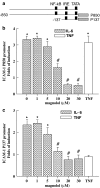
Magnolol inhibits IL-6-induced ICAM-1 promoter activity. Promoter construct (P850 or P137) of ICAM-1 containing luciferase as a reporter gene was cotransfected with pSV-_β_-galactosidase plasmid into ECs. ECs were pretreated with Mag at indicated dose for 1 h following with stimulation of IL-6 (10 ng ml−1) for 16 h in the presence of Mag. Luciferase activities after normalizing with _β_-galactosidase were expressed as folds of induction. Folds of induction are shown in the treated ECs as compared with those of untreated controls. Results are shown as mean±s.e. from three independent experiments. *P<0.05 vs control cells; #P<0.05 vs IL-6-treated cells.
Figure 3
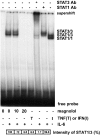
Magnolol treatment suppresses IL-6-induced Stat3 binding to IRE. ECs were pretreated with Mag at indicated dose for 1 h following with stimulation of IL-6 (10 ng ml−1) for 20 min in the presence of Mag. Nuclear protein was extracted and electrophoretic mobility shift assay (EMSA) was performed using radiolabeled oligonucleotides containing consensus IRE-binding sequence. Supershift of EMSA was performed by preincubating nuclear extracts with antibody to Stat3 or Stat1. For Stat3 activation, ECs treated with IL-6 or TNF-α (10 ng ml−1) were used as positive or negative controls, respectively. IFN-_γ_-treated ECs (1 ng ml−1) were used as positive controls for Stat1 activation. Relative intensity of each STAT1/3 band was expressed as percentage as compared with IL-6 treated cells. Data shown are the representative from three independent experiments with similar results.
Figure 4
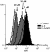
Magnolol decreases IL-6-induced ICAM-1 expression on endothelial surface. ECs pretreated with Mag for 1 h were followed with the stimulation of IL-6 (10 ng ml−1) for 24 h in the presence of Mag. ECs were resuspended and incubated in PBS with FITC-conjugated monoclonal anti-human ICAM-1 antibody. ECs were washed and fixed and analyzed using a fluorescence-activated cell sorter (FACScan, Becton Dickinson, NJ, U.S.A.) using 104 cells per sample. ICAM-1 expression on cell surface is represented by mean fluorescence intensity (MFI). Result is a representative of three independent experiments with similar results.
Figure 5
Magnolol pretreatment inhibits IL-6-induced monocyte adhesion to ECs. ECs were incubated with or without Mag for 1 h prior to IL-6 (10 ng ml−1) treatment for 24 h. Treated ECs were then incubated with 3H-labeled THP-1 monocytic cells for 1 h. Adherent THP-1 cells were lysed, and the radioactivity was counted. Results are shown as folds of induction of radioactivity from experimental groups compared with those of untreated controls. ECs treated with TNF-α (10 ng ml−1) for 24 h were used as a positive control. Data are shown as mean±s.e.m. from five separate experiments. *P<0.05 vs IL-6-treated ECs.
Figure 6
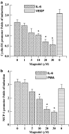
Magnolol treatment suppresses IL-6-induced cyclin D1 and MCP-1 promoter activities. Promoter construct of cyclin D1 (1745 bps) or MCP-1 (540 bps) containing luciferase as a reporter gene was cotransfected with pSV-_β_-galactosidase plasmid into ECs. ECs were pretreated with Mag at indicated concentration for 1 h followed with the stimulation of IL-6 (10 ng ml−1) for 16 h in the presence of Mag. ECs treated with VEGF (10 ng ml−1) or PMA (100 ng ml−1) were used as positive controls for cyclin D1 and MCP-1, respectively. Luciferase activities were normalized with _β_-galactosidase and results were expressed as mean±s.e. from three independent experiments. *P<0.05 vs IL-6-treated ECs without Mag.
Similar articles
- ICAM-1 induction by TNFalpha and IL-6 is mediated by distinct pathways via Rac in endothelial cells.
Wung BS, Ni CW, Wang DL. Wung BS, et al. J Biomed Sci. 2005;12(1):91-101. doi: 10.1007/s11373-004-8170-z. J Biomed Sci. 2005. PMID: 15864742 - Resveratrol suppresses IL-6-induced ICAM-1 gene expression in endothelial cells: effects on the inhibition of STAT3 phosphorylation.
Wung BS, Hsu MC, Wu CC, Hsieh CW. Wung BS, et al. Life Sci. 2005 Dec 12;78(4):389-97. doi: 10.1016/j.lfs.2005.04.052. Epub 2005 Sep 16. Life Sci. 2005. PMID: 16150460 - Chalcone inhibits the activation of NF-kappaB and STAT3 in endothelial cells via endogenous electrophile.
Liu YC, Hsieh CW, Wu CC, Wung BS. Liu YC, et al. Life Sci. 2007 Mar 20;80(15):1420-30. doi: 10.1016/j.lfs.2006.12.040. Epub 2007 Feb 2. Life Sci. 2007. PMID: 17320913 - Emerging translational approaches to target STAT3 signalling and its impact on vascular disease.
Dutzmann J, Daniel JM, Bauersachs J, Hilfiker-Kleiner D, Sedding DG. Dutzmann J, et al. Cardiovasc Res. 2015 Jun 1;106(3):365-74. doi: 10.1093/cvr/cvv103. Epub 2015 Mar 17. Cardiovasc Res. 2015. PMID: 25784694 Free PMC article. Review. - Targeted inhibition of STAT3 as a potential treatment strategy for atherosclerosis.
Chen Q, Lv J, Yang W, Xu B, Wang Z, Yu Z, Wu J, Yang Y, Han Y. Chen Q, et al. Theranostics. 2019 Aug 14;9(22):6424-6442. doi: 10.7150/thno.35528. eCollection 2019. Theranostics. 2019. PMID: 31588227 Free PMC article. Review.
Cited by
- Signal transducer and activator of transcription-3, inflammation, and cancer: how intimate is the relationship?
Aggarwal BB, Kunnumakkara AB, Harikumar KB, Gupta SR, Tharakan ST, Koca C, Dey S, Sung B. Aggarwal BB, et al. Ann N Y Acad Sci. 2009 Aug;1171:59-76. doi: 10.1111/j.1749-6632.2009.04911.x. Ann N Y Acad Sci. 2009. PMID: 19723038 Free PMC article. Review. - Cadmium induces lung inflammation independent of lung cell proliferation: a molecular approach.
Kundu S, Sengupta S, Chatterjee S, Mitra S, Bhattacharyya A. Kundu S, et al. J Inflamm (Lond). 2009 Jun 12;6:19. doi: 10.1186/1476-9255-6-19. J Inflamm (Lond). 2009. PMID: 19523218 Free PMC article. - Magnolia extract is effective for the chemoprevention of oral cancer through its ability to inhibit mitochondrial respiration at complex I.
Zhang Q, Cheng G, Pan J, Zielonka J, Xiong D, Myers CR, Feng L, Shin SS, Kim YH, Bui D, Hu M, Bennett B, Schmainda K, Wang Y, Kalyanaraman B, You M. Zhang Q, et al. Cell Commun Signal. 2020 Apr 7;18(1):58. doi: 10.1186/s12964-020-0524-2. Cell Commun Signal. 2020. PMID: 32264893 Free PMC article. - Medicinal Herbs Effective Against Atherosclerosis: Classification According to Mechanism of Action.
Kim JY, Shim SH. Kim JY, et al. Biomol Ther (Seoul). 2019 May 1;27(3):254-264. doi: 10.4062/biomolther.2018.231. Biomol Ther (Seoul). 2019. PMID: 30917628 Free PMC article. Review. - The anti-inflammatory effect of a magnoliae cortex and Zea mays L. extract mixture in a canine model of ligature-induced periodontitis.
Kim SE, Hwang SY, Park YH, Davis WC, Park KT. Kim SE, et al. BMC Vet Res. 2024 Sep 28;20(1):437. doi: 10.1186/s12917-024-04243-0. BMC Vet Res. 2024. PMID: 39342169 Free PMC article.
References
- CALDENHOVEN E., COFFER P., YUAN J., VAN DE STOLPE A., HORN F., KRUIJER W., VAN DER SAAG P.T. Stimulation of the human intercellular adhesion molecule-1 promoter by interleukin-6 and interferon-gamma involves binding of distinct factors to a palindromic response element. J. Biol. Chem. 1994;269:21146–21154. - PubMed
- CHEN C.C., CHOW M.P., HUANG W.C., LIN Y.C., CHANG Y.J. Flavonoids inhibit tumor necrosis factor-alpha-induced up-regulation of intercellular adhesion molecule-1 (ICAM-1) in respiratory epithelial cells through activator protein-1 and nuclear factor-kappaB: structure-activity relationships. Mol. Pharmacol. 2004;66:683–693. - PubMed
- CHENG J.J., WUNG B.S., CHAO Y.J., WANG D.L. Cyclic strain enhances adhesion of monocytes to endothelial cells by increasing intercellular adhesion molecule-1 expression. Hypertension. 1996;28:386–391. - PubMed
- CHUNG C.D., LIAO J., LIU B., RAO X., JAY P., BERTA P., SHUAI K. Specific inhibition of Stat3 signal transduction by PIAS3. Science. 1997;278:1803–1805. - PubMed
Publication types
MeSH terms
Substances
LinkOut - more resources
Full Text Sources
Research Materials
Miscellaneous


