Methylation-dependent silencing of CST6 in primary human breast tumors and metastatic lesions - PubMed (original) (raw)
Methylation-dependent silencing of CST6 in primary human breast tumors and metastatic lesions
Ashley G Rivenbark et al. Exp Mol Pathol. 2007 Oct.
Abstract
CST6 is a breast tumor suppressor gene that is expressed in normal breast epithelium, but is epigenetically silenced as a consequence of promoter hypermethylation in metastatic breast cancer cell lines. In the current study, we investigated the expression and methylation status of CST6 in primary breast tumors and lymph node metastases. 25/45 (56%) primary tumors and 17/20 (85%) lymph node metastases expressed significantly lower levels of cystatin M compared to normal breast tissue. Bisulfite sequencing demonstrated CST6 promoter hypermethylation in 11/23 (48%) neoplastic lesions analyzed, including 3/11 (27%) primary tumors and 8/12 (67%) lymph node metastases. In most cases (12/23, 52%), the expression of cystatin M directly reflected CST6 promoter methylation status. In remaining lesions (8/23, 35%) loss of cystatin M was not associated with CST6 promoter hypermethylation, indicating that other mechanisms can account for loss of CST6 expression. These results show that methylation-dependent silencing of CST6 occurs in a subset of primary breast cancers, but more frequently in metastatic lesions, possibly reflecting progression-related genomic events. To examine this possibility, primary breast tumors and matched lymph node metastases were analyzed. In 2/3 (67%) patients, primary tumors were positive for cystatin M and negative for CST6 promoter methylation, and matched metastatic lesions lacked cystatin M expression and CST6 was hypermethylated. This observation suggests that progression-related epigenetic alterations in CST6 gene expression can accompany metastatic spread from a primary tumor site. Overall, the results of the current investigation suggest that methylation-dependent epigenetic silencing of CST6 represents an important mechanism for loss of CST6 during breast tumorigenesis and/or progression to metastasis.
Figures
Fig. 1
Immunohistochemical analysis of cystatin M expression in human primary breast tumors. (A) Panels show H&E and cystatin M immunostaining in the same tumors. Normal breast (NB1) and primary tumors P3 and P4 show positive staining for cystatin M. Tumor P1 shows reduced cystatin M staining compared to NB1. (B) Panels show cytokeratin 18 (CK18) and cystatin M immunostaining in the same tumors. All tumors show strong staining for CK18. Tumors P22 and P30 exhibit positive cystatin M immunostaining. Tumors P35 and P44 show reduced cystatin M staining. (Original objective lens magnification 10x).
Fig. 2
Immunohistochemical analysis of cystatin M expression in lymph node metastases. (A) Panels show H&E and cystatin M immunostaining in the same lymph nodes. Lymph node P1N1 shows positive staining for cystatin M. Lymph nodes P2N1, P4N1, LNM2, and LNM5 show reduced cystatin M immunostaining. (B) Panels show cytokeratin 18 (CK18) and cystatin M immunostaining in the same lymph nodes. All metastatic lesions show strong staining for CK18 and exhibit reduced cystatin M immunostaining. (Original objective lens magnification 10x).
Fig. 3
Immunohistochemical analysis of cystatin M expression in matched primary breast tumors and lymph node metastases. Representative examples of matched pairs of primary breast tumors (top panel) and lymph node metastasis (bottom panel) are shown. Primary breast tumor P1 shows reduced cystatin M immunostaining compared to its matched lymph node P1N1. Primary breast tumor P3 and lymph node metastasis P3N1 both show positive staining for cystatin M. Primary breast tumor P4 shows positive staining for cystatin M compared to its matched lymph node metastasis P4N1. Primary breast tumor P5 and lymph node metastasis P5N1 both show negative staining for cystatin M. (Original objective lens magnification 10x).
Fig. 4
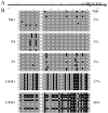
Methylation analysis of the CST6 proximal promoter and exon 1 in representative primary breast tumors and lymph node metastases. (A) The distribution of CpG dinucleotides proximal to the transcription start site in the promoter (0 to −1400 nucleotides) and exon 1 (0 to +294 nucleotides) of CST6 are depicted schematically (vertical lines indicate the relative position of individual CpG dinucleotides). Methylation analysis was performed on a region of the promoter spanning from −118 to +242 (indicated by a solid horizontal line), which contains 46 CpG dinucleotides and encompasses a large CpG island. (B) All clones analyzed for methylation of the CST6 promoter and exon 1 (46 CpGs) are shown for representative primary breast tumor and lymph node metastases examples. Black circles correspond to methylated CpGs and open circles correspond to unmethylated CpGs. TMI values for the entire promoter/exon 1 region (46 CpGs) are given for each primary breast tumor and lymph node metastases. NB1, P4, and P3 express cystatin M, while LNM1 and LNM5 lack cystatin M protein expression.
Fig. 5
Correlation analysis of cystatin M expression and CST6 methylation status in primary breast tumors and lymph node metastases. Panels show cystatin M immunostaining (on left) and a summary of the methylation analysis of the CST6 promoter/exon 1 (46 CpGs) is show on the right. Black circles correspond to fully (100%) methylated CpGs, gray circles correspond to CpGs with intermediate methylation, and open circles correspond to unmethylated CpGs. TMI values for the entire promoter/exon 1 region (46 CpGs) are given for each tissue sample. P4 and P3 primary breast tumors, and lymph node metastasis P3N1 express cystatin M. Metastatic lesions P4N1, LNM1, and LNM5 show reduced expression of cystatin M. (Original objective lens magnification 10x).
Similar articles
- Epigenetic silencing of the tumor suppressor cystatin M occurs during breast cancer progression.
Ai L, Kim WJ, Kim TY, Fields CR, Massoll NA, Robertson KD, Brown KD. Ai L, et al. Cancer Res. 2006 Aug 15;66(16):7899-909. doi: 10.1158/0008-5472.CAN-06-0576. Cancer Res. 2006. PMID: 16912163 - DNA methylation-dependent silencing of CST6 in human breast cancer cell lines.
Rivenbark AG, Jones WD, Coleman WB. Rivenbark AG, et al. Lab Invest. 2006 Dec;86(12):1233-42. doi: 10.1038/labinvest.3700485. Epub 2006 Oct 16. Lab Invest. 2006. PMID: 17043665 - Invasion suppressor cystatin E/M (CST6): high-level cell type-specific expression in normal brain and epigenetic silencing in gliomas.
Qiu J, Ai L, Ramachandran C, Yao B, Gopalakrishnan S, Fields CR, Delmas AL, Dyer LM, Melnick SJ, Yachnis AT, Schwartz PH, Fine HA, Brown KD, Robertson KD. Qiu J, et al. Lab Invest. 2008 Sep;88(9):910-25. doi: 10.1038/labinvest.2008.66. Epub 2008 Jul 7. Lab Invest. 2008. PMID: 18607344 Free PMC article. - Epigenetic regulation of cystatins in cancer.
Rivenbark AG, Coleman WB. Rivenbark AG, et al. Front Biosci (Landmark Ed). 2009 Jan 1;14(2):453-62. doi: 10.2741/3254. Front Biosci (Landmark Ed). 2009. PMID: 19273077 Review. - Cystatin M/E (Cystatin 6): A Janus-Faced Cysteine Protease Inhibitor with Both Tumor-Suppressing and Tumor-Promoting Functions.
Lalmanach G, Kasabova-Arjomand M, Lecaille F, Saidi A. Lalmanach G, et al. Cancers (Basel). 2021 Apr 14;13(8):1877. doi: 10.3390/cancers13081877. Cancers (Basel). 2021. PMID: 33919854 Free PMC article. Review.
Cited by
- Exploring the intrinsic differences among breast tumor subtypes defined using immunohistochemistry markers based on the decision tree.
Li Y, Tang XQ, Bai Z, Dai X. Li Y, et al. Sci Rep. 2016 Oct 27;6:35773. doi: 10.1038/srep35773. Sci Rep. 2016. PMID: 27786176 Free PMC article. - Integrated epigenetics of human breast cancer: synoptic investigation of targeted genes, microRNAs and proteins upon demethylation treatment.
Radpour R, Barekati Z, Kohler C, Schumacher MM, Grussenmeyer T, Jenoe P, Hartmann N, Moes S, Letzkus M, Bitzer J, Lefkovits I, Staedtler F, Zhong XY. Radpour R, et al. PLoS One. 2011;6(11):e27355. doi: 10.1371/journal.pone.0027355. Epub 2011 Nov 4. PLoS One. 2011. PMID: 22076154 Free PMC article. - A Pan-Cancer Analysis of Cystatin E/M Reveals Its Dual Functional Effects and Positive Regulation of Epithelial Cell in Human Tumors.
Xu D, Ding S, Cao M, Yu X, Wang H, Qiu D, Xu Z, Bi X, Mu Z, Li K. Xu D, et al. Front Genet. 2021 Sep 17;12:733211. doi: 10.3389/fgene.2021.733211. eCollection 2021. Front Genet. 2021. PMID: 34603393 Free PMC article. - Human mammary cancer progression model recapitulates methylation events associated with breast premalignancy.
Dumont N, Crawford YG, Sigaroudinia M, Nagrani SS, Wilson MB, Buehring GC, Turashvili G, Aparicio S, Gauthier ML, Fordyce CA, McDermott KM, Tlsty TD. Dumont N, et al. Breast Cancer Res. 2009;11(6):R87. doi: 10.1186/bcr2457. Epub 2009 Dec 8. Breast Cancer Res. 2009. PMID: 19995452 Free PMC article. - Aberrant promoter CpG methylation and its translational applications in breast cancer.
Xiang TX, Yuan Y, Li LL, Wang ZH, Dan LY, Chen Y, Ren GS, Tao Q. Xiang TX, et al. Chin J Cancer. 2013 Jan;32(1):12-20. doi: 10.5732/cjc.011.10344. Epub 2011 Nov 4. Chin J Cancer. 2013. PMID: 22059908 Free PMC article. Review.
References
- Ai L, et al. Epigenetic Silencing of the Tumor Suppressor Cystatin M Occurs during Breast Cancer Progression. Cancer Res. 2006;66:7899–7909. - PubMed
- Cromer A, et al. Identification of genes associated with tumorigenesis and metastatic potential of hypopharyngeal cancer by microarray analysis. Oncogene. 2004;23:2484–98. - PubMed
- Deng G, et al. Promoter methylation inhibits APC gene expression by causing changes in chromatin conformation and interfering with the binding of transcription factor CCAAT-binding factor. Cancer Res. 2004;64:2692–8. - PubMed
- Douglas DB, et al. Hypermethylation of a small CpGuanine-rich region correlates with loss of activator protein-2alpha expression during progression of breast cancer. Cancer Res. 2004;64:1611–20. - PubMed
Publication types
MeSH terms
Substances
LinkOut - more resources
Full Text Sources
Other Literature Sources
Medical
Research Materials
Miscellaneous


