The human keratins: biology and pathology - PubMed (original) (raw)
Review
The human keratins: biology and pathology
Roland Moll et al. Histochem Cell Biol. 2008 Jun.
Abstract
The keratins are the typical intermediate filament proteins of epithelia, showing an outstanding degree of molecular diversity. Heteropolymeric filaments are formed by pairing of type I and type II molecules. In humans 54 functional keratin genes exist. They are expressed in highly specific patterns related to the epithelial type and stage of cellular differentiation. About half of all keratins--including numerous keratins characterized only recently--are restricted to the various compartments of hair follicles. As part of the epithelial cytoskeleton, keratins are important for the mechanical stability and integrity of epithelial cells and tissues. Moreover, some keratins also have regulatory functions and are involved in intracellular signaling pathways, e.g. protection from stress, wound healing, and apoptosis. Applying the new consensus nomenclature, this article summarizes, for all human keratins, their cell type and tissue distribution and their functional significance in relation to transgenic mouse models and human hereditary keratin diseases. Furthermore, since keratins also exhibit characteristic expression patterns in human tumors, several of them (notably K5, K7, K8/K18, K19, and K20) have great importance in immunohistochemical tumor diagnosis of carcinomas, in particular of unclear metastases and in precise classification and subtyping. Future research might open further fields of clinical application for this remarkable protein family.
Figures
Fig. 1
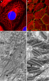
Cytoskeleton of epithelial cells. a Immunofluorescence staining of keratin K18 (red, nuclei stained in blue by DAPI) in PLC (liver carcinoma) cells in vitro. b Keratin filaments (in red) and the desmosomal component desmoplakin (in green) are labeled in cultured keratinocytes of line HaCaT. c Electron microscopic image of tonofilament (keratin) bundles (arrowhead) of HaCaT keratinocytes. d Keratin intermediate filaments (black arrowhead) insert at desmosomes (white arrowhead) at cell–cell contact sites of keratinocytes of the epidermal stratum spinosum (electron microscopy)
Fig. 2
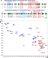
Human keratins. a Organization of the human keratin genes in the genome. The type I and type II keratin gene subdomains are located on chromosomes 17 and 12, respectively. The type I keratin K18 is located in the type II cluster on chromosome 17 (arrow). b Two-dimensional catalog of the human keratin proteins according to molecular weights (MW) and isoelectric points (IEP) as calculated from amino acid sequences. The keratin genes are designated according to the new keratin nomenclature (Schweizer et al. 2006)
Fig. 3
Keratins in simple epithelia and adenocarcinomas (paraffin sections of human tissues; avidin–biotin complex peroxidase staining). Keratin K20 is a characteristic and prominent keratin of the foveolar epithelium of the gastric (a) and colorectal (b) mucosa. The K7−/K20+ phenotype of the normal mucosa is mostly maintained in primary and metastatic colorectal adenocarcinomas. This is shown here for a skin metastasis (on the head) of a poorly differentiated adenocarcinoma: the tumor cells are negative for K7 (c) but positive for K20 (d, left portion of the figure), even including a tumor cell cluster that has invaded a lymphatic capillary (d, right upper corner). This phenotype is strongly suggestive of colorectal origin. A primary tumor in the right colon was detected later. Liver metastasis of a ductal adenocarcinoma of the pancreas with typical keratin pattern, showing extended expression of K7 (e) and staining of scattered tumor cells for K20 (f). Magnifications: a, d ×80; b, c, e, f ×140
Fig. 4
Keratins in stratified squamous epithelia and squamous cell carcinomas (paraffin sections of human tissues; avidin–biotin complex peroxidase staining). In the epidermis as an example of a normal stratified squamous epithelium, the basal cell layer contains abundant keratin K5 (a) whereas the differentiating suprabasal compartment strongly stains for K10 (b; note the negative basal cell layer). Lymph node metastasis of a squamous cell carcinoma of the head and neck region, expressing K5 (c; more intensely in the peripheral tumor cell layers) as well as K6 (d; particularly strongly in central tumor cells) as signs of their keratinocyte origin. Keratin K5 is also maintained in a lymph node micrometastasis of a squamous cell carcinoma of the head and neck region (e) and in a lymph node metastasis of an undifferentiated nasopharyngeal carcinoma with dissociated growth pattern of the tumor cells (f), in these examples being a diagnostically helpful feature. Magnifications: a, b ×160; c–e ×80; f ×140
Fig. 5
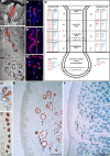
Keratin K77 in eccrine sweat glands and adnexal tumors. K77 mRNA and protein by in situ hybridization (ISH, a, a′) and indirect immunofluorescence (IIF, b-b′′) microscopy. This keratin is specifically expressed in the luminal cells (lc) of the intraglandular (igd), the intradermal (idd), the sweat duct ridge (sdr) and the intraepidermal/acrosyringial (ied/ac) duct of eccrine sweat glands of plantar skin. Peripheral duct cells are negative. The secretory portions (asterisks) are K77-negative. sb str. basale, ss str. spinosum, sg str. granulosum, sc str. corneum, gl glandular region with secretory and intraglandular duct portions. c Expression scheme of keratins of the eccrine sweat gland. d-d′′ Immunoperoxidase staining of K77 in the luminal cells of the eccrine sweat gland duct of the foot sole epidermis. K77 is also expressed in the tubular structures (arrows) of eccrine tumors such as syringoma (e) and cylindroma (f). Bars 100 μm
Fig. 6
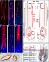
Immunofluorescence labeling of hair follicle-specific and hair keratins. The hair follicle-specific epithelial keratin K75 (a) is specifically found in the hair companion layer (cl) and in the medulla (med) of sexual (e.g. beard) hairs. K71 (b) is expressed in all compartments and K72 (c) in the cuticle (icu) of the hair inner root sheath (IRS). The hair keratin K85 (d) expression is found from the hair matrix to the upper cortex and the hair cuticle (cu), whereas K82 (e) is restricted to the hair cuticle. K86 (f) is an example for hair keratins expressed in the mid-to-upper hair cortex (co). g Hair keratin K81 is also expressed in the upper transitional cells of pilomatricoma. co cortex, dp dermal papilla, ORS outer root sheath. h, i Summary schemes of the expression of all hair and hair follicle-specific keratins in the human hair follicle. **K37 is found in the cortex of vellus hairs and medulla of sexual hairs. *K38 is heterogeneously expressed in the cortex. The keratin genes are designated according to the new keratin nomenclature (Schweizer et al. 2006)
Similar articles
- Expression of keratin K14 in the epidermis and hair follicle: insights into complex programs of differentiation.
Coulombe PA, Kopan R, Fuchs E. Coulombe PA, et al. J Cell Biol. 1989 Nov;109(5):2295-312. doi: 10.1083/jcb.109.5.2295. J Cell Biol. 1989. PMID: 2478566 Free PMC article. - Type II keratins precede type I keratins during early embryonic development.
Lu H, Hesse M, Peters B, Magin TM. Lu H, et al. Eur J Cell Biol. 2005 Aug;84(8):709-18. doi: 10.1016/j.ejcb.2005.04.001. Eur J Cell Biol. 2005. PMID: 16180309 - Distribution of keratins in normal endothelial cells and a spectrum of vascular tumors: implications in tumor diagnosis.
Miettinen M, Fetsch JF. Miettinen M, et al. Hum Pathol. 2000 Sep;31(9):1062-7. doi: 10.1053/hupa.2000.9843. Hum Pathol. 2000. PMID: 11014572 - Keratin intermediate filaments in the colon: guardians of epithelial homeostasis.
Polari L, Alam CM, Nyström JH, Heikkilä T, Tayyab M, Baghestani S, Toivola DM. Polari L, et al. Int J Biochem Cell Biol. 2020 Dec;129:105878. doi: 10.1016/j.biocel.2020.105878. Epub 2020 Nov 2. Int J Biochem Cell Biol. 2020. PMID: 33152513 Review. - Toward unraveling the complexity of simple epithelial keratins in human disease.
Omary MB, Ku NO, Strnad P, Hanada S. Omary MB, et al. J Clin Invest. 2009 Jul;119(7):1794-805. doi: 10.1172/JCI37762. Epub 2009 Jul 1. J Clin Invest. 2009. PMID: 19587454 Free PMC article. Review.
Cited by
- Anaplastic thyroid carcinoma: vimentin segregates at the invasive front of tumors in a murine xenograft model.
Miraglia A, Giannotti L, De Nuccio F, Treglia AS, Maffia M, Lofrumento DD, Di Jeso B, Nicolardi G. Miraglia A, et al. Histochem Cell Biol. 2024 Nov 18;163(1):6. doi: 10.1007/s00418-024-02329-2. Histochem Cell Biol. 2024. PMID: 39557701 - Viral infection and antiviral immunity in the oral cavity.
Hickman HD, Moutsopoulos NM. Hickman HD, et al. Nat Rev Immunol. 2024 Nov 12. doi: 10.1038/s41577-024-01100-x. Online ahead of print. Nat Rev Immunol. 2024. PMID: 39533045 Review. - Downregulation of the keratins CK13 and CK14 does not significantly affect cell viability of human urinary bladder carcinoma cells.
Scherping A, Schinlauer A, Czapiewski P, Garbers C. Scherping A, et al. Contemp Oncol (Pozn). 2024;28(3):227-234. doi: 10.5114/wo.2024.144215. Epub 2024 Oct 15. Contemp Oncol (Pozn). 2024. PMID: 39512531 Free PMC article. - The relationship between keratin 18 and epithelial-derived tumors: as a diagnostic marker, prognostic marker, and its role in tumorigenesis.
Yan J, Yang A, Tu S. Yan J, et al. Front Oncol. 2024 Oct 22;14:1445978. doi: 10.3389/fonc.2024.1445978. eCollection 2024. Front Oncol. 2024. PMID: 39502314 Free PMC article. Review.
References
- {'text': '', 'ref_index': 1, 'ids': [{'type': 'PubMed', 'value': '12121235', 'is_inner': True, 'url': 'https://pubmed.ncbi.nlm.nih.gov/12121235/'}\]}
- Abrahams NA, Ormsby AH, Brainard J (2002) Validation of cytokeratin 5/6 as an effective substitute for keratin 903 in the differentiation of benign from malignant glands in prostate needle biopsies. Histopathology 41(1):35–41 - PubMed
- {'text': '', 'ref_index': 1, 'ids': [{'type': 'PubMed', 'value': '14502799', 'is_inner': True, 'url': 'https://pubmed.ncbi.nlm.nih.gov/14502799/'}\]}
- Abrahams NA, Bostwick DG, Ormsby AH, Qian J, Brainard JA (2003) Distinguishing atrophy and high-grade prostatic intraepithelial neoplasia from prostatic adenocarcinoma with and without previous adjuvant hormone therapy with the aid of cytokeratin 5/6. Am J Clin Pathol 120(3):368–376 - PubMed
- {'text': '', 'ref_index': 1, 'ids': [{'type': 'PubMed', 'value': '2874659', 'is_inner': True, 'url': 'https://pubmed.ncbi.nlm.nih.gov/2874659/'}\]}
- Altmannsberger M, Dirk T, Droese M, Weber K, Osborn M (1986) Keratin polypeptide distribution in benign and malignant breast tumors: subdivision of ductal carcinomas using monoclonal antibodies. Virchows Arch B Cell Pathol Incl Mol Pathol 51(3):265–275 - PubMed
- {'text': '', 'ref_index': 1, 'ids': [{'type': 'PMC', 'value': 'PMC1167052', 'is_inner': False, 'url': 'https://pmc.ncbi.nlm.nih.gov/articles/PMC1167052/'}, {'type': 'PubMed', 'value': '2428612', 'is_inner': True, 'url': 'https://pubmed.ncbi.nlm.nih.gov/2428612/'}\]}
- Bader BL, Magin TM, Hatzfeld M, Franke WW (1986) Amino acid sequence and gene organization of cytokeratin no. 19, an exceptional tail-less intermediate filament protein. EMBO J 5(8):1865–1875 - PMC - PubMed
- {'text': '', 'ref_index': 1, 'ids': [{'type': 'PubMed', 'value': '2465399', 'is_inner': True, 'url': 'https://pubmed.ncbi.nlm.nih.gov/2465399/'}\]}
- Balaton AJ, Nehama-Sibony M, Gotheil C, Callard P, Baviera EE (1988) Distinction between hepatocellular carcinoma, cholangiocarcinoma, and metastatic carcinoma based on immunohistochemical staining for carcinoembryonic antigen and for cytokeratin 19 on paraffin sections. J Pathol 156(4):305–310 - PubMed
Publication types
MeSH terms
Substances
LinkOut - more resources
Full Text Sources
Other Literature Sources
Research Materials
Miscellaneous


