Fructose induces the inflammatory molecule ICAM-1 in endothelial cells - PubMed (original) (raw)
Fructose induces the inflammatory molecule ICAM-1 in endothelial cells
Olena Glushakova et al. J Am Soc Nephrol. 2008 Sep.
Abstract
Epidemiologic studies have linked fructose intake with the metabolic syndrome, and it was recently reported that fructose induces an inflammatory response in the rat kidney. Here, we examined whether fructose directly stimulates endothelial inflammatory processes by upregulating the inflammatory molecule intercellular adhesion molecule-1 (ICAM-1). When human aortic endothelial cells were stimulated with physiologic concentrations of fructose, ICAM-1 mRNA and protein expression increased in a time- and dosage-dependent manner, which was independent of NF-kappaB activation. Fructose reduced endothelial nitric oxide (NO) levels and caused a transient reduction in endothelial NO synthase expression. The administration of an NO donor inhibited fructose-induced ICAM-1 expression, whereas blocking NO synthase enhanced it, suggesting that NO inhibits endothelial ICAM-1 expression. Furthermore, fructose resulted in decreased intracellular ATP; administration of exogenous ATP blocked fructose-induced ICAM-1 expression and increased NO levels. Consistent with the in vitro studies, dietary intake of fructose at physiologic dosages increased both serum ICAM-1 concentration and endothelial ICAM-1 expression in the rat kidney. These data suggest that fructose induces inflammatory changes in vascular cells at physiologic concentrations.
Figures
Figure 1.
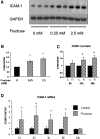
Fructose induces ICAM-1 expression in HAEC. (A) Fructose dosage-dependently induces ICAM-1 protein expression at 48 h by Western blot analysis. (B) Quantification by image analysis demonstrates the significant increase of ICAM-1 expression by 0.25 and 2.5 mM fructose at 48 h. a_P_ < 0.05 versus fructose 0 mM. (C) Fructose-induced ICAM-1 protein level is significantly increased at 24 and 48 h compared with control (C; 0 mM fructose). a_P_ < 0.05 versus control at each time point. (D) ICAM-1 mRNA expression. Real-time PCR demonstrated that ICAM-1 mRNA expression is significantly induced as early as 1 h after exposure to fructose, and it is sustained by 8 h. a_P_ < 0.05 versus control at each time point.
Figure 2.
Glucose does not stimulate ICAM-1 expression in HAEC. In contrast to fructose, glucose (0.25 mM) does not induce ICAM-1 expression at 24 h in HAEC. a_P_ < 0.05 versus control; b_P_ < 0.05 versus fructose at 24 h.
Figure 3.
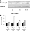
Fructose does not activate NF-κB on endothelial cell. (A) Western blotting demonstrated that 1 mM fructose did not phosphorylate NF-κB p65 on HAEC as opposed to rat TNF-α (10 ng/ml) at 4 h. TNF-α was used as a positive control for p65 activation. (B) Quantification of the ratio of phosphorylated p65/total p65 at various time points using NIH image. C, control; Fr, 1 mM fructose.
Figure 4.
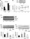
Role of eNO on ICAM-1 expression. (A) NO levels in the culture medium is significantly lower in response to fructose (Fr) than control (C) at 2 and 8 h. a_P_ < 0.01 versus control at 2 h; b_P_ < 0.05 versus control at 8 h. (B) Total eNOS protein expression is significantly reduced by 1 mM fructose at 4 h, but it returns to normal level after 8 h. b_P_ < 0.05 versus control. (C) Immunoblotting for dimer and monomer of eNOS protein. A total of 1 mM fructose administration does not result in the monomer formation of eNOS protein. M, molecular marker; DN, denatured protein as positive control for monomer. (D) Increased ICAM-1 expression in response to fructose is inhibited by 10−5M NONOate (NONO) at 24 h. Quantification by image analyzer shows that NONOate significantly reduces ICAM-1 expression induced by 1 mM fructose at 24 h. (E) L-NAME (10−3 M) significantly enhances ICAM-1 expression induced by 1 mM fructose at 24 h. b_P_ < 0.05 versus control; c_P_ < 0.05 versus fructose.
Figure 5.
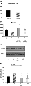
Role of ATP on NO availability and ICAM-1 expression in HAEC. (A) Fructose (1 mM) decreases intracellular ATP content in HAEC at 4 h. (B) Exogenous ATP (100 μM and 1 mM) significantly blocks the reduction of NO level in response to 1 mM fructose at 4 h. (C) ATP (10 μM) prevents an induction of ICAM-1 expression in response to fructose at 24 h. (D) Quantification demonstrated that ATP significantly inhibits fructose-induced ICAM-1 expression in HAEC.
Figure 6.
Endothelial MCP-1 and VCAM expressions in response to fructose. (A) Total MCP-1 protein level in culture medium is divided by total cell protein. Fructose at 2.5 and 25.0 mM significantly increased MCP-1 protein level in culture medium at 48 h. a_P_ < 0.05 versus fructose at 0 mM. (B) Time course of MCP-1 protein expression in response to 1 mM fructose. Data are shown as arbitrary units. (C) Time course of MCP-1 mRNA level in response to 1 mM fructose. Data are shown as arbitrary units. (D) VCAM-1 protein and mRNA expression. The immunoblotting shows no VCAM expression in control and fructose stimulation as opposed to a stimulation with 10 ng/ml rat TNF-α (positive control). Samples 1 through 3, control; samples 4 through 6, 1 mM fructose; samples 7 and 8, TNF-α. (Bottom) Time course of VCAM-1 mRNA expression under fructose stimulation or control. Data are shown as arbitrary units.
Figure 7.
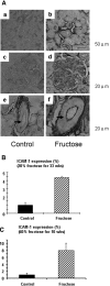
(A) Immunohistochemistry for ICAM-1 in 20% fructose-fed rat kidney. (a and b) Tubulointerstitium. (c and d) Glomerulus. (e and f) Arcuate artery. Bar = 20 μm. Compared with rats on control diet (a, c, and e), ICAM-1 expression (brown color) was induced in endothelial cell in the peritubular capillary (b), glomerulus (d), and arcuate artery (f; arrow) in 20% fructose-fed rat kidney. (B) Quantification of renal ICAM-1 expression in rat with 20% fructose diet for 33 weeks. (C) Quantification of renal ICAM-1 expression in rat with 60% fructose diet for 10 weeks.
Similar articles
- A new ATP-sensitive potassium channel opener protects endothelial function in cultured aortic endothelial cells.
Wang H, Long C, Duan Z, Shi C, Jia G, Zhang Y. Wang H, et al. Cardiovasc Res. 2007 Feb 1;73(3):497-503. doi: 10.1016/j.cardiores.2006.10.007. Epub 2006 Oct 14. Cardiovasc Res. 2007. PMID: 17116295 - Effects of Chinese yellow wine on nitric oxide synthase and intercellular adhesion molecule-1 expressions in rat vascular endothelial cells.
Zhao F, Ji Z, Chi J, Tang W, Zhai X, Meng L, Guo H. Zhao F, et al. Acta Cardiol. 2016 Feb;71(1):27-34. doi: 10.2143/AC.71.1.3132094. Acta Cardiol. 2016. PMID: 26853250 - Thiram activates NF-kappaB and enhances ICAM-1 expression in human microvascular endothelial HMEC-1 cells.
Kurpios-Piec D, Grosicka-Maciąg E, Woźniak K, Kowalewski C, Kiernozek E, Szumiło M, Rahden-Staroń I. Kurpios-Piec D, et al. Pestic Biochem Physiol. 2015 Feb;118:82-9. doi: 10.1016/j.pestbp.2014.12.003. Epub 2014 Dec 8. Pestic Biochem Physiol. 2015. PMID: 25752435
Cited by
- Association between serum uric acid and nonalcoholic fatty liver disease in the US population.
Shih MH, Lazo M, Liu SH, Bonekamp S, Hernaez R, Clark JM. Shih MH, et al. J Formos Med Assoc. 2015 Apr;114(4):314-20. doi: 10.1016/j.jfma.2012.11.014. Epub 2013 Jan 11. J Formos Med Assoc. 2015. PMID: 25839764 Free PMC article. - High-fructose diet during adolescent development increases neuroinflammation and depressive-like behavior without exacerbating outcomes after stroke.
Harrell CS, Zainaldin C, McFarlane D, Hyer MM, Stein D, Sayeed I, Neigh GN. Harrell CS, et al. Brain Behav Immun. 2018 Oct;73:340-351. doi: 10.1016/j.bbi.2018.05.018. Epub 2018 May 19. Brain Behav Immun. 2018. PMID: 29787857 Free PMC article. - H(2)S inhibits oscillatory shear stress-induced monocyte binding to endothelial cells via nitric oxide production.
Go YM, Lee HR, Park H. Go YM, et al. Mol Cells. 2012 Nov;34(5):449-55. doi: 10.1007/s10059-012-0200-5. Epub 2012 Oct 30. Mol Cells. 2012. PMID: 23124382 Free PMC article. - Effects of long-term dehydration and quick rehydration on the camel kidney: pathological changes and modulation of the expression of solute carrier proteins and aquaporins.
Damir HA, Ali MA, Adem MA, Amir N, Ali OM, Tariq S, Adeghate E, Greenwood MP, Lin P, Alvira-Iraizoz F, Gillard B, Murphy D, Adem A. Damir HA, et al. BMC Vet Res. 2024 Aug 16;20(1):367. doi: 10.1186/s12917-024-04215-4. BMC Vet Res. 2024. PMID: 39148099 Free PMC article. - A Higher Fructose Intake Is Associated with Greater Albuminuria in Subjects with Type 2 Diabetes Mellitus.
Gómez-Sámano MÁ, Almeda-Valdes P, Cuevas-Ramos D, Navarro-Flores MF, Espinosa-Salazar HD, Martínez-Saavedra M, León-Domínguez JA, Enríquez-Estrada VM, López-González AL, Sarmiento-Moreno AL, Rivera-González LA, Juárez-León ÓA, Pérez-González B, Ávila-Palacios Y, Sigala-Pedroza L, Huerta-Ávila E, Vargas-Álvarez MA, Sánchez-Jaimes C, Cárdenas-Vera M, Mehta R, López-Flores A La Torre MA, Manjarrez-Martínez I, Brito-Córdova GX, Zuarth-Vázquez JM, Vega-Beyhart A, López-Carrasco G, Johnson RJ, Gómez-Pérez FJ. Gómez-Sámano MÁ, et al. Int J Nephrol. 2018 Oct 17;2018:5459439. doi: 10.1155/2018/5459439. eCollection 2018. Int J Nephrol. 2018. PMID: 30416829 Free PMC article.
References
- Weiss R, Dziura J, Burgert TS, Tamborlane WV, Taksali SE, Yeckel CW, Allen K, Lopes M, Savoye M, Morrison J, Sherwin RS, Caprio S: Obesity and the metabolic syndrome in children and adolescents. N Engl J Med 350: 2362–2374, 2004 - PubMed
- Nakagawa T, Tuttle KR, Short RA, Johnson RJ: Hypothesis: Fructose-induced hyperuricemia as a causal mechanism for the epidemic of the metabolic syndrome. Nat Clin Pract Nephrol 1: 80–86, 2005 - PubMed
- Bray GA, Nielsen SJ, Popkin BM: Consumption of high-fructose corn syrup in beverages may play a role in the epidemic of obesity. Am J Clin Nutr 79: 537–543, 2004 - PubMed
- Beck-Nielsen H, Pedersen O, Lindskov HO: Impaired cellular insulin binding and insulin sensitivity induced by high-fructose feeding in normal subjects. Am J Clin Nutr 33: 273–278, 1980 - PubMed
- Faeh D, Minehira K, Schwarz JM, Periasamy R, Park S, Tappy L: Effect of fructose overfeeding and fish oil administration on hepatic de novo lipogenesis and insulin sensitivity in healthy men. Diabetes 54: 1907–1913, 2005 - PubMed
Publication types
MeSH terms
Substances
LinkOut - more resources
Full Text Sources
Medical
Miscellaneous
