Maintenance of intratumoral androgens in metastatic prostate cancer: a mechanism for castration-resistant tumor growth - PubMed (original) (raw)
Maintenance of intratumoral androgens in metastatic prostate cancer: a mechanism for castration-resistant tumor growth
R Bruce Montgomery et al. Cancer Res. 2008.
Abstract
Therapy for advanced prostate cancer centers on suppressing systemic androgens and blocking activation of the androgen receptor (AR). Despite anorchid serum androgen levels, nearly all patients develop castration-resistant disease. We hypothesized that ongoing steroidogenesis within prostate tumors and the maintenance of intratumoral androgens may contribute to castration-resistant growth. Using mass spectrometry and quantitative reverse transcription-PCR, we evaluated androgen levels and transcripts encoding steroidogenic enzymes in benign prostate tissue, untreated primary prostate cancer, metastases from patients with castration-resistant prostate cancer, and xenografts derived from castration-resistant metastases. Testosterone levels within metastases from anorchid men [0.74 ng/g; 95% confidence interval (95% CI), 0.59-0.89] were significantly higher than levels within primary prostate cancers from untreated eugonadal men (0.23 ng/g; 95% CI, 0.03-0.44; P < 0.0001). Compared with primary prostate tumors, castration-resistant metastases displayed alterations in genes encoding steroidogenic enzymes, including up-regulated expression of FASN, CYP17A1, HSD3B1, HSD17B3, CYP19A1, and UGT2B17 and down-regulated expression of SRD5A2 (P < 0.001 for all). Prostate cancer xenografts derived from castration-resistant tumors maintained similar intratumoral androgen levels when passaged in castrate compared with eugonadal animals. Metastatic prostate cancers from anorchid men express transcripts encoding androgen-synthesizing enzymes and maintain intratumoral androgens at concentrations capable of activating AR target genes and maintaining tumor cell survival. We conclude that intracrine steroidogenesis may permit tumors to circumvent low levels of circulating androgens. Maximal therapeutic efficacy in the treatment of castration-resistant prostate cancer will require novel agents capable of inhibiting intracrine steroidogenic pathways within the prostate tumor microenvironment.
Figures
Figure 1
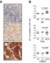
Expression of androgen receptor and PSA in castration-resistant metastases. A) Immunohistochemical analysis of AR and PSA expression in metastatic lymph node foci of prostate adenocarcinoma. Protein expression is reflected as brown chromogen reactivity: (i) hematoxylin and eosin staining demonstrating characteristics of adenocarcinoma; (ii) AR staining of the same metastasis as in (i) with abundant nuclear AR expression; (iii) PSA staining of the same metastasis as in (i) with abundant cytoplasmic PSA expression (all images at 10x magnification). B) Transcript levels for AR, PSA and FKBP5 in the benign prostate (BP), cancer prostate (CP) and castration-resistant metastatic tumor (Mets) samples. Cycle thresholds (Ct) for each gene were normalized to the housekeeping gene RPL13A in the same sample. The y-axis is the _RPL13A_-normalized Ct, more positive numbers reflect higher transcript abundance. Unpaired two sample t-tests were used to compare the mean Ct's for each gene between the cancer prostate (CP) and metastatic tumor (Mets) samples. P values < 0.05 were considered significant.
Figure 2
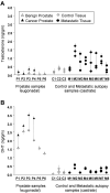
Quantitation of tissue androgens in primary and castration-resistant metastatic prostate tumors. A) Testosterone and B) DHT levels were evaluated by mass spectrometry in paired benign and cancer prostate tissues from 4 eugonadal patients undergoing prostatectomy (P1-P4); in benign prostate tissue from 2 patients undergoing cystoprostatectomy for bladder cancer (P5-P6); and in multiple metastatic tumor deposits obtained at autopsy from each of 8 patients with castration-resistant prostate cancer (M1-M8). Control tissues not involved by tumor were simultaneously obtained from a subset of patients during the autopsy procedure (C1-C3). Each tissue sample was subdivided into triplicate samples that were separately processed; the data points represent the mean of each triplicate.
Figure 3
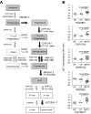
Expression of steroidogenic enzyme transcripts in primary and metastatic prostate tumors. A) The enzymatic pathways mediating the sequential biosynthesis and metabolism of Testosterone and DHT from cholesterol and progestin precursors were evaluated by quantitative RT-PCR. Bold arrows in A denote several of the key metabolic steps (colored enzymes) for which transcript levels were significantly altered in the castration-resistant prostate cancer (CRPC) metastases versus primary prostate tumors. B) Representative dot plots for the key metabolic enzymes highlighted in panel A (HSD3B1, CYP17A1, AKR1C3, SRD5A2 and UGT2B17). Transcript levels for the indicated enzymes were evaluated in the benign prostate (BP), cancer prostate (CP) and metastatic tumor (Mets) samples; cycle thresholds (Ct) for each gene were normalized to expression of the housekeeping gene RPL13A in the same sample. The y-axis is the _RPL13A_-normalized Ct, where more positive numbers reflect higher transcript abundance. Unpaired two sample t-tests were used to compare the mean Ct's for each gene between the cancer prostate (CP) and metastatic tumor (Mets) samples. P values < 0.05 were considered significant.
Figure 4
Unsupervised hierarchical clustering of primary prostate tissues and castration-resistant metastases based on expression of steroidogenic enzyme transcripts. A) The dendrogram depicts the relationship of the different tissue samples based on relative expression of the indicated genes listed in panel b. Metastatic tumor deposits are color coded according to patient of origin. B) The heatmap depicts the mean-centered expression of each gene relative to the average _RPL13A_-normalized cycle threshold for each gene across all samples. The scale is from bright green (lowest expression) to black (equivalent expression) to bright red (highest expression). Gray squares denote samples for which no transcript was detectable.
Figure 5
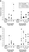
Androgen Levels In LuCaP Prostate Cancer Xenografts Grown in Castrate and Intact Mice. A) Testosterone and B) DHT levels were measured by mass spectrometry in castration-sensitive (CS) and castration-resistant (CR) variants of the indicated xenografts. Two to five CS and CR tumors of each line were passaged in non-castrate (intact) or castrate male SCID mice respectively, as indicated. The LuCaP 35 CS→CR samples are xenografts initially passaged as castration-sensitive in intact mice, which then responded to castration with castration-resistant growth. Each data point represents the mean value for an individual xenograft harvested from one mouse; samples were subdivided and assayed in duplicate. Androgen levels were also evaluated in normal kidney and muscle tissues simultaneously obtained from each set of castrate and intact animals. Mean androgen levels for each CS and CR xenografts are presented in Supplemental Table 2.
Similar articles
- Dominant-negative androgen receptor inhibition of intracrine androgen-dependent growth of castration-recurrent prostate cancer.
Titus MA, Zeithaml B, Kantor B, Li X, Haack K, Moore DT, Wilson EM, Mohler JL, Kafri T. Titus MA, et al. PLoS One. 2012;7(1):e30192. doi: 10.1371/journal.pone.0030192. Epub 2012 Jan 17. PLoS One. 2012. PMID: 22272301 Free PMC article. - Distinct patterns of dysregulated expression of enzymes involved in androgen synthesis and metabolism in metastatic prostate cancer tumors.
Mitsiades N, Sung CC, Schultz N, Danila DC, He B, Eedunuri VK, Fleisher M, Sander C, Sawyers CL, Scher HI. Mitsiades N, et al. Cancer Res. 2012 Dec 1;72(23):6142-52. doi: 10.1158/0008-5472.CAN-12-1335. Epub 2012 Sep 12. Cancer Res. 2012. PMID: 22971343 Free PMC article. - Osteoblasts promote castration-resistant prostate cancer by altering intratumoral steroidogenesis.
Hagberg Thulin M, Nilsson ME, Thulin P, Céraline J, Ohlsson C, Damber JE, Welén K. Hagberg Thulin M, et al. Mol Cell Endocrinol. 2016 Feb 15;422:182-191. doi: 10.1016/j.mce.2015.11.013. Epub 2015 Nov 14. Mol Cell Endocrinol. 2016. PMID: 26586211 - New agents and strategies for the hormonal treatment of castration-resistant prostate cancer.
Sharifi N. Sharifi N. Expert Opin Investig Drugs. 2010 Jul;19(7):837-46. doi: 10.1517/13543784.2010.494178. Expert Opin Investig Drugs. 2010. PMID: 20524793 Review. - Key targets of hormonal treatment of prostate cancer. Part 1: the androgen receptor and steroidogenic pathways.
Vis AN, Schröder FH. Vis AN, et al. BJU Int. 2009 Aug;104(4):438-48. doi: 10.1111/j.1464-410X.2009.08695.x. Epub 2009 Jun 24. BJU Int. 2009. PMID: 19558559 Review.
Cited by
- A novel prostate cancer therapeutic strategy using icaritin-activated arylhydrocarbon-receptor to co-target androgen receptor and its splice variants.
Sun F, Indran IR, Zhang ZW, Tan MH, Li Y, Lim ZL, Hua R, Yang C, Soon FF, Li J, Xu HE, Cheung E, Yong EL. Sun F, et al. Carcinogenesis. 2015 Jul;36(7):757-68. doi: 10.1093/carcin/bgv040. Epub 2015 Apr 23. Carcinogenesis. 2015. PMID: 25908644 Free PMC article. - Recent Advances in Epigenetic Biomarkers and Epigenetic Targeting in Prostate Cancer.
Kumaraswamy A, Welker Leng KR, Westbrook TC, Yates JA, Zhao SG, Evans CP, Feng FY, Morgan TM, Alumkal JJ. Kumaraswamy A, et al. Eur Urol. 2021 Jul;80(1):71-81. doi: 10.1016/j.eururo.2021.03.005. Epub 2021 Mar 27. Eur Urol. 2021. PMID: 33785255 Free PMC article. Review. - Can Environmental Manipulation Help Suppress Cancer? Non-Linear Competition Among Tumor Cells in Periodically Changing Conditions.
Babajanyan SG, Koonin EV, Cheong KH. Babajanyan SG, et al. Adv Sci (Weinh). 2020 Jul 1;7(16):2000340. doi: 10.1002/advs.202000340. eCollection 2020 Aug. Adv Sci (Weinh). 2020. PMID: 32832349 Free PMC article. - Metabolic changes during prostate cancer development and progression.
Beier AK, Puhr M, Stope MB, Thomas C, Erb HHH. Beier AK, et al. J Cancer Res Clin Oncol. 2023 May;149(5):2259-2270. doi: 10.1007/s00432-022-04371-w. Epub 2022 Sep 23. J Cancer Res Clin Oncol. 2023. PMID: 36151426 Free PMC article. Review. - Human castration resistant prostate cancer rather prefer to decreased 5α-reductase activity.
Kosaka T, Miyajima A, Nagata H, Maeda T, Kikuchi E, Oya M. Kosaka T, et al. Sci Rep. 2013;3:1268. doi: 10.1038/srep01268. Sci Rep. 2013. PMID: 23429215 Free PMC article.
References
- Huggins C, Hodges CV. Studies on prostatic cancer: I. The effect of castration, of estrogen and of androgen injection on serum phosphatases in metastatic carcinoma of the prostate. 1941. J Urol. 2002;168:9–12. - PubMed
- Small EJ, Ryan CJ. The Case for Secondary Hormonal Therapies in the Chemotherapy Age. The Journal of Urology. Innovations and Challenges in Prostate Cancer: Recommendations for Defining and Treating High Risk Disease. 2006;176:S66–S71. - PubMed
- Chen CD, Welsbie DS, Tran C, et al. Molecular determinants of resistance to antiandrogen therapy. Nat Med. 2004;10:33–9. - PubMed
- Feldman BJ, Feldman D. The development of androgen-independent prostate cancer. Nat Rev Cancer. 2001;1:34–45. - PubMed
- Visakorpi T, Hyytinen E, Koivisto P, et al. In vivo amplification of the androgen receptor gene and progression of human prostate cancer. Nat Genet. 1995;9:401–6. - PubMed
Publication types
MeSH terms
Substances
Grants and funding
- RR00163/RR/NCRR NIH HHS/United States
- K23CA122820/CA/NCI NIH HHS/United States
- P50CA97186/CA/NCI NIH HHS/United States
- K23 CA122820/CA/NCI NIH HHS/United States
- R01 DK65204/DK/NIDDK NIH HHS/United States
- P30CA15704/CA/NCI NIH HHS/United States
- R01 DK065204/DK/NIDDK NIH HHS/United States
- P30 CA015704/CA/NCI NIH HHS/United States
- P51 RR000163/RR/NCRR NIH HHS/United States
- P50 CA097186/CA/NCI NIH HHS/United States
- P50 CA097186-060005/CA/NCI NIH HHS/United States
- K01 RR000163/RR/NCRR NIH HHS/United States
LinkOut - more resources
Full Text Sources
Other Literature Sources
Medical
Molecular Biology Databases
Research Materials
Miscellaneous


