Autoimmune liver serology: current diagnostic and clinical challenges - PubMed (original) (raw)
Review
Autoimmune liver serology: current diagnostic and clinical challenges
Dimitrios-P Bogdanos et al. World J Gastroenterol. 2008.
Abstract
Liver-related autoantibodies are crucial for the correct diagnosis and classification of autoimmune liver diseases (AiLD), namely autoimmune hepatitis types 1 and 2 (AIH-1 and 2), primary biliary cirrhosis (PBC), and the sclerosing cholangitis variants in adults and children. AIH-1 is specified by anti-nuclear antibody (ANA) and smooth muscle antibody (SMA). AIH-2 is specified by antibody to liver kidney microsomal antigen type-1 (anti-LKM1) and anti-liver cytosol type 1 (anti-LC1). SMA, ANA and anti-LKM antibodies can be present in de-novo AIH following liver transplantation. PBC is specified by antimitochondrial antibodies (AMA) reacting with enzymes of the 2-oxo-acid dehydrogenase complexes (chiefly pyruvate dehydrogenase complex E2 subunit) and disease-specific ANA mainly reacting with nuclear pore gp210 and nuclear body sp100. Sclerosing cholangitis presents as at least two variants, first the classical primary sclerosing cholangitis (PSC) mostly affecting adult men wherein the only (and non-specific) reactivity is an atypical perinuclear antineutrophil cytoplasmic antibody (p-ANCA), also termed perinuclear anti-neutrophil nuclear antibodies (p-ANNA) and second the childhood disease called autoimmune sclerosing cholangitis (ASC) with serological features resembling those of type 1 AIH. Liver diagnostic serology is a fast-expanding area of investigation as new purified and recombinant autoantigens, and automated technologies such as ELISAs and bead assays, become available to complement (or even compete with) traditional immunofluorescence procedures. We survey for the first time global trends in quality assurance impacting as it does on (1) manufacturers/purveyors of kits and reagents, (2) diagnostic service laboratories that fulfill clinicians' requirements, and (3) the end-user, the physician providing patient care, who must properly interpret test results in the overall clinical context.
Figures
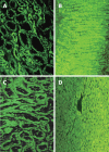
Figure 1
Immunofluorescence of anti-mitochondrial (A and B), and anti-liver kidney microsomal antibody (anti-LKM1) (C and D). AMA stain (A) stronger the smaller, distal tubules while anti-LKM1 the proximal tubules of the rat kidney (C). These specificities are frequently misdiagnosed, especially when only the kidney substrate is used and the sections do not contain both proximal and distal tubules. Thus, the use of rat stomach (B) and liver (D) is strongly recommended to prevent misinterpretation; AMA characteristically stain the gastric parietal cells while anti-LKM1 stain the rat liver but not the stomach.
Similar articles
- [Autoimmune hepatitis and overlap syndrome: diagnosis].
Berg PA, Klein R. Berg PA, et al. Praxis (Bern 1994). 2002 Aug 21;91(34):1339-46. doi: 10.1024/0369-8394.91.34.1339. Praxis (Bern 1994). 2002. PMID: 12233264 Review. German. - The clinical usage and definition of autoantibodies in immune-mediated liver disease: A comprehensive overview.
Terziroli Beretta-Piccoli B, Mieli-Vergani G, Vergani D. Terziroli Beretta-Piccoli B, et al. J Autoimmun. 2018 Dec;95:144-158. doi: 10.1016/j.jaut.2018.10.004. Epub 2018 Oct 23. J Autoimmun. 2018. PMID: 30366656 Review. - Antineutrophil nuclear antibodies (ANNA) in primary biliary cirrhosis: their prevalence and antigen specificity.
Orth T, Gerken G, Meyer Zum Büschenfelde KH, Mayet WJ. Orth T, et al. Z Gastroenterol. 1997 Feb;35(2):113-21. Z Gastroenterol. 1997. PMID: 9066101 - Clinical significance of autoantibodies in autoimmune hepatitis.
Liberal R, Mieli-Vergani G, Vergani D. Liberal R, et al. J Autoimmun. 2013 Oct;46:17-24. doi: 10.1016/j.jaut.2013.08.001. Epub 2013 Sep 7. J Autoimmun. 2013. PMID: 24016388 Review. - [Autoimmune liver diseases and their overlap syndromes].
Strassburg CP. Strassburg CP. Praxis (Bern 1994). 2006 Sep 6;95(36):1363-81. doi: 10.1024/1661-8157.95.36.1363. Praxis (Bern 1994). 2006. PMID: 16989180 Review. German.
Cited by
- Autoimmune Hepatitis: A Diagnostic and Therapeutic Overview.
Mercado LA, Gil-Lopez F, Chirila RM, Harnois DM. Mercado LA, et al. Diagnostics (Basel). 2024 Feb 9;14(4):382. doi: 10.3390/diagnostics14040382. Diagnostics (Basel). 2024. PMID: 38396421 Free PMC article. Review. - Antibody against apolipoprotein-A1, non-alcoholic fatty liver disease and cardiovascular risk: a translational study.
Pagano S, Bakker SJL, Juillard C, Vossio S, Moreau D, Brandt KJ, Mach F, Dullaart RPF, Vuilleumier N. Pagano S, et al. J Transl Med. 2023 Oct 5;21(1):694. doi: 10.1186/s12967-023-04569-7. J Transl Med. 2023. PMID: 37798764 Free PMC article. - Evaluation of immunoserological detection of anti-liver kidney microsomal, anti-soluble liver antigen and anti-mitochondrial antibodies.
Campos-Murguia A, Henjes N, Loges S, Wedemeyer H, Jaeckel E, Taubert R, Engel B. Campos-Murguia A, et al. Sci Rep. 2023 Jun 20;13(1):10038. doi: 10.1038/s41598-023-37095-z. Sci Rep. 2023. PMID: 37340049 Free PMC article. - Primary Biliary Cholangitis: Promising Emerging Innovative Therapies and Their Impact on GLOBE Scores.
Sohal A, Kowdley KV. Sohal A, et al. Hepat Med. 2023 Jun 8;15:63-77. doi: 10.2147/HMER.S361077. eCollection 2023. Hepat Med. 2023. PMID: 37312929 Free PMC article. Review. - Role of autoantibodies in the clinical management of primary biliary cholangitis.
Rigopoulou EI, Bogdanos DP. Rigopoulou EI, et al. World J Gastroenterol. 2023 Mar 28;29(12):1795-1810. doi: 10.3748/wjg.v29.i12.1795. World J Gastroenterol. 2023. PMID: 37032725 Free PMC article. Review.
References
- Bogdanos DP, Baum H, Vergani D. Antimitochondrial and other autoantibodies. Clin Liver Dis. 2003;7:759–777, vi. - PubMed
- Invernizzi P, Lleo A, Podda M. Interpreting serological tests in diagnosing autoimmune liver diseases. Semin Liver Dis. 2007;27:161–172. - PubMed
- Czaja AJ, Homburger HA. Autoantibodies in liver disease. Gastroenterology. 2001;120:239–249. - PubMed
- Selmi C, Mackay IR, Gershwin ME. The immunological milieu of the liver. Semin Liver Dis. 2007;27:129–139. - PubMed
- Alvarez F, Berg PA, Bianchi FB, Bianchi L, Burroughs AK, Cancado EL, Chapman RW, Cooksley WG, Czaja AJ, Desmet VJ, et al. International Autoimmune Hepatitis Group Report: review of criteria for diagnosis of autoimmune hepatitis. J Hepatol. 1999;31:929–938. - PubMed
Publication types
MeSH terms
Substances
LinkOut - more resources
Full Text Sources
Other Literature Sources
Miscellaneous
