Cellular senescence and organismal ageing in the absence of p21(CIP1/WAF1) in ku80(-/-) mice - PubMed (original) (raw)
Cellular senescence and organismal ageing in the absence of p21(CIP1/WAF1) in ku80(-/-) mice
Bo Zhao et al. EMBO Rep. 2009 Jan.
Erratum in
- EMBO Rep. 2011 Mar;12(3):283
Abstract
Ku80 is important in the repair of DNA double-strand breaks by its essential function in non-homologous end-joining. The absence of Ku80 causes the accumulation of DNA damage and leads to premature ageing in mice. We showed that mouse embryonic fibroblasts (MEFs) from ku80(-/-) mice senesced rapidly with elevated levels of p53 and p21. Deletion of p21 delayed the early senescence phenotype in ku80(-/-) MEFs, despite an otherwise intact response of p53. In contrast to ku80(-/-)p53(-/-) mice, which die rapidly primarily from lymphomas, there was no significant increase in tumorigenesis in ku80(-/-)p21(-/-) mice. However, ku80(-/-)p21(-/-) mice showed no improvement with respect to rough fur coat or osteopaenia, and even showed a shortened lifespan compared with ku80(-/-) mice. These results show that the increased lifespan of ku80(-/-) MEFs owing to the loss of p21 is not associated with an improvement of the premature ageing phenotypes of ku80(-/-) mice observed at the organismal level.
Conflict of interest statement
The authors declare that they have no conflict of interest.
Figures
Figure 1
Early senescence of _ku80_−/− mouse embryonic fibroblasts correlates with elevated levels of p53 and p21. (A) Growth curves of MEFs starting at PD8 (which reflects time zero on the x axis), with genotypes as indicated. (B) Quantification of BrdU/SA-β-gal of WT and _ku80_−/− MEFs at PD8. Mean±s.d. reflects the results of three separate experiments from individual isolates. (C) Western blot showing levels of p53 and p21 in WT, _ku80_−/−, _p53_−/− (PD12) and _p21_−/− (PD6) MEFs either untreated (−) or treated (+) with doxorubicin (Doxo; 100 ng/ml) for 20 h. (D) Quantifications of the levels of p53 and p21 were normalized to β-actin, and to basal levels of p53 and p21 in WT MEFs, respectively. Mean±s.d. reflects the results of three separate experiments from individual isolates. For all the experiments, at least two sets of isolates for each genotype were assayed and similar results were obtained. BrdU, 5-bromodeoxyuridine; MEF, mouse embryonic fibroblast; PD, population doubling; SA-β-gal, senescence-associated β-galactosidase; WT, wild type.
Figure 2
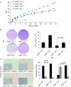
Effects of p21 deletion on growth properties of _ku80_−/− mouse embryonic fibroblasts. (A) Growth curves of MEFs starting at PD8, with genotypes as indicated. Of note is that _p21_−/−, WT and _ku80_−/−_p21_−/− MEFs showed sub-populations associated with a loss of p53, which bypassed senescence and eventually took over the culture, as reflected by the accelerated phase on their growth curves. (B) Saturation densities of MEFs. MEFs (1 × 10⁴) at PD4 were plated in six 60-mm petri dishes and the medium was changed every 3 days. At 1 week, three dishes were stained with crystal violet, and representative images are shown on the left. Cell numbers from parallel dishes are shown on the right. Mean±s.d. reflects results of 3 separate experiments from individual MEFs. (C) Representative images of BrdU (brown)/SA-β-gal (blue) double staining and cell morphology after 22 days in culture; PDs are extrapolated from growth curves. Quantifications for BrdU or SA-β-gal staining are shown on the right. Mean±s.d. reflects the results of three separate experiments from individual isolates. For all the experiments, at least two sets of isolates for each genotype were assayed and similar results were obtained. BrdU, 5-bromodeoxyuridine; MEF, mouse embryonic fibroblast; PD, population doubling; SA-β-gal, senescence-associated β-galactosidase; WT, wild type.
Figure 3
Cell-cycle profiles and p53 responses in _ku80_−/−_p21_−/− mouse embryonic fibroblasts. Representative cell-cycle profiles of (A) Ku80 +/+ and _ku80_−/− MEFs with or without p21 at PD10, and (B) in WT and _ku80_−/−_p21_−/− MEFs at PD26. Mean±s.d. reflects the results of three separate experiments from individual MEFs. Two sets of MEFs for each genotype were assayed with similar results (*P<0.05). (C) Western blot analysis of the response of p53 in _ku80_−/−_p21_−/− MEFs with increasing PDs. Cells were either untreated (−) or treated (+) with doxorubicin (Doxo; 300 ng/ml) for 20 h, and levels of p53 and p19 are shown; β-actin was used as a loading control. MEF, mouse embryonic fibroblast; PD, population doubling; WT, wild type.
Figure 4
Effects of p21 deletion on ageing phenotypes of _ku80_−/− mice. (A) Survival curves (100% × number of mice alive after each week/total number of mice at 2 weeks) for mice with each genotype. (B) Comparison of the appearance of 32-week-old mice with each genotype; note the rough fur coat and kyphosis in _ku80_−/− and _ku80_−/−_p21_−/− mice. (C) Comparison of the distal femoral epiphysis in mice with each genotype. The cortical wall and part of the diaphysis are also shown. Chondrocyte numbers in the growth plates from two mice between 32 and 34 weeks old for each genotype were counted, and the quantification is shown on the right. Scale bar 200μm.
Figure 5
Effects of p21 deletion on lymphoid tissue in _ku80_−/− mice. (A) Spleen/body weight ratio from mice with various genetic backgrounds at 16 weeks. (B) HE staining of spleen tissue from each group; note the loss of splenic follicles in _ku80_−/− compared with Ku80 +/+ mice. (C) TUNEL staining of spleen tissue was performed on paraffin sections (three mice for each genotype). Quantification of three high-power fields (HP) per spleen is shown on the right. HE, haematoxylin and eosin; TUNEL, TdT-mediated dUTP nick-end labelling.
Similar articles
- Effects of p21 deletion in mouse models of premature aging.
Benson EK, Zhao B, Sassoon DA, Lee SW, Aaronson SA. Benson EK, et al. Cell Cycle. 2009 Jul 1;8(13):2002-4. doi: 10.4161/cc.8.13.8997. Epub 2009 Jul 12. Cell Cycle. 2009. PMID: 19535900 Free PMC article. - Unlike p53, p27 failed to exhibit an anti-tumor genetic interaction with Ku80.
Holcomb VB, Vogel H, Hasty P. Holcomb VB, et al. Cell Cycle. 2009 Aug;8(15):2463-6. doi: 10.4161/cc.8.15.9249. Epub 2009 Aug 11. Cell Cycle. 2009. PMID: 19597334 - Ku80 functions as a tumor suppressor in hepatocellular carcinoma by inducing S-phase arrest through a p53-dependent pathway.
Wei S, Xiong M, Zhan DQ, Liang BY, Wang YY, Gutmann DH, Huang ZY, Chen XP. Wei S, et al. Carcinogenesis. 2012 Mar;33(3):538-47. doi: 10.1093/carcin/bgr319. Epub 2012 Jan 5. Carcinogenesis. 2012. PMID: 22226916 - DNA repair, genome stability, and aging.
Lombard DB, Chua KF, Mostoslavsky R, Franco S, Gostissa M, Alt FW. Lombard DB, et al. Cell. 2005 Feb 25;120(4):497-512. doi: 10.1016/j.cell.2005.01.028. Cell. 2005. PMID: 15734682 Review.
Cited by
- The yin-yang of DNA damage response: roles in tumorigenesis and cellular senescence.
Li X, Xu H, Xu C, Lin M, Song X, Yi F, Feng Y, Coughlan KA, Cho WC, Kim SS, Cao L. Li X, et al. Int J Mol Sci. 2013 Jan 25;14(2):2431-48. doi: 10.3390/ijms14022431. Int J Mol Sci. 2013. PMID: 23354477 Free PMC article. - Therapeutic and space radiation exposure of mouse brain causes impaired DNA repair response and premature senescence by chronic oxidant production.
Suman S, Rodriguez OC, Winters TA, Fornace AJ Jr, Albanese C, Datta K. Suman S, et al. Aging (Albany NY). 2013 Aug;5(8):607-22. doi: 10.18632/aging.100587. Aging (Albany NY). 2013. PMID: 23928451 Free PMC article. - Loss of a single allele for Ku80 leads to progenitor dysfunction and accelerated aging in skeletal muscle.
Didier N, Hourdé C, Amthor H, Marazzi G, Sassoon D. Didier N, et al. EMBO Mol Med. 2012 Sep;4(9):910-23. doi: 10.1002/emmm.201101075. Epub 2012 Aug 23. EMBO Mol Med. 2012. PMID: 22915554 Free PMC article. - Bax deficiency extends the survival of Ku70 knockout mice that develop lung and heart diseases.
Ngo J, Matsuyama M, Kim C, Poventud-Fuentes I, Bates A, Siedlak SL, Lee HG, Doughman YQ, Watanabe M, Liner A, Hoit B, Voelkel N, Gerson S, Hasty P, Matsuyama S. Ngo J, et al. Cell Death Dis. 2015 Mar 26;6(3):e1706. doi: 10.1038/cddis.2015.11. Cell Death Dis. 2015. PMID: 25811803 Free PMC article. - Effects of p21 deletion in mouse models of premature aging.
Benson EK, Zhao B, Sassoon DA, Lee SW, Aaronson SA. Benson EK, et al. Cell Cycle. 2009 Jul 1;8(13):2002-4. doi: 10.4161/cc.8.13.8997. Epub 2009 Jul 12. Cell Cycle. 2009. PMID: 19535900 Free PMC article.
References
- Brown JP, Wei W, Sedivy JM (1997) Bypass of senescence after disruption of p21CIP1/WAF1 gene in normal diploid human fibroblasts. Science 277: 831–834 - PubMed
- Brugarolas J, Chandrasekaran C, Gordon JI, Beach D, Jacks T, Hannon GJ (1995) Radiation-induced cell cycle arrest compromised by p21 deficiency. Nature 377: 552–557 - PubMed
- Bunz F, Dutriaux A, Lengauer C, Waldman T, Zhou S, Brown JP, Sedivy JM, Kinzler KW, Vogelstein B (1998) Requirement for p53 and p21 to sustain G2 arrest after DNA damage. Science 282: 1497–1501 - PubMed
- Campisi J (2005) Senescent cells, tumor suppression, and organismal aging: good citizens, bad neighbors. Cell 120: 513–522 - PubMed
Publication types
MeSH terms
Substances
Grants and funding
- 1R01CA127247/CA/NCI NIH HHS/United States
- R01 CA085214/CA/NCI NIH HHS/United States
- 5R01CA085214/CA/NCI NIH HHS/United States
- R01 CA085681/CA/NCI NIH HHS/United States
- 5P01CA080058/CA/NCI NIH HHS/United States
- P01 CA080058/CA/NCI NIH HHS/United States
- 2R01CA085681/CA/NCI NIH HHS/United States
- R01 CA127247/CA/NCI NIH HHS/United States
LinkOut - more resources
Full Text Sources
Medical
Molecular Biology Databases
Research Materials
Miscellaneous


