Epigenetics and T helper 1 differentiation - PubMed (original) (raw)
Review
Epigenetics and T helper 1 differentiation
Thomas M Aune et al. Immunology. 2009 Mar.
Abstract
Naïve T helper cells differentiate into two subsets, T helper 1 and 2, which either transcribe the Ifng gene and silence the Il4 gene or transcribe the Il4 gene and silence the Ifng gene, respectively. This process is an essential feature of the adaptive immune response to a pathogen and the development of long-lasting immunity. The 'histone code' hypothesis proposes that formation of stable epigenetic histone marks at a gene locus that activate or repress transcription is essential for cell fate determinations, such as T helper 1/T helper 2 cell fate decisions. Activation and silencing of the Ifng gene are achieved through the creation of stable epigenetic histone marks spanning a region of genomic DNA over 20 times greater than the gene itself. Key transcription factors that drive the T helper 1 lineage decision, signal transducer and activator 4 (STAT4) and T-box expressed in T cells (T-bet), play direct roles in the formation of activating histone marks at the Ifng locus. Conversely, STAT6 and GATA binding protein 3, transcription factors essential for the T helper 2 cell lineage decision, establish repressive histone marks at the Ifng locus. Functional studies demonstrate that multiple genomic elements up to 50 kilobases from Ifng play critical roles in its proper transcriptional regulation. Studies of three-dimensional chromatin conformation indicate that these distal regulatory elements may loop towards Ifng to regulate its transcription. We speculate that these complex mechanisms have evolved to tightly control levels of interferon-gamma production, given that too little or too much production would be very deleterious to the host.
Figures
Figure 1
The Ifng locus. (a) Schematic and functional properties of two human IFNG transgenes. The upper transgene is an ∼8·6 kilobase (kb) fragment containing the human IFNG gene and 2·0–2·5 of upstream and downstream sequence. The lower transgene is an ∼190 kb bacterial artificial chromosome with the human IFNG gene and flanking sequence. (b) Positions of evolutionarily conserved non-coding DNA elements relative to the Ifng gene from the dcode website (
). (c) Positions of conserved non-coding sequences (CNS), filled circles, relative to the mouse (upper) and human (lower) Ifng/IFNG genes. Lines connect representative individual CNS within the mouse and human loci.
Figure 2
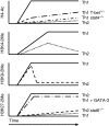
Kinetics of histone modifications at the Ifng locus during T helper 1 (Th1)/Th2 differentiation. Initiation of Th1 and Th2 differentiation programmes induces formation of markedly different histone ‘marks’ at the Ifng locus. Both signal transducer and activator 4 (STAT4) and T-box expressed in T cells (T-bet) play critical roles in directing the formation of histone 4 (H4)-acetylation marks in developing Th1 cells and STAT6 and GATA-binding protein 3 (GATA-3) play critical roles in directing the formation of H3K27-methylation marks in developing Th2 cells. Changes in H3K9-methylation marks during differentiation illustrate the dynamic nature of the histone code in developing effector T cells.
Figure 3
Changes in the three-dimensional (3-D) conformation of the Ifng locus may recruit distal conserved non-coding sequences (CNS) to the gene to regulate transcription. Distal evolutionarily conserved DNA elements are occupied by transcription factors (TF) after initiation of T helper type 1 (Th1)/Th2 differentiation programmes. These transcription factors can tether enzymes that catalyse histone modifications, chromatin remodelling and other functions to these DNA elements. Changes in three-dimensional conformation of the locus may serve to localize these DNA elements and their associated proteins to the Ifng gene. CNS, conserved non-coding sequences.
Similar articles
- T-bet regulates Th1 responses through essential effects on GATA-3 function rather than on IFNG gene acetylation and transcription.
Usui T, Preiss JC, Kanno Y, Yao ZJ, Bream JH, O'Shea JJ, Strober W. Usui T, et al. J Exp Med. 2006 Mar 20;203(3):755-66. doi: 10.1084/jem.20052165. Epub 2006 Mar 6. J Exp Med. 2006. PMID: 16520391 Free PMC article. - T-bet dependent removal of Sin3A-histone deacetylase complexes at the Ifng locus drives Th1 differentiation.
Chang S, Collins PL, Aune TM. Chang S, et al. J Immunol. 2008 Dec 15;181(12):8372-81. doi: 10.4049/jimmunol.181.12.8372. J Immunol. 2008. PMID: 19050254 Free PMC article. - Regulation of the Ifng locus in the context of T-lineage specification and plasticity.
Balasubramani A, Mukasa R, Hatton RD, Weaver CT. Balasubramani A, et al. Immunol Rev. 2010 Nov;238(1):216-32. doi: 10.1111/j.1600-065X.2010.00961.x. Immunol Rev. 2010. PMID: 20969595 Free PMC article. Review. - Transcriptional mechanisms that regulate T helper 1 cell differentiation.
Oestreich KJ, Weinmann AS. Oestreich KJ, et al. Curr Opin Immunol. 2012 Apr;24(2):191-5. doi: 10.1016/j.coi.2011.12.004. Epub 2012 Jan 10. Curr Opin Immunol. 2012. PMID: 22240120 Free PMC article. Review.
Cited by
- IFN-γ, should not be ignored in SLE.
Liu W, Zhang S, Wang J. Liu W, et al. Front Immunol. 2022 Aug 10;13:954706. doi: 10.3389/fimmu.2022.954706. eCollection 2022. Front Immunol. 2022. PMID: 36032079 Free PMC article. Review. - Cutting edge: influence of Tmevpg1, a long intergenic noncoding RNA, on the expression of Ifng by Th1 cells.
Collier SP, Collins PL, Williams CL, Boothby MR, Aune TM. Collier SP, et al. J Immunol. 2012 Sep 1;189(5):2084-8. doi: 10.4049/jimmunol.1200774. Epub 2012 Jul 30. J Immunol. 2012. PMID: 22851706 Free PMC article. - Lineage-specific adjacent IFNG and IL26 genes share a common distal enhancer element.
Collins PL, Henderson MA, Aune TM. Collins PL, et al. Genes Immun. 2012 Sep;13(6):481-8. doi: 10.1038/gene.2012.22. Epub 2012 May 24. Genes Immun. 2012. PMID: 22622197 Free PMC article. - IκBζ is essential for natural killer cell activation in response to IL-12 and IL-18.
Miyake T, Satoh T, Kato H, Matsushita K, Kumagai Y, Vandenbon A, Tani T, Muta T, Akira S, Takeuchi O. Miyake T, et al. Proc Natl Acad Sci U S A. 2010 Oct 12;107(41):17680-5. doi: 10.1073/pnas.1012977107. Epub 2010 Sep 27. Proc Natl Acad Sci U S A. 2010. PMID: 20876105 Free PMC article. - Early Th1 cell differentiation is marked by a Tfh cell-like transition.
Nakayamada S, Kanno Y, Takahashi H, Jankovic D, Lu KT, Johnson TA, Sun HW, Vahedi G, Hakim O, Handon R, Schwartzberg PL, Hager GL, O'Shea JJ. Nakayamada S, et al. Immunity. 2011 Dec 23;35(6):919-31. doi: 10.1016/j.immuni.2011.11.012. Immunity. 2011. PMID: 22195747 Free PMC article.
References
- Mosmann TR, Coffman RL. TH1 and TH2 cells: different patterns of lymphokine secretion lead to different functional properties. Annu Rev Immunol. 1989;7:145–73. - PubMed
- Seder RA, Paul WE. Acquisition of lymphokine-producing phenotype by CD4+ T cells. Annu Rev Immunol. 1994;12:635–73. - PubMed
- Murphy KM, Reiner SL. The lineage decisions of helper T cells. Nat Rev Immunol. 2002;2:933–44. - PubMed
- Thierfelder WE, van Deursen JM, Yamamoto K, et al. Requirement for Stat4 in interleukin-12-mediated responses of natural killer and T cells. Nature. 1996;382:171–4. - PubMed
- Kaplan MH, Schindler U, Smiley ST, Grusby MJ. Stat6 is required for mediating responses to IL-4 and for the development of Th2 cells. Immunity. 1996;4:313–9. - PubMed
Publication types
MeSH terms
Substances
LinkOut - more resources
Full Text Sources
Other Literature Sources
Research Materials
Miscellaneous


