miR-331-3p regulates ERBB-2 expression and androgen receptor signaling in prostate cancer - PubMed (original) (raw)
miR-331-3p regulates ERBB-2 expression and androgen receptor signaling in prostate cancer
Michael R Epis et al. J Biol Chem. 2009.
Abstract
MicroRNAs (miRNAs) are short, non-coding RNAs that regulate gene expression and are aberrantly expressed in human cancer. The ERBB-2 tyrosine kinase receptor is frequently overexpressed in prostate cancer and is associated with disease progression and poor survival. We have identified two specific miR-331-3p target sites within the ERBB-2 mRNA 3'-untranslated region and show that miR-331-3p expression is decreased in prostate cancer tissue relative to normal adjacent prostate tissue. Transfection of multiple prostate cancer cell lines with miR-331-3p reduced ERBB-2 mRNA and protein expression and blocked downstream phosphatidylinositol 3-kinase/AKT signaling. Furthermore, miR-331-3p transfection blocked the androgen receptor signaling pathway in prostate cancer cells, reducing activity of an androgen-stimulated prostate-specific antigen promoter and blocking prostate-specific antigen expression. Our findings provide insight into the regulation of ERBB-2 expression in cancer and suggest that miR-331-3p has the capacity to regulate signaling pathways critical to the development and progression of prostate cancer cells.
Figures
FIGURE 1.
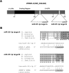
Identification of two specific miR-331-3p target sites within the ERBB-2 mRNA 3′-UTR. A, schematic representation of the ERBB-2 mRNA with two 3′-UTR miR-331-3p binding sites (A and B) predicted by TargetScan. The miR-331-3p seed sequence is underlined. B, sequence alignment of the predicted ERBB-2 3′-UTR miR-331-3p target sites showing conservation between human, mouse, rat, and dog. The miR-331-3p seed sequence (CCAGGGG) is shown in bold and underlined, and conserved nucleotides are shaded. Stars indicate nucleotides conserved across all four species.
FIGURE 2.
miR-331-3p regulates ERBB-2 expression in prostate cancer cell lines. A, immunoblotting detection of ERBB-2 and β-actin expression using protein extracts harvested from LNCaP, 22RV1, and DU145 cells 3 days after transfection with miR-331-3p or the miR-NC precursor. B, qRT-PCR analysis of _ERB_B-2 mRNA expression in LNCaP, 22RV1, and DU145 cells 24 h after transfection with miR-331-3p or miR-NC. ERBB-2 RNA expression was normalized to GAPDH RNA expression, and is shown as a ratio of miR-331-3p-transfected cells to miR-NC-transfected cells using the 2−ΔΔCT method and GenEx statistical software. Data are representative of three independent experiments. Asterisk indicates a significant difference from miR-NC-transfected control cells (p < 0.03). Error bars represent confidence intervals (CI = 0.95).
FIGURE 3.
miR-331-3p expression is reduced in prostate tumor relative to normal adjacent tissue and is inversely correlated with ERBB-2 mRNA expression. A, qRT-PCR analysis of the pri-miR-331-3p expression in normal adjacent prostate tissue (NAT) RNA versus prostate tumor (T) RNA. Total RNA was reverse transcribed and miR-331-3p expression determined by qRT-PCR. Data were normalized to GAPDH expression and relative tumor miR-331-3p expression was calculated. Asterisk indicates a significant difference between pri-miR-331-3p expression in NAT versus tumor (p < 0.0001). B, qRT-PCR analysis for mature miR-331-3p in NAT versus tumor. Total RNA was reverse transcribed and miR-331-3p expression determined by the TaqMan miRNA qRT-PCR assay. Data were normalized to U44 and U6 small nuclear RNA expression and relative miR-331-3p expression was calculated. Asterisk indicates a significant difference between mature miR-331-3p expression in tumor versus NAT (p < 0.00001). C, qRT-PCR analysis of ERBB-2 mRNA expression in NAT versus tumor RNA. Total RNA was reverse transcribed and _ERB_B-2 and GAPDH expression determined by qRT-PCR. Data were normalized to GAPDH RNA expression and tumor ERBB-2 was expressed relative to NAT ERBB-2. Asterisk indicates a significant difference between ERBB-2 expression in tumor versus NAT (p < 0.0001). Error bars are as described in the legend to Fig. 2.
FIGURE 4.
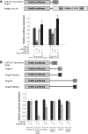
The 3′-UTR of ERBB-2 mRNA is a direct target of miR-331-3p via two miR-331-3p target sites. A, schematic representation of firefly luciferase reporter constructs for full-length, wild type ERBB-2 3′-UTR and perfect miR-331-3p target. 22RV1 cells were co-transfected with pmiR-REPORT constructs and miR-NC (1 n
m
), miR-331-3p (1 n
m
), LNA-NC (10 n
m
), or LNA-miR-331-3p (10 n
m
). B, schematic representation of firefly luciferase reporter constructs of wild type and mutant ERBB-2 3′-UTR miR-331-3p-A and -B target sites and perfect -3p target. 22RV1 cells were co-transfected with pmiR-REPORT and CMV-Renilla constructs, and miR-NC or miR-331-3p (1 n
m
), and assayed for firefly and Renilla luciferase activities after 24 h. Relative luciferase expression values are expressed as a ratio of miR-NC to LNA. Asterisk indicates significant difference between miR-NC transfected control cells (p < 0.05). All data are representative of at least three independent experiments. Error bars are as described in the legend to Fig. 2.
FIGURE 5.
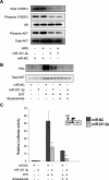
miR-331-3p decreases ERBB-2 protein expression and signaling, and blocks PSA expression and promoter activity in LNCaP cells. A, LNCaP cells were transfected with miR-NC or miR-331-3p (30 n
m
) for 48 h and serum starved for 24 h thereafter, followed by stimulation ± heregulin (HRG; 50 ng/ml) for 20 min. Cell lysates were analyzed for total ERBB-2, phospho-ERBB-2, AR, total AKT, and phospho-AKT expression by immunoblotting. B, LNCaP cells were transfected with miR-331-3p for 48 h and treated ± DHT (10 n
m
) and ± bicalutamide (10 μ
m
). Total PSA expression was determined by immunoblotting. C, LNCaP cells were co-transfected with a PSA-luciferase vector (PSA-LUC) and thymidine kinase-Renilla vector and with miR-NC or miR-331-3p (1 n
m
). Relative luciferase expression (firefly normalized to Renilla) values are expressed as a ratio of miR-NC-transfected cells (±S.D.). Asterisk indicates significant difference between miR-NC transfected control cells (p < 0.05). Error bars represent confidence intervals (CI = 0.95).
FIGURE 6.
miR-331-3p blocks AR signaling via inhibition of ERBB-2 expression and AKT activity in prostate cancer cells. AR antagonists such as bicalutamide bind to the AR and prevent its activation and expression of AR target genes, such as PSA. Nevertheless, AR signaling may persist in prostate cancer cells despite AR blockade, in part via increased expression of the ERBB-2 receptor tyrosine kinase and subsequent activation of the PI3K/AKT pathway, which causes AR phosphorylation and promotes expression of AR target genes. miR-331-3p directly targets the ERBB-2 mRNA 3′-UTR to regulate ERBB-2 protein expression, thereby reducing PI3K/AKT signaling and AR signaling. The combination of an AR antagonist (bicalutamide) and miR-331-3p effectively blocks AR signaling (PSA expression and PSA promoter activity) in LNCaP prostate cancer cells. (+) indicates activation step of pathway and (−) indicates inhibition of pathway component.
Similar articles
- miR-103a-2-5p/miR-30c-1-3p inhibits the progression of prostate cancer resistance to androgen ablation therapy via targeting androgen receptor variant 7.
Chen W, Yao G, Zhou K. Chen W, et al. J Cell Biochem. 2019 Aug;120(8):14055-14064. doi: 10.1002/jcb.28680. Epub 2019 Apr 8. J Cell Biochem. 2019. PMID: 30963631 - The RNA-binding protein HuR opposes the repression of ERBB-2 gene expression by microRNA miR-331-3p in prostate cancer cells.
Epis MR, Barker A, Giles KM, Beveridge DJ, Leedman PJ. Epis MR, et al. J Biol Chem. 2011 Dec 2;286(48):41442-41454. doi: 10.1074/jbc.M111.301481. Epub 2011 Oct 4. J Biol Chem. 2011. PMID: 21971048 Free PMC article. - [miR-141-3p regulates the expression of androgen receptor by targeting its 3'UTR in prostate cancer LNCaP cells].
Wang C, Ouyang Y, Lu M, Wei J, Zhang H. Wang C, et al. Xi Bao Yu Fen Zi Mian Yi Xue Za Zhi. 2015 Jun;31(6):736-9. Xi Bao Yu Fen Zi Mian Yi Xue Za Zhi. 2015. PMID: 26062412 Chinese. - Multifaceted Function of MicroRNA-299-3p Fosters an Antitumor Environment Through Modulation of Androgen Receptor and VEGFA Signaling Pathways in Prostate Cancer.
Ganapathy K, Staklinski S, Hasan MF, Ottman R, Andl T, Berglund AE, Park JY, Chakrabarti R. Ganapathy K, et al. Sci Rep. 2020 Mar 20;10(1):5167. doi: 10.1038/s41598-020-62038-3. Sci Rep. 2020. PMID: 32198489 Free PMC article. - Cancer Stem Cells and Androgen Receptor Signaling: Partners in Disease Progression.
Quintero JC, Díaz NF, Rodríguez-Dorantes M, Camacho-Arroyo I. Quintero JC, et al. Int J Mol Sci. 2023 Oct 11;24(20):15085. doi: 10.3390/ijms242015085. Int J Mol Sci. 2023. PMID: 37894767 Free PMC article. Review.
Cited by
- MicroRNA-125b down-regulation mediates endometrial cancer invasion by targeting ERBB2.
Shang C, Lu YM, Meng LR. Shang C, et al. Med Sci Monit. 2012 Apr;18(4):BR149-55. doi: 10.12659/msm.882617. Med Sci Monit. 2012. PMID: 22460089 Free PMC article. - Minireview: The roles of small RNA pathways in reproductive medicine.
Hawkins SM, Buchold GM, Matzuk MM. Hawkins SM, et al. Mol Endocrinol. 2011 Aug;25(8):1257-79. doi: 10.1210/me.2011-0099. Epub 2011 May 5. Mol Endocrinol. 2011. PMID: 21546411 Free PMC article. Review. - MicroRNA and HER2-overexpressing cancer.
Wang SE, Lin RJ. Wang SE, et al. Microrna. 2013;2(2):137-47. doi: 10.2174/22115366113029990011. Microrna. 2013. PMID: 25070783 Free PMC article. Review. - ARTIK-52 induces replication-dependent DNA damage and p53 activation exclusively in cells of prostate and breast cancer origin.
Fleyshman D, Cheney P, Ströse A, Mudambi S, Safina A, Commane M, Purmal A, Morgan K, Wang NJ, Gray J, Spellman PT, Issaeva N, Gurova K. Fleyshman D, et al. Cell Cycle. 2016;15(3):455-70. doi: 10.1080/15384101.2015.1127478. Epub 2015 Dec 22. Cell Cycle. 2016. PMID: 26694952 Free PMC article. - microRNAs Mediated Regulation of the Ribosomal Proteins and its Consequences on the Global Translation of Proteins.
Reza AMMT, Yuan YG. Reza AMMT, et al. Cells. 2021 Jan 8;10(1):110. doi: 10.3390/cells10010110. Cells. 2021. PMID: 33435549 Free PMC article. Review.
References
- Jemal A., Siegel R., Ward E., Murray T., Xu J., Thun M. J. (2007) CA Cancer J. Clin. 57, 43–66 - PubMed
- Craft N., Shostak Y., Carey M., Sawyers C. L. (1999) Nat. Med. 5, 280–285 - PubMed
- Festuccia C., Gravina G. L., Muzi P., Pomante R., Ventura L., Vessella R. L., Vicentini C., Bologna M. (2007) Endocr. Relat. Cancer 14, 601–611 - PubMed
Publication types
MeSH terms
Substances
LinkOut - more resources
Full Text Sources
Other Literature Sources
Medical
Research Materials
Miscellaneous


