Intratumoral CD8(+) T/FOXP3 (+) cell ratio is a predictive marker for survival in patients with colorectal cancer - PubMed (original) (raw)
Intratumoral CD8(+) T/FOXP3 (+) cell ratio is a predictive marker for survival in patients with colorectal cancer
Hiroyuki Suzuki et al. Cancer Immunol Immunother. 2010 May.
Abstract
The human immune system consists of a balance between immune surveillance against non-self antigens and tolerance of self-antigens. CD8(+) T cells and CD4(+) regulatory T cells (Tregs) are the main players for immune surveillance and tolerance, respectively. We examined immunohistochemically the immunological balance at the tumor site using 94 surgically resected colorectal cancer tissues. Forkhead box P3 (FOXP3)(+) cells were considered to be Tregs in the present study. The number of intratumoral FOXP3(+) cells (itFOXP3(+) cells) was positively correlated with lymph node metastases (P = 0.030). itCD8(+) T/itFOXP3(+) cell ratio negatively correlated with pathological stages (P = 0.048). Next, relationship between the number of itCD8(+) T cells or itFOXP3(+) cells and survival prognosis in 94 patients who underwent a curative resection was analyzed. Only itCD8(+) T/itFOXP3(+) cell ratio positively correlated with disease-free survival (0.023) and overall survival (P = 0.010). Multivariate analysis indicated that itCD8(+) T/itFOXP3(+) cell ratio is an independent prognostic factor (P = 0.035) of overall survival. The number of itFOXP3(+) cells positively correlated with transforming growth factor-beta TGF-beta production at the tumor site (P = 0.020). In conclusion, itCD8(+) T/itFOXP3(+) cell ratio is a predictive marker for both disease-free survival time and overall survival time in patients with colorectal cancer. Importantly, itCD8(+) T/itFOXP3(+) cell ratio may be an independent prognostic factor. And, tumor-producing TGF-beta may contribute to the increased number of itFOXP3(+) cells.
Figures
Fig. 1
Immunohistochemical staining shows that FOXP3+ lymphocytes are distinguishable from CD8+ lymphocytes in colorectal cancer. a FOXP3 positive cells are stained brown, as shown by asterisks (×400). b CD8 positive cells are stained red, as shown by arrows (×400). c Double staining of FOXP3 (asterisks, in cell nucleus) and CD8 (arrows, on cell membrane) (×400). d The correlation between CD8+ T cells and FOXP3+ cells is shown (Mann–Whitney U test). P values less than 0.05 were considered significant (color figure online)
Fig. 2
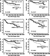
The itCD8+ T/itFOXP3+ cell ratio predicts poor disease-free survival and overall survival in 94 colorectal cancer patients. itCD8+ T cells (a and b) and itFOXP3+ cells (c and d) correlated with neither disease-free survival nor with overall survival. Disease-free survival and overall survival of the patients showing high an itCD8+ T/itFOXP3+ cell ratio were significantly better compared with those of the patients showing a low itCD8+ T/itFOXP3+ cell ratio (e and f). P values less than 0.05 were considered significant
Fig. 3
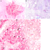
Representative pictures of immunohistochemical double staining. a A high TGF-β-expressing specimen (red) (×400). b A low TGF-β-expressing specimen are shown (×400). c There are a high number of FOXP3+ cells (brown) indicated by asterisks, in the high TGF-β expressing area (×400) (color figure online)
Comment in
- Risk factors. CD8+:FOXP3+ cell ratio is a novel survival marker for colorectal cancer.
Kirk R. Kirk R. Nat Rev Clin Oncol. 2010 Jun;7(6):299. doi: 10.1038/nrclinonc.2010.79. Nat Rev Clin Oncol. 2010. PMID: 20527684 No abstract available.
Similar articles
- CD4/CD8 + T cells, DC subsets, Foxp3, and IDO expression are predictive indictors of gastric cancer prognosis.
Li F, Sun Y, Huang J, Xu W, Liu J, Yuan Z. Li F, et al. Cancer Med. 2019 Dec;8(17):7330-7344. doi: 10.1002/cam4.2596. Epub 2019 Oct 20. Cancer Med. 2019. PMID: 31631566 Free PMC article. - Tumor-infiltrating lymphocytes, tumor characteristics, and recurrence in patients with early breast cancer.
Kim ST, Jeong H, Woo OH, Seo JH, Kim A, Lee ES, Shin SW, Kim YH, Kim JS, Park KH. Kim ST, et al. Am J Clin Oncol. 2013 Jun;36(3):224-31. doi: 10.1097/COC.0b013e3182467d90. Am J Clin Oncol. 2013. PMID: 22495453 - Prognostic value of intra-tumoral CD8+ /FoxP3+ lymphocyte ratio in patients with resected colorectal cancer liver metastasis.
Sideras K, Galjart B, Vasaturo A, Pedroza-Gonzalez A, Biermann K, Mancham S, Nigg AL, Hansen BE, Stoop HA, Zhou G, Verhoef C, Sleijfer S, Sprengers D, Kwekkeboom J, Bruno MJ. Sideras K, et al. J Surg Oncol. 2018 Jul;118(1):68-76. doi: 10.1002/jso.25091. Epub 2018 Jun 7. J Surg Oncol. 2018. PMID: 29878369 Free PMC article. - T cell subsets and colorectal cancer: discerning the good from the bad.
Scurr M, Gallimore A, Godkin A. Scurr M, et al. Cell Immunol. 2012 Sep;279(1):21-4. doi: 10.1016/j.cellimm.2012.08.004. Epub 2012 Sep 14. Cell Immunol. 2012. PMID: 23041206 Review. - FOXP3+ Tregs: heterogeneous phenotypes and conflicting impacts on survival outcomes in patients with colorectal cancer.
Zhuo C, Xu Y, Ying M, Li Q, Huang L, Li D, Cai S, Li B. Zhuo C, et al. Immunol Res. 2015 Mar;61(3):338-47. doi: 10.1007/s12026-014-8616-y. Immunol Res. 2015. PMID: 25608795 Review.
Cited by
- Superior antitumor immune response achieved with proton over photon immunoradiotherapy is amplified by the nanoradioenhancer NBTXR3.
Hu Y, Paris S, Sahoo N, Wang Q, Wang Q, Barsoumian HB, Huang A, Da Silva J, Bienassis C, Leyton CSK, Voss TA, Masrorpour F, Riad T, Leuschner C, Puebla-Osorio N, Gandhi S, Nguyen QN, Wang J, Cortez MA, Welsh JW. Hu Y, et al. J Nanobiotechnology. 2024 Oct 1;22(1):597. doi: 10.1186/s12951-024-02855-0. J Nanobiotechnology. 2024. PMID: 39354474 Free PMC article. - DNA Methylation-Derived Immune Cell Proportions and Cancer Risk in Black Participants.
Semancik CS, Zhao N, Koestler DC, Boerwinkle E, Bressler J, Buchsbaum RJ, Kelsey KT, Platz EA, Michaud DS. Semancik CS, et al. Cancer Res Commun. 2024 Oct 1;4(10):2714-2723. doi: 10.1158/2767-9764.CRC-24-0257. Cancer Res Commun. 2024. PMID: 39324671 Free PMC article. - Prognostic value of tumor-infiltrating lymphocytes and PD-L1 expression in esophageal squamous cell carcinoma.
Hu J, Toyozumi T, Murakami K, Endo S, Matsumoto Y, Otsuka R, Shiraishi T, Iida S, Morishita H, Makiyama T, Nishioka Y, Uesato M, Hayano K, Nakano A, Matsubara H. Hu J, et al. Cancer Med. 2024 Sep;13(17):e70179. doi: 10.1002/cam4.70179. Cancer Med. 2024. PMID: 39264227 Free PMC article. - Inferring Characteristics of the Tumor Immune Microenvironment of Patients with HNSCC from Single-Cell Transcriptomics of Peripheral Blood.
Cao Y, Chang T, Schischlik F, Wang K, Sinha S, Hannenhalli S, Jiang P, Ruppin E. Cao Y, et al. Cancer Res Commun. 2024 Sep 1;4(9):2335-2348. doi: 10.1158/2767-9764.CRC-24-0092. Cancer Res Commun. 2024. PMID: 39113621 Free PMC article. - In situ analysis of CCR8+ regulatory T cells in lung cancer: suppression of GzmB+ CD8+ T cells and prognostic marker implications.
Hayashi Y, Ueyama A, Funaki S, Jinushi K, Higuchi N, Morihara H, Hirata M, Nagira Y, Saito T, Kawashima A, Iwahori K, Shintani Y, Wada H. Hayashi Y, et al. BMC Cancer. 2024 May 23;24(1):627. doi: 10.1186/s12885-024-12363-x. BMC Cancer. 2024. PMID: 38783281 Free PMC article.
References
Publication types
MeSH terms
Substances
LinkOut - more resources
Full Text Sources
Medical
Research Materials


