Characterization of TEM1/endosialin in human and murine brain tumors - PubMed (original) (raw)
Characterization of TEM1/endosialin in human and murine brain tumors
Eleanor B Carson-Walter et al. BMC Cancer. 2009.
Abstract
Background: TEM1/endosialin is an emerging microvascular marker of tumor angiogenesis. We characterized the expression pattern of TEM1/endosialin in astrocytic and metastatic brain tumors and investigated its role as a therapeutic target in human endothelial cells and mouse xenograft models.
Methods: In situ hybridization (ISH), immunohistochemistry (IH) and immunofluorescence (IF) were used to localize TEM1/endosialin expression in grade II-IV astrocytomas and metastatic brain tumors on tissue microarrays. Changes in TEM1/endosialin expression in response to pro-angiogenic conditions were assessed in human endothelial cells grown in vitro. Intracranial U87MG glioblastoma (GBM) xenografts were analyzed in nude TEM1/endosialin knockout (KO) and wildtype (WT) mice.
Results: TEM1/endosialin was upregulated in primary and metastatic human brain tumors, where it localized primarily to the tumor vasculature and a subset of tumor stromal cells. Analysis of 275 arrayed grade II-IV astrocytomas demonstrated TEM1/endosialin expression in 79% of tumors. Robust TEM1/endosialin expression occurred in 31% of glioblastomas (grade IV astroctyomas). TEM1/endosialin expression was inversely correlated with patient age. TEM1/endosialin showed limited co-localization with CD31, alphaSMA and fibronectin in clinical specimens. In vitro, TEM1/endosialin was upregulated in human endothelial cells cultured in matrigel. Vascular Tem1/endosialin was induced in intracranial U87MG GBM xenografts grown in mice. Tem1/endosialin KO vs WT mice demonstrated equivalent survival and tumor growth when implanted with intracranial GBM xenografts, although Tem1/endosialin KO tumors were significantly more vascular than the WT counterparts.
Conclusion: TEM1/endosialin was induced in the vasculature of high-grade brain tumors where its expression was inversely correlated with patient age. Although lack of TEM1/endosialin did not suppress growth of intracranial GBM xenografts, it did increase tumor vascularity. The cellular localization of TEM1/endosialin and its expression profile in primary and metastatic brain tumors support efforts to therapeutically target this protein, potentially via antibody mediated drug delivery strategies.
Figures
Figure 1
Localization of TEM1/endosialin expression. A. Representative RT-PCR for TEM1/endosialin (TEM1) in whole cell lysates from two GBMs (T1, T2), two metastatic brain tumors (T3, T4) and two non-neoplastic controls (N1, N2). Expression was normalized to von Willebrand Factor (vWF), an endothelial positive control. B. Combined quantitative RT-PCR results for TEM1/endosialin in two controls (N), five GBMs (GBM) and five metastatic tumors (Met). Error bars, standard error of the mean. *p = 0.05. C. TEM1/endosialin was visualized by immunohistochemistry (IHC) using the TEM1/33 custom antibody or in situ hybridization (ISH) in glioblastoma (GBM), metastatic brain tumor (MET) and non-neoplastic brain tissues (Normal). Staining for vWF was used as an endothelial positive control. Original magnification for whole spot, 40×; detail, 100×.
Figure 2
Immunofluorescence for TEM1/endosialin: co-localization in tumor specimens. TEM1 and CD31 (top panel), αSMA (middle panel) and fibronectin (FN, bottom panel) co-localize to microvessels in human GBMs, but demonstrate limited cellular overlap. Anti-TEM1 (TEM1/33), red. Anti-CD31, anti-αSMA and anti-FN, green. Nuclei, blue. Scale bar, 20 μm.
Figure 3
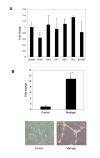
Expression of TEM1/endosialin by cultured endothelial cells. A. Expression of TEM1/endosialin after treatment of endothelial cells with pro-angiogenic growth factors. Cells were treated for 48 hours (see Methods) and analyzed by quantitative RT-PCR. Error bars, standard error of the mean. No significant change in TEM1/endosialin was detected. B. Induction of TEM1/endosialin after growth of endothelial cells in matrigel. Cells were grown on plastic (Control) or in matrigel (Matrigel) for 6 hours then analyzed by quantitative RT-PCR. Error bars, standard error of the mean. *p = 0.05. After 6 hours on matrigel and prior to harvest, HMVEC cells demonstrate branching tubulogenesis (lower right) while cells grown on plastic do not (lower left).
Figure 4
Tem1/endosialin expression in mouse brain tumor models. A. Induction of Tem1/endosialin in intracranial U87MG xenografts grown in nude mice. ISH with mouse specific riboprobes detected Tem1/endosialin (Tem1) in tumor (T) but not normal mouse brain (NL). Probes for mouse vWF were used as a positive endothelial control (vWF). Original magnification, 200×. B. Survival of nude TEM1/endosialin WT and KO mice after intracranial injection with 105 U87MG glioblastoma cells. Animals sacrificed after 100 days showed no evidence of tumor take. C. Tumor vessels stained for vWF in intracranial xenografts from nude TEM1/endosialin WT or KO mice. Size bar, 50 μm. D. Comparison of vessel numbers from nude TEM1/endosialin WT and KO tumors. Ten non-overlapping high power fields were counted per animal. Error bars, standard error of the mean. *p = 0.001.
Similar articles
- Interaction of endosialin/TEM1 with extracellular matrix proteins mediates cell adhesion and migration.
Tomkowicz B, Rybinski K, Foley B, Ebel W, Kline B, Routhier E, Sass P, Nicolaides NC, Grasso L, Zhou Y. Tomkowicz B, et al. Proc Natl Acad Sci U S A. 2007 Nov 13;104(46):17965-70. doi: 10.1073/pnas.0705647104. Epub 2007 Nov 6. Proc Natl Acad Sci U S A. 2007. PMID: 17986615 Free PMC article. - Endosialin: molecular and functional links to tumor angiogenesis.
Kontsekova S, Polcicova K, Takacova M, Pastorekova S. Kontsekova S, et al. Neoplasma. 2016;63(2):183-92. doi: 10.4149/202_15090N474. Neoplasma. 2016. PMID: 26774137 Review. - Endosialin (Tem1) is a marker of tumor-associated myofibroblasts and tumor vessel-associated mural cells.
Christian S, Winkler R, Helfrich I, Boos AM, Besemfelder E, Schadendorf D, Augustin HG. Christian S, et al. Am J Pathol. 2008 Feb;172(2):486-94. doi: 10.2353/ajpath.2008.070623. Epub 2008 Jan 10. Am J Pathol. 2008. PMID: 18187565 Free PMC article. - Human endothelial precursor cells express tumor endothelial marker 1/endosialin/CD248.
Bagley RG, Rouleau C, St Martin T, Boutin P, Weber W, Ruzek M, Honma N, Nacht M, Shankara S, Kataoka S, Ishida I, Roberts BL, Teicher BA. Bagley RG, et al. Mol Cancer Ther. 2008 Aug;7(8):2536-46. doi: 10.1158/1535-7163.MCT-08-0050. Mol Cancer Ther. 2008. PMID: 18723498 - Endosialin in Cancer: Expression Patterns, Mechanistic Insights, and Therapeutic Approaches.
Lu S, Gan L, Lu T, Zhang K, Zhang J, Wu X, Han D, Xu C, Liu S, Yang F, Qin W, Wen W. Lu S, et al. Theranostics. 2024 Jan 1;14(1):379-391. doi: 10.7150/thno.89495. eCollection 2024. Theranostics. 2024. PMID: 38164138 Free PMC article. Review.
Cited by
- CD248 facilitates tumor growth via its cytoplasmic domain.
Maia M, DeVriese A, Janssens T, Moons M, Lories RJ, Tavernier J, Conway EM. Maia M, et al. BMC Cancer. 2011 May 8;11:162. doi: 10.1186/1471-2407-11-162. BMC Cancer. 2011. PMID: 21549007 Free PMC article. - CD93 promotes β1 integrin activation and fibronectin fibrillogenesis during tumor angiogenesis.
Lugano R, Vemuri K, Yu D, Bergqvist M, Smits A, Essand M, Johansson S, Dejana E, Dimberg A. Lugano R, et al. J Clin Invest. 2018 Aug 1;128(8):3280-3297. doi: 10.1172/JCI97459. Epub 2018 Jun 25. J Clin Invest. 2018. PMID: 29763414 Free PMC article. - A differential role for CD248 (Endosialin) in PDGF-mediated skeletal muscle angiogenesis.
Naylor AJ, McGettrick HM, Maynard WD, May P, Barone F, Croft AP, Egginton S, Buckley CD. Naylor AJ, et al. PLoS One. 2014 Sep 22;9(9):e107146. doi: 10.1371/journal.pone.0107146. eCollection 2014. PLoS One. 2014. PMID: 25243742 Free PMC article. - Vascular targeting of nanocarriers: perplexing aspects of the seemingly straightforward paradigm.
Howard M, Zern BJ, Anselmo AC, Shuvaev VV, Mitragotri S, Muzykantov V. Howard M, et al. ACS Nano. 2014 May 27;8(5):4100-32. doi: 10.1021/nn500136z. Epub 2014 May 7. ACS Nano. 2014. PMID: 24787360 Free PMC article. Review. - Tumor endothelial marker 1-specific DNA vaccination targets tumor vasculature.
Facciponte JG, Ugel S, De Sanctis F, Li C, Wang L, Nair G, Sehgal S, Raj A, Matthaiou E, Coukos G, Facciabene A. Facciponte JG, et al. J Clin Invest. 2014 Apr;124(4):1497-511. doi: 10.1172/JCI67382. Epub 2014 Mar 18. J Clin Invest. 2014. PMID: 24642465 Free PMC article.
References
- St Croix B, Rago C, Velculescu V, Traverso G, Romans KE, Montgomery E, Lal A, Riggins GJ, Lengauer C, Vogelstein B. Genes expressed in human tumor endothelium. Science (New York, NY) 2000;289(5482):1197–1202. - PubMed
- Christian S, Ahorn H, Koehler A, Eisenhaber F, Rodi HP, Garin-Chesa P, Park JE, Rettig WJ, Lenter MC. Molecular cloning and characterization of endosialin, a C-type lectin-like cell surface receptor of tumor endothelium. The Journal of biological chemistry. 2001;276(10):7408–7414. doi: 10.1074/jbc.M009604200. - DOI - PubMed
- Rettig WJ, Garin-Chesa P, Healey JH, Su SL, Jaffe EA, Old LJ. Identification of endosialin, a cell surface glycoprotein of vascular endothelial cells in human cancer. Proceedings of the National Academy of Sciences of the United States of America. 1992;89(22):10832–10836. doi: 10.1073/pnas.89.22.10832. - DOI - PMC - PubMed
Publication types
MeSH terms
Substances
LinkOut - more resources
Full Text Sources
Other Literature Sources
Medical
Research Materials
Miscellaneous


