The origin and pathogenesis of epithelial ovarian cancer: a proposed unifying theory - PubMed (original) (raw)
The origin and pathogenesis of epithelial ovarian cancer: a proposed unifying theory
Robert J Kurman et al. Am J Surg Pathol. 2010 Mar.
Abstract
Ovarian cancer is the most lethal gynecologic malignancy. Efforts at early detection and new therapeutic approaches to reduce mortality have been largely unsuccessful, because the origin and pathogenesis of epithelial ovarian cancer are poorly understood. Despite numerous studies that have carefully scrutinized the ovaries for precursor lesions, none have been found. This has led to the proposal that ovarian cancer develops de novo. Studies have shown that epithelial ovarian cancer is not a single disease but is composed of a diverse group of tumors that can be classified based on distinctive morphologic and molecular genetic features. One group of tumors, designated type I, is composed of low-grade serous, low-grade endometrioid, clear cell, mucinous and transitional (Brenner) carcinomas. These tumors generally behave in an indolent fashion, are confined to the ovary at presentation and, as a group, are relatively genetically stable. They lack mutations of TP53, but each histologic type exhibits a distinctive molecular genetic profile. Moreover, the carcinomas exhibit a shared lineage with the corresponding benign cystic neoplasm, often through an intermediate (borderline tumor) step, supporting the morphologic continuum of tumor progression. In contrast, another group of tumors, designated type II, is highly aggressive, evolves rapidly and almost always presents in advanced stage. Type II tumors include conventional high-grade serous carcinoma, undifferentiated carcinoma, and malignant mixed mesodermal tumors (carcinosarcoma). They displayTP53 mutations in over 80% of cases and rarely harbor the mutations that are found in the type I tumors. Recent studies have also provided cogent evidence that what have been traditionally thought to be primary ovarian tumors actually originate in other pelvic organs and involve the ovary secondarily. Thus, it has been proposed that serous tumors arise from the implantation of epithelium (benign or malignant) from the fallopian tube. Endometrioid and clear cell tumors have been associated with endometriosis that is regarded as the precursor of these tumors. As it is generally accepted that endometriosis develops from endometrial tissue by retrograde menstruation, it is reasonable to assume that the endometrium is the source of these ovarian neoplasms. Finally, preliminary data suggest that mucinous and transitional (Brenner) tumors arise from transitional-type epithelial nests at the tubal-mesothelial junction by a process of metaplasia. Appreciation of these new concepts will allow for a more rationale approach to screening, treatment, and prevention that potentially can have a significant impact on reducing the mortality of this devastating disease.
Figures
Figure 1
Serous tubal intraepithelial carcinoma (STIC). A. High magnification. Hematoxylin and eosin stain. B. Immunohistochemical stain for p53. An asterisk defines the boundary of the lesion.
Figure 2
Transfer of normal tubal epithelium to the ovary. A. Anatomical relationship of fallopian tube to the ovary at the time of ovulation. The fimbria envelops the ovary. B. Ovulation. The ovarian surface ruptures with expulsion and transfer of the oocyte to the fimbria. The fimbria is in intimate contact with the ovary at the site of rupture. C. Tubal epithelial cells from the fimbria are dislodged and implant on the denuded surface of the ovary resulting in the formation of an inclusion cyst.
Figure 3
Proposed development of low-grade (LG) and high-grade (HG) serous carcinoma. A. One mechanism involves normal tubal epithelium that is shed from the fimbria, which implants on the ovary to form an inclusion cyst. Depending on whether there is a mutation of KRAS/BRAF/ERRB2 or TP53 a low-grade or high-grade serous carcinoma develops respectively. Low-grade serous carcinoma often develops from a serous borderline tumor (SBT), which in turn arises from a serous cystadenoma. Another mechanism involves exfoliation of malignant cells from a serous tubal intraepithelial carcinoma (STIC) that implants on the ovarian surface resulting in the development of a high-grade serous carcinoma. B. A schematic representation of direct dissemination or shedding of STIC cells onto the ovarian surface where the carcinoma cells ultimately establish a tumor mass that is presumably arising from the ovary. Of note there may be stages of tumor progression that precede the formation of a STIC.
Figure 4
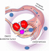
Proposed development of low-grade endometrioid and clear cell carcinoma. Endometrial tissue, by a process of retrograde menstruation, implants on the ovarian surface to form an endometriotic cyst from which a low-grade endometrioid or clear cell carcinoma can develop. EMC: low-grade endometrioid carcinoma of the ovary; CCC: clear cell carcinoma of the ovary.
Figure 5
Comparison of the immunohistochemical staining pattern for ovarian surface epithelium (mesothelium), normal fallopian tube epithelium, and high-grade serous carcinoma. PAX8 is a marker of mullerian-type epithelium such as fallopian tube epithelium and calretinin is a marker of mesothelium.
Similar articles
- Molecular pathogenesis and extraovarian origin of epithelial ovarian cancer--shifting the paradigm.
Kurman RJ, Shih IeM. Kurman RJ, et al. Hum Pathol. 2011 Jul;42(7):918-31. doi: 10.1016/j.humpath.2011.03.003. Hum Pathol. 2011. PMID: 21683865 Free PMC article. Review. - Precursors and pathogenesis of ovarian carcinoma.
Lim D, Oliva E. Lim D, et al. Pathology. 2013 Apr;45(3):229-42. doi: 10.1097/PAT.0b013e32835f2264. Pathology. 2013. PMID: 23478230 Review. - Origin and molecular pathogenesis of ovarian high-grade serous carcinoma.
Kurman RJ. Kurman RJ. Ann Oncol. 2013 Dec;24 Suppl 10:x16-21. doi: 10.1093/annonc/mdt463. Ann Oncol. 2013. PMID: 24265397 Review. - Recent concepts of ovarian carcinogenesis: type I and type II.
Koshiyama M, Matsumura N, Konishi I. Koshiyama M, et al. Biomed Res Int. 2014;2014:934261. doi: 10.1155/2014/934261. Epub 2014 Apr 23. Biomed Res Int. 2014. PMID: 24868556 Free PMC article. Review. - Pathogenesis of ovarian cancer: lessons from morphology and molecular biology and their clinical implications.
Kurman RJ, Shih IeM. Kurman RJ, et al. Int J Gynecol Pathol. 2008 Apr;27(2):151-60. doi: 10.1097/PGP.0b013e318161e4f5. Int J Gynecol Pathol. 2008. PMID: 18317228 Free PMC article. Review.
Cited by
- Ovarian Cancer: From Precursor Lesion Identification to Population-Based Prevention Programs.
Sowamber R, Lukey A, Huntsman D, Hanley G. Sowamber R, et al. Curr Oncol. 2023 Nov 29;30(12):10179-10194. doi: 10.3390/curroncol30120741. Curr Oncol. 2023. PMID: 38132375 Free PMC article. Review. - Insulin Resistance: The Increased Risk of Cancers.
Szablewski L. Szablewski L. Curr Oncol. 2024 Feb 13;31(2):998-1027. doi: 10.3390/curroncol31020075. Curr Oncol. 2024. PMID: 38392069 Free PMC article. Review. - Associations between Diabetes Mellitus and Selected Cancers.
Pliszka M, Szablewski L. Pliszka M, et al. Int J Mol Sci. 2024 Jul 8;25(13):7476. doi: 10.3390/ijms25137476. Int J Mol Sci. 2024. PMID: 39000583 Free PMC article. Review. - Tumour Versus Germline BRCA Testing in Ovarian Cancer: A Single-Site Institution Experience in the United Kingdom.
Akaev I, Rahimi S, Onifade O, Gardner FJE, Castells-Rufas D, Jones E, Acharige S, Yeoh CC. Akaev I, et al. Diagnostics (Basel). 2021 Mar 19;11(3):547. doi: 10.3390/diagnostics11030547. Diagnostics (Basel). 2021. PMID: 33808557 Free PMC article. - GEICO (Spanish Group for Investigation on Ovarian Cancer) treatment guidelines in ovarian cancer 2012.
González Martín A, Redondo A, Jurado M, De Juan A, Romero I, Bover I, Del Campo JM, Cervantes A, García Y, López-Guerrero JA, Mendiola C, Palacios J, Rubio MJ, Poveda Velasco A. González Martín A, et al. Clin Transl Oncol. 2013 Jul;15(7):509-25. doi: 10.1007/s12094-012-0995-8. Epub 2013 Mar 7. Clin Transl Oncol. 2013. PMID: 23468275 Free PMC article.
References
- Bell DA, Scully RE. Early de novo ovarian carcinoma. A study of fourteen cases. Cancer. 1994;73:1859–64. - PubMed
- Bulun SE. Endometriosis. N Engl J Med. 2009;360:268–79. - PubMed
- Callahan MJ, Crum CP, Medeiros F, et al. Primary fallopian tube malignancies in BRCA-positive women undergoing surgery for ovarian cancer risk reduction. J Clin Oncol. 2007;25:3985–90. - PubMed
- Carcangiu ML, Radice P, Manoukian S, et al. Atypical epithelial proliferation in fallopian tubes in prophylactic salpingo-oophorectomy specimens from BRCA1 and BRCA2 germline mutation carriers. Int J Gynecol Pathol. 2004;23:35–40. - PubMed
Publication types
MeSH terms
Substances
Grants and funding
- R01 CA116184-04/CA/NCI NIH HHS/United States
- R01CA116184/CA/NCI NIH HHS/United States
- R01 CA116184/CA/NCI NIH HHS/United States
- R01CA129080/CA/NCI NIH HHS/United States
- R01CA103937/CA/NCI NIH HHS/United States
LinkOut - more resources
Full Text Sources
Other Literature Sources
Medical
Research Materials
Miscellaneous


