Oncogenic KRAS modulates mitochondrial metabolism in human colon cancer cells by inducing HIF-1α and HIF-2α target genes - PubMed (original) (raw)
Oncogenic KRAS modulates mitochondrial metabolism in human colon cancer cells by inducing HIF-1α and HIF-2α target genes
Sang Y Chun et al. Mol Cancer. 2010.
Abstract
Background: Activating KRAS mutations are important for cancer initiation and progression; and have recently been shown to cause primary resistance to therapies targeting the epidermal growth factor receptor. Therefore, strategies are currently in development to overcome treatment resistance due to oncogenic KRAS. The hypoxia-inducible factors-1α and -2α (HIF-1α and HIF-2α) are activated in cancer due to dysregulated ras signaling.
Methods: To understand the individual and combined roles of HIF-1α and HIF-2α in cancer metabolism and oncogenic KRAS signaling, we used targeted homologous recombination to disrupt the oncogenic KRAS, HIF-1α, and HIF-2α gene loci in HCT116 colon cancer cells to generate isogenic HCT116WT KRAS, HCT116HIF-1α-/-, HCT116HIF-2α-/-, and HCT116HIF-1α-/-HIF-2α-/- cell lines.
Results: Global gene expression analyses of these cell lines reveal that HIF-1α and HIF-2α work together to modulate cancer metabolism and regulate genes signature overlapping with oncogenic KRAS. Cancer cells with disruption of both HIF-1α and HIF-2α or oncogenic KRAS showed decreased aerobic respiration and ATP production, with increased ROS generation.
Conclusion: Our findings suggest novel strategies for treating tumors with oncogenic KRAS mutations.
Figures
Figure 1
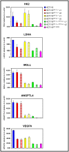
Analyses of genes regulated by oncogenic KRAS, HIF-1α, HIF-2α, and both HIF-1α and HIF-2α. Expression of target genes in HCT116, HCT116HIF-1α-/-, HCT116HIF-2α-/-, HCT116HIF-1α-/-HIF-2α-/-, and HCT116WT KRAS cell lines. HK2, hexokinase 2; LDHA, lactate dehydrogenase A; MGLL, monoglyceride lipase; ANGPTL4, angiopoietin-like protein 4; and VEGFA, vascular endothelial growth factor A relative to β-actin were measured by real-time reverse transcription-PCR (n = 5). Bars, stdev. p < 0.05 by Student's t test comparing knockout cells with parental HCT116 cells. c, clone.
Figure 2
Analyses of metabolism gene set regulated by oncogenic KRAS and HIF. A, heatmap analysis comparing the following metabolism gene sets: HCT116 vs HCT116HIF-1α-/-, HCT116 vs HCT116HIF-2α-/-, HCT116 vs HCT116HIF-1α-/-HIF-2α-/-, and HCT116 vs HCT116WT KRAS cells. Expression values are averages of three samples. B, Venn diagrams analyzing the extent of overlap of the following metabolism gene sets: 1) HCT116 vs HCT116HIF-1α-/-, HCT116 vs HCT116HIF-2α-/-, and HCT116 vs HCT116HIF-1α-/-HIF-2α-/-; 2) HCT116 vs HCT116HIF-1α-/-HIF-2α-/- and HCT116 vs HCT116WT KRAS. Each circle represents a single gene set; and the number of genes common between the gene sets is denoted within the overlaps of the circles. C, Clonogenic survival assay of HCT116, HCT116HIF-1α-/-, HCT116HIF-2α-/-, HCT116HIF-1α-/-HIF-2α-/-, and HCT116WT KRAS cells. c, clone.
Figure 3
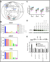
Analyses of target genes regulating phospholipids synthesis. A, overview of mitochondrial respiration and the contributory role of mitochondrial phospholipids synthesis. B, expression of genes regulating phospholipids synthesis in HCT116, HCT116HIF-1α-/-, HCT116HIF-2α-/-, HCT116HIF-1α-/-HIF-2α-/-, and HCT116WT KRAS cell lines. c, clone. ACSL5, acyl-CoA synthetase 5; AGPAT7, 1-acyl-sn-glycerol-3-phosphate acyltransferase 7; and PCK2, phosphoenolpyruvate carboxykinase 2 relative to β-actin were measured by real-time reverse transcription-PCR (n = 5). Bars, stdev. p < 0.05 by Student's t test comparing knockout cells with parental HCT116 cells. C, Expression of ACSL5, AGPAT7, and PCK2 in primary colon cancers. Gene expression, relative to β-ACTIN, in normal mucosa and primary tumor was determined by real-time RT-PCR. Log expression values were graphed as box and whiskers plots; showing median expression (horizontal line), surrounded by the first and third quartiles of those values (box), and the extreme values (whiskers). Hypothesis testing was performed using the Wilcoxon signed-rank test, with * = p < 0.05 considered statistically significant, comparing tumor to mucosa. D, western blot of ACSL5 in cell lines, with antibody to α-tubulin as loading control. c, clone. E, ACSL5 promoter activity in HCT116, HCT116HIF-1α-/-, HCT116HIF-2α-/-, and HCT116HIF-1α-/-HIF-2α-/- cells (n = 5). Diamond represents HRE site. The pGL3pro construct is a minimal promoter Firefly luciferase reporter. The ACSL5-pGL3pro construct contains an 1883 bp fragment of the 5' untranslated region of the ACSL5 gene with the HRE site. This construct is subjected to site-directed mutagenesis at the HRE site to generate the ACSL5(-HRE)-pGL3pro. Bars, stdev. p < 0.05 by Student's t test comparing transfection with pGL3pro construct versus ACSL5-pGL3pro or ACSL5(-HRE)-pGL3pro constructs.
Figure 4
Quantitation of total cellular (A) phosphatidyl choline (PC) and (B) cardiolipin (CL) levels in HCT116, HCT116HIF-1α-/-HIF-2α-/-, HCT116WT KRAS cells, HCT116 cells transduced with control scramble shRNA, and HCT116 cells transduced with ACSL5 shRNA (n = 3). Bars, stdev. p < 0.05 by Student's t test comparing knockout cells or shRNA transduced cells with parental HCT116 cells. Four lentiviral constructs carrying ACSL5 shRNA were tested for effectiveness in suppressing ACSL5 expression in comparison to the lentiviral construct carrying control scramble shRNA (Additional file 3, Fig. S3). Clone H45551 was chosen for subsequent experiments as it was the most effective (Additional file 3, Fig. S3).
Figure 5
Effects of genetic inactivation of oncogenic KRAS or both HIF-1α and HIF-2α on mitochondrial metabolism. A, change in cellular O2 consumption; B, change in MTT reduction; and C, change in intracellular ATP level in HCT116HIF-1α-/-HIF-2α-/- and HCT116WT KRAS relative to HCT116 cells (n = 5). Bars, stdev. p < 0.05 by Student's t test comparing knockout cells with parental HCT116 cells. D, cellular and mitochondrial ROS levels in HCT116, HCT116HIF-1α-/-HIF-2α-/-, and HCT116WT KRAS cells, as measured by the fluorescent dyes CM-H2DCFDA and MitoSOX.
Figure 6
Effects of ACSL5 gene knockdown on mitochondrial metabolism and tumorigenesis. A, change in cellular O2 consumption; and B, change in intracellular ATP level in HCT116 and LOVO cells transduced with ACSL5 shRNA relative to control scramble shRNA (n = 5). Bars, stdev. p < 0.05 by Student's t test comparing cells transduced with ACSL5 shRNA versus control scramble shRNA. C, cellular and mitochondrial ROS levels in HCT116 cells transduced with ACSL5 shRNA versus control scramble shRNA, as measured by the fluorescent dyes CM-H2DCFDA and MitoSOX.
Similar articles
- Oncogenic KRAS and BRAF differentially regulate hypoxia-inducible factor-1alpha and -2alpha in colon cancer.
Kikuchi H, Pino MS, Zeng M, Shirasawa S, Chung DC. Kikuchi H, et al. Cancer Res. 2009 Nov 1;69(21):8499-506. doi: 10.1158/0008-5472.CAN-09-2213. Epub 2009 Oct 20. Cancer Res. 2009. PMID: 19843849 Free PMC article. - Multidimensional Screening Platform for Simultaneously Targeting Oncogenic KRAS and Hypoxia-Inducible Factors Pathways in Colorectal Cancer.
Bousquet MS, Ma JJ, Ratnayake R, Havre PA, Yao J, Dang NH, Paul VJ, Carney TJ, Dang LH, Luesch H. Bousquet MS, et al. ACS Chem Biol. 2016 May 20;11(5):1322-31. doi: 10.1021/acschembio.5b00860. Epub 2016 Mar 3. ACS Chem Biol. 2016. PMID: 26938486 Free PMC article. - Increased activation of the hypoxia-inducible factor pathway in varicose veins.
Lim CS, Kiriakidis S, Paleolog EM, Davies AH. Lim CS, et al. J Vasc Surg. 2012 May;55(5):1427-39. doi: 10.1016/j.jvs.2011.10.111. Epub 2012 Jan 24. J Vasc Surg. 2012. PMID: 22277691 - Multiplicity of hypoxia-inducible transcription factors and their connection to the circadian clock in the zebrafish.
Pelster B, Egg M. Pelster B, et al. Physiol Biochem Zool. 2015 Mar-Apr;88(2):146-57. doi: 10.1086/679751. Epub 2015 Jan 14. Physiol Biochem Zool. 2015. PMID: 25730270 Review. - Combination therapy targeting cancer metabolism.
Wenger JB, Chun SY, Dang DT, Luesch H, Dang LH. Wenger JB, et al. Med Hypotheses. 2011 Feb;76(2):169-72. doi: 10.1016/j.mehy.2010.09.008. Epub 2010 Oct 13. Med Hypotheses. 2011. PMID: 20947261 Free PMC article. Review.
Cited by
- Antimigratory Effect of Lipophilic Cations Derived from Gallic and Gentisic Acid and Synergistic Effect with 5-Fluorouracil on Metastatic Colorectal Cancer Cells: A New Synthesis Route.
Suárez-Rozas C, Jara JA, Cortés G, Rojas D, Araya-Valdés G, Molina-Berrios A, González-Herrera F, Fuentes-Retamal S, Aránguiz-Urroz P, Campodónico PR, Maya JD, Vivar R, Catalán M. Suárez-Rozas C, et al. Cancers (Basel). 2024 Aug 27;16(17):2980. doi: 10.3390/cancers16172980. Cancers (Basel). 2024. PMID: 39272835 Free PMC article. - Monoacylglycerol lipase exerts dual control over endocannabinoid and fatty acid pathways to support prostate cancer.
Nomura DK, Lombardi DP, Chang JW, Niessen S, Ward AM, Long JZ, Hoover HH, Cravatt BF. Nomura DK, et al. Chem Biol. 2011 Jul 29;18(7):846-56. doi: 10.1016/j.chembiol.2011.05.009. Chem Biol. 2011. PMID: 21802006 Free PMC article. - The Metabolic Landscape of RAS-Driven Cancers from biology to therapy.
Mukhopadhyay S, Vander Heiden MG, McCormick F. Mukhopadhyay S, et al. Nat Cancer. 2021 Mar;2(3):271-283. doi: 10.1038/s43018-021-00184-x. Epub 2021 Mar 24. Nat Cancer. 2021. PMID: 33870211 Free PMC article. Review. - Deconvoluting the context-dependent role for autophagy in cancer.
White E. White E. Nat Rev Cancer. 2012 Apr 26;12(6):401-10. doi: 10.1038/nrc3262. Nat Rev Cancer. 2012. PMID: 22534666 Free PMC article. Review. - Analysis of context-specific KRAS-effector (sub)complexes in Caco-2 cells.
Ternet C, Junk P, Sevrin T, Catozzi S, Wåhlén E, Heldin J, Oliviero G, Wynne K, Kiel C. Ternet C, et al. Life Sci Alliance. 2023 Mar 9;6(5):e202201670. doi: 10.26508/lsa.202201670. Print 2023 May. Life Sci Alliance. 2023. PMID: 36894174 Free PMC article.
References
- Bos JL. ras oncogenes in human cancer: a review. Cancer Res. 1989;49(17):4682–9. - PubMed
- Friday BB, Adjei AA. K-ras as a target for cancer therapy. Biochim Biophys Acta. 2005;1756(2):127–44. - PubMed
- Weinberg RA. ras Oncogenes and the molecular mechanisms of carcinogenesis. Blood. 1984;64(6):1143–5. - PubMed
Publication types
MeSH terms
Substances
LinkOut - more resources
Full Text Sources
Other Literature Sources
Research Materials
Miscellaneous


