Management of esophageal stricture after complete circular endoscopic submucosal dissection for superficial esophageal squamous cell carcinoma - PubMed (original) (raw)
Management of esophageal stricture after complete circular endoscopic submucosal dissection for superficial esophageal squamous cell carcinoma
Hajime Isomoto et al. BMC Gastroenterol. 2011.
Abstract
Background: Endoscopic submucosal dissection (ESD) permits removal of esophageal epithelial neoplasms en bloc, but is associated with esophageal stenosis, particularly when ESD involves the entire circumference of the esophageal lumen. We examined the effectiveness of systemic steroid administration for control of postprocedural esophageal stricture after complete circular ESD.
Methods: Seven patients who underwent wholly circumferential ESD for superficially extended esophageal squamous cell carcinoma were enrolled in this study. In 3 patients, prophylactic endoscopic balloon dilatation (EBD) was started on the third post-ESD day and was performed twice a week for 8 weeks. In 4 patients, oral prednisolone was started with 30 mg daily on the third post-ESD day, tapered gradually (daily 30, 30, 25, 25, 20, 15, 10, 5 mg for 7 days each), and then discontinued at 8 weeks. EBD was used as needed whenever patients complained of dysphagia.
Results: En bloc ESD with tumor-free margins was safely achieved in all cases. Patients in the prophylactic EBD group required a mean of 32.7 EBD sessions; the postprocedural stricture was dilated up to 18 mm in diameter in these patients. On the other hand, systemic steroid administration substantially reduced or eliminated the need for EBD. Corticosteroid therapy was not associated with any adverse events. Post-ESD esophageal stricture after complete circular ESD was persistent, requiring multiple EBD sessions.
Conclusions: Use of oral prednisolone administration may be an effective treatment strategy for reducing post-ESD esophageal stricture after complete circular ESD.
Figures
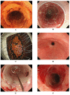
Figure 1
In Case 3, complete circular endoscopic submucosal dissection (ESD) was achieved, and endoscopic balloon dilatation (EBD) was performed preemptively. Nevertheless, he required total 48 sessions to relieve his dysphagia. A. Chromoendoscopy with an iodine solution reveals the iodine-unstained area spreading to involve nearly the entire circumference of the esophagus (Case 3, Table 1). Wholly circumferential ESD was performed. B. Artificial ulcer immediately after complete circular resection. C. The tumor was removed en bloc with tumor-free lateral and basal margins, and histopathological assessment revealed intramucosal invasive squamous cell carcinoma (m2). Repeat esophagoscopy revealed persistent esophageal stricture (D) despite 16 sessions (twice a week, for 8 weeks) of EBD (E), which was started on the third postoperative day. Temporary improvement of the stricture was achieved with EBD (F), but this patient required 48 EBD sessions.
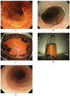
Figure 2
In Case 5, complete circular ESD was achieved, and oral prednisolone was given. He has not required any EBD sessions without no postprocedural stricture and the related dysphagia. A. Chromoendoscopy with iodine staining revealed a discolored area spreading to involve nearly the entire circumference of the esophagus in the middle thoracic esophagus (Case 5, Table 1), and wholly circumferential, endoscopic submucosal dissection was performed. B. Artificial ulcer immediately after complete circular resection. Complete circular resection was achieved (C), and the tumor was removed en bloc with tumor-free lateral and basal margins (D). Histopathological assessment revealed intramucosal invasive squamous cell carcinoma (m2). Oral prednisolone (30 mg) was initiated on the third postoperative day, tapered, and then discontinued 8 weeks later. E. Follow-up endoscopy 6 months later revealed no postprocedural stricture without EBD.
Similar articles
- Usefulness of oral prednisolone in the treatment of esophageal stricture after endoscopic submucosal dissection for superficial esophageal squamous cell carcinoma.
Yamaguchi N, Isomoto H, Nakayama T, Hayashi T, Nishiyama H, Ohnita K, Takeshima F, Shikuwa S, Kohno S, Nakao K. Yamaguchi N, et al. Gastrointest Endosc. 2011 Jun;73(6):1115-21. doi: 10.1016/j.gie.2011.02.005. Epub 2011 Apr 14. Gastrointest Endosc. 2011. PMID: 21492854 - Control of severe strictures after circumferential endoscopic submucosal dissection for esophageal carcinoma: oral steroid therapy with balloon dilation or balloon dilation alone.
Sato H, Inoue H, Kobayashi Y, Maselli R, Santi EG, Hayee B, Igarashi K, Yoshida A, Ikeda H, Onimaru M, Aoyagi Y, Kudo SE. Sato H, et al. Gastrointest Endosc. 2013 Aug;78(2):250-7. doi: 10.1016/j.gie.2013.01.008. Epub 2013 Feb 27. Gastrointest Endosc. 2013. PMID: 23453294 - Effect of oral prednisolone on esophageal stricture after complete circular endoscopic submucosal dissection for superficial esophageal squamous cell carcinoma: a case report.
Yamaguchi N, Isomoto H, Shikuwa S, Nakayama T, Hayashi T, Ohnita K, Takeshima F, Kohno S, Nakao K. Yamaguchi N, et al. Digestion. 2011;83(4):291-5. doi: 10.1159/000321093. Epub 2011 Feb 1. Digestion. 2011. PMID: 21282955 - Management of complications associated with endoscopic submucosal dissection/ endoscopic mucosal resection for esophageal cancer.
Isomoto H, Yamaguchi N, Minami H, Nakao K. Isomoto H, et al. Dig Endosc. 2013 Mar;25 Suppl 1:29-38. doi: 10.1111/j.1443-1661.2012.01388.x. Epub 2013 Jan 24. Dig Endosc. 2013. PMID: 23368404 Review. - Prevention of Esophageal Stricture After Endoscopic Submucosal Dissection: A Systematic Review.
Yu JP, Liu YJ, Tao YL, Ruan RW, Cui Z, Zhu SW, Shi W. Yu JP, et al. World J Surg. 2015 Dec;39(12):2955-64. doi: 10.1007/s00268-015-3193-3. World J Surg. 2015. PMID: 26335901 Review.
Cited by
- Endoscopic submucosal dissection--current success and future directions.
Yamamoto H. Yamamoto H. Nat Rev Gastroenterol Hepatol. 2012 Sep;9(9):519-29. doi: 10.1038/nrgastro.2012.97. Epub 2012 Jun 5. Nat Rev Gastroenterol Hepatol. 2012. PMID: 22664591 Review. - Esophageal Stricture Prevention after Endoscopic Submucosal Dissection.
Jain D, Singhal S. Jain D, et al. Clin Endosc. 2016 May;49(3):241-56. doi: 10.5946/ce.2015.099. Epub 2016 Mar 7. Clin Endosc. 2016. PMID: 26949124 Free PMC article. Review. - A patient-like swine model of gastrointestinal fibrotic strictures for advancing therapeutics.
Li L, Itani MI, Salimian KJ, Li Y, Gutierrez OB, Hu H, Fayad G, Donet JA, Joo MK, Ensign LM, Kumbhari V, Selaru FM. Li L, et al. Sci Rep. 2021 Jun 25;11(1):13344. doi: 10.1038/s41598-021-92628-8. Sci Rep. 2021. PMID: 34172773 Free PMC article. - Ten-year experience of esophageal endoscopic submucosal dissection of superficial esophageal neoplasms in a single center.
Park HC, Kim DH, Gong EJ, Na HK, Ahn JY, Lee JH, Jung KW, Choi KD, Song HJ, Lee GH, Jung HY, Kim JH. Park HC, et al. Korean J Intern Med. 2016 Nov;31(6):1064-1072. doi: 10.3904/kjim.2015.210. Epub 2016 Sep 13. Korean J Intern Med. 2016. PMID: 27618866 Free PMC article. - Endoscopic balloon dilation and submucosal injection of triamcinolone acetonide in the treatment of esophageal stricture: A single-center retrospective study.
Qi L, He W, Yang J, Gao Y, Chen J. Qi L, et al. Exp Ther Med. 2018 Dec;16(6):5248-5252. doi: 10.3892/etm.2018.6858. Epub 2018 Oct 12. Exp Ther Med. 2018. PMID: 30542481 Free PMC article.
References
- Oyama T, Tomori A, Hotta K, Morita S, Kominato K, Tanaka M, Miyata Y. Endoscopic submucosal dissection of early esophageal cancer. Clin Gastroenterol Hepatol. 2005;3(Suppl 1):S67–S70. - PubMed
- Fujishiro M, Yahagi N, Kakushima N, Kodashima S, Muraki Y, Ono S, Yamamichi N, Tateishi A, Shimizu Y, Oka M, Ogura K, Kawabe T, Ichinose M, Omata M. Endoscopic submucosal dissection of esophageal squamous cell neoplasms. Clin Gastroenterol Hepatol. 2006;4:688–694. doi: 10.1016/j.cgh.2006.03.024. - DOI - PubMed
MeSH terms
Substances
LinkOut - more resources
Full Text Sources
Medical
Research Materials
Miscellaneous

