Nebulization with γ-tocopherol ameliorates acute lung injury after burn and smoke inhalation in the ovine model - PubMed (original) (raw)
doi: 10.1097/SHK.0b013e3182459482.
Perenlei Enkhbaatar, Linda E Sousse, Hiroyuki Sakurai, Sebastian W Rehberg, Sven Asmussen, Edward R Kraft, Charlotte L Wright, Eva Bartha, Robert A Cox, Hal K Hawkins, Lillian D Traber, Maret G Traber, Csaba Szabo, David N Herndon, Daniel L Traber
Affiliations
- PMID: 22266978
- PMCID: PMC3306540
- DOI: 10.1097/SHK.0b013e3182459482
Nebulization with γ-tocopherol ameliorates acute lung injury after burn and smoke inhalation in the ovine model
Yusuke Yamamoto et al. Shock. 2012 Apr.
Abstract
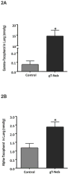
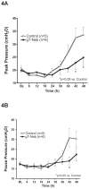
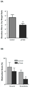
We hypothesize that the nebulization of γ-tocopherol (g-T) in the airway of our ovine model of acute respiratory distress syndrome will effectively improve pulmonary function following burn and smoke inhalation after 96 h. Adult ewes (n = 14) were subjected to 40% total body surface area burn and were insufflated with 48 breaths of cotton smoke under deep anesthesia, in a double-blind comparative study. A customized aerosolization device continuously delivered g-T in ethanol with each breath from 3 to 48 h after the injury (g-T group, n = 6), whereas the control group (n = 5) was nebulized with only ethanol. Animals were weaned from the ventilator when possible. All animals were killed after 96 h, with the exception of one untreated animal that was killed after 64 h. Lung g-T concentration significantly increased after g-T nebulization compared with the control group (38.5 ± 16.8 vs. 0.39 ± 0.46 nmol/g, P < 0.01). The PaO(2)/FIO(2) ratio was significantly higher after treatment with g-T compared with the control group (310 ± 152 vs. 150 ± 27.0, P < 0.05). The following clinical parameters were improved with g-T treatment: pulmonary shunt fraction, peak and pause pressures, lung bloodless wet-to-dry weight ratios (2.9 ± 0.87 vs. 4.6 ± 1.4, P < 0.05), and bronchiolar obstruction (2.0% ± 1.1% vs. 4.6% ± 1.7%, P < 0.05). Nebulization of g-T, carried by ethanol, improved pulmonary oxygenation and markedly reduced the time necessary for assisted ventilation in burn- and smoke-injured sheep. Delivery of g-T into the lungs may be a safe, novel, and efficient approach for management of acute lung injury patients who have sustained oxidative damage to the airway.
Figures
Figure 1
Protocol for Weaning from Mechanical Ventilation. Weaning from ventilator was initiated if PaO2/FiO2 ratio is above 250 at 48 or more hours after injury.
Figure 2
Gamma- and Alpha-Tocopherol Concentrations after Burn and Smoke Inhalation Injury with and without Gamma-Tocopherol Nebulization. After a 40% TBSA full thickness burn and a smoke inhalation injury, sheep were killed after 96 hours and lung homogenate was used to indirectly measure oxidative stress. Results were compared to injured animals that were nebulized with vitamin E. Oxidative stress was evaluated by measuring ROS and RNS scavenger gamma-tocopherol in the (A) lung and (C) liver, and ROS scavenger alpha-tocopherol in the (B) lung and (D) liver. Lung oxidative stress was attenuated post-injury with vitamin E treatment based on the associated increases in tocopherols. Data are shown as means ± SEM. *p < 0.05 versus injured animals sacrificed after 96 hours.
Figure 3
Pulmonary Gas Exchange after Burn and Smoke Inhalation Injury with and without Gamma-Tocopherol Nebulization. After a 40% TBSA full thickness burn and a smoke inhalation injury, sheep were killed after 96 hours. Results were compared to injured animals that were nebulized with vitamin E. Pulmonary gas exchange was evaluated by measuring (A) PaO2/FiO2, and (B) pulmonary shunt fraction. Pulmonary gas exchange significantly improved with vitamin E treatment. Data are shown as means ± SEM. * P < 0.05 versus injured animals sacrificed after 96 hours.
Figure 4
Peak and Pause Pressures after Burn and Smoke Inhalation Injury with and without Gamma-Tocopherol. After a 40% TBSA full thickness burn and a smoke inhalation injury, sheep were killed after 96 hours. Results were compared to injured animals that were nebulized with vitamin E. Compliance was evaluated by measuring (A) peak pressure, and (B) pause pressure in the first 48 hours after injury. Peak and pause pressure both significantly decreased and improved with vitamin E treatment after 42 and 48 hours. Data are shown as means ± SEM. * P < 0.05 versus injured animals sacrificed after 96 hours.
Figure 5
Obstruction Scores of Bronchi and Bronchioles and Lung Wet-to-Dry Ratio after Burn and Smoke Inhalation Injury with and without Gamma-Tocopherol. After a 40% TBSA full thickness burn and a smoke inhalation injury, sheep were killed after 96 hours. Results were compared to injured animals that were nebulized with vitamin E. Pulmonary pathophysiology was evaluated by measuring (A) bloodless wet-to-dry ratio, and (B) obstruction score. Both obstruction of the bronchioles and bronchi and wet-to-dry ratio significantly decreased and improved with vitamin E treatment after 42 and 48 hours. Data are shown as means ± SEM. *p < 0.05 versus injured animals sacrificed after 96 hours.
Figure 6
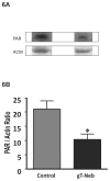
Poly (ADP Ribose)-Polymerase (PAR) Activation Significantly Decreases with vitamin E Treatment after Burn and Smoke Inhalation Injury. After a 40% TBSA full thickness burn and a smoke inhalation injury, sheep were killed after 96 hours. Representative Western blot analysis of the (A) poly(ADP-ribosylated) (PAR) proteins indicating the PARP activity in sheep lung. The poly(ADP-ribosylation) of the proteins was significantly decreased by 96 hours with vitamin E treatment after burn and smoke inhalation. Equality of protein loading was confirmed by the expression of β-actin. (B) Densitometric evaluation of PAR/actin ratios. Data are shown as means ± SEM. *p<0.05 vs. injured animals.
Similar articles
- γ-tocopherol nebulization decreases oxidative stress, arginase activity, and collagen deposition after burn and smoke inhalation in the ovine model.
Yamamoto Y, Sousse LE, Enkhbaatar P, Kraft ER, Deyo DJ, Wright CL, Taylor A, Traber MG, Cox RA, Hawkins HK, Rehberg SW, Traber LD, Herndon DN, Traber DL. Yamamoto Y, et al. Shock. 2012 Dec;38(6):671-6. doi: 10.1097/SHK.0b013e3182758759. Shock. 2012. PMID: 23160521 Free PMC article. - gamma-Tocopherol nebulization by a lipid aerosolization device improves pulmonary function in sheep with burn and smoke inhalation injury.
Hamahata A, Enkhbaatar P, Kraft ER, Lange M, Leonard SW, Traber MG, Cox RA, Schmalstieg FC, Hawkins HK, Whorton EB, Horvath EM, Szabo C, Traber LD, Herndon DN, Traber DL. Hamahata A, et al. Free Radic Biol Med. 2008 Aug 15;45(4):425-33. doi: 10.1016/j.freeradbiomed.2008.04.037. Epub 2008 May 3. Free Radic Biol Med. 2008. PMID: 18503777 Free PMC article. - Continuous nebulized albuterol attenuates acute lung injury in an ovine model of combined burn and smoke inhalation.
Palmieri TL, Enkhbaatar P, Bayliss R, Traber LD, Cox RA, Hawkins HK, Herndon DN, Greenhalgh DG, Traber DL. Palmieri TL, et al. Crit Care Med. 2006 Jun;34(6):1719-24. doi: 10.1097/01.CCM.0000217215.82821.C5. Crit Care Med. 2006. PMID: 16607229 - Inhaled anticoagulation regimens for the treatment of smoke inhalation-associated acute lung injury: a systematic review.
Miller AC, Elamin EM, Suffredini AF. Miller AC, et al. Crit Care Med. 2014 Feb;42(2):413-9. doi: 10.1097/CCM.0b013e3182a645e5. Crit Care Med. 2014. PMID: 24158173 Free PMC article. Review. - Nebulized anticoagulants for acute lung injury - a systematic review of preclinical and clinical investigations.
Tuinman PR, Dixon B, Levi M, Juffermans NP, Schultz MJ. Tuinman PR, et al. Crit Care. 2012 Dec 12;16(2):R70. doi: 10.1186/cc11325. Crit Care. 2012. PMID: 22546487 Free PMC article. Review.
Cited by
- Vitamin E beyond Its Antioxidant Label.
Ungurianu A, Zanfirescu A, Nițulescu G, Margină D. Ungurianu A, et al. Antioxidants (Basel). 2021 Apr 21;10(5):634. doi: 10.3390/antiox10050634. Antioxidants (Basel). 2021. PMID: 33919211 Free PMC article. Review. - SOCS-1 Suppresses Inflammation Through Inhibition of NALP3 Inflammasome Formation in Smoke Inhalation-Induced Acute Lung Injury.
Zhang L, Xu C, Chen X, Shi Q, Su W, Zhao H. Zhang L, et al. Inflammation. 2018 Aug;41(4):1557-1567. doi: 10.1007/s10753-018-0802-y. Inflammation. 2018. PMID: 29907905 Free PMC article. - Implications of Vitamins in COVID-19 Prevention and Treatment through Immunomodulatory and Anti-Oxidative Mechanisms.
Toledano JM, Moreno-Fernandez J, Puche-Juarez M, Ochoa JJ, Diaz-Castro J. Toledano JM, et al. Antioxidants (Basel). 2021 Dec 21;11(1):5. doi: 10.3390/antiox11010005. Antioxidants (Basel). 2021. PMID: 35052509 Free PMC article. Review. - Inhalation Injury in the Burned Patient.
Foncerrada G, Culnan DM, Capek KD, González-Trejo S, Cambiaso-Daniel J, Woodson LC, Herndon DN, Finnerty CC, Lee JO. Foncerrada G, et al. Ann Plast Surg. 2018 Mar;80(3 Suppl 2):S98-S105. doi: 10.1097/SAP.0000000000001377. Ann Plast Surg. 2018. PMID: 29461292 Free PMC article. Review. - Compiling Evidence for EVALI: A Scoping Review of In Vivo Pulmonary Effects After Inhaling Vitamin E or Vitamin E Acetate.
Feldman R, Stanton M, Suelzer EM. Feldman R, et al. J Med Toxicol. 2021 Jul;17(3):278-288. doi: 10.1007/s13181-021-00823-w. Epub 2021 Feb 2. J Med Toxicol. 2021. PMID: 33528766 Free PMC article.
References
- Bartha E, Solti I, Kereskai L, Lantos J, Plozer E, Magyar K, Szabados E, Kalai T, Hideg K, Halmosi R, Sumegi B, Toth K. PARP inhibition delays transition of hypertensive cardiopathy to heart failure in spontaneously hypertensive rats. Cardiovasc Res. 2009;83:501–510. - PubMed
- Boxman D. Alcohol vapor in the emergency treatment of acute pulmonary edema. Journal of the American Osteopathic Association. 1958;57:659–661. - PubMed
- Brochard L, Mancebo J, Elliott MW. Noninvasive ventilation for acute respiratory failure. Eur Respir J. 2002;19:712–721. - PubMed
- Cook D, De Jonghe B, Brochard L, Brun-Buisson C. Influence of airway management on ventilator-associated pneumonia: evidence from randomized trials. JAMA. 1998;279:781–787. - PubMed
Publication types
MeSH terms
Substances
Grants and funding
- GM66312-01/GM/NIGMS NIH HHS/United States
- R01 GM060688-01A1/GM/NIGMS NIH HHS/United States
- P01 GM066312-01A2/GM/NIGMS NIH HHS/United States
- R01 GM060688/GM/NIGMS NIH HHS/United States
- T32 GM008256/GM/NIGMS NIH HHS/United States
- GM60668/GM/NIGMS NIH HHS/United States
- P01 GM066312/GM/NIGMS NIH HHS/United States
LinkOut - more resources
Full Text Sources
Other Literature Sources
Medical
Miscellaneous


