Ezrin phosphorylation on tyrosine 477 regulates invasion and metastasis of breast cancer cells - PubMed (original) (raw)
Ezrin phosphorylation on tyrosine 477 regulates invasion and metastasis of breast cancer cells
Hannah Mak et al. BMC Cancer. 2012.
Abstract
Background: The membrane cytoskeletal crosslinker, ezrin, a member of the ERM family of proteins, is frequently over-expressed in human breast cancers, and is required for motility and invasion of epithelial cells. Our group previously showed that ezrin acts co-operatively with the non-receptor tyrosine kinase, Src, in deregulation of cell-cell contacts and scattering of epithelial cells. In particular, ezrin phosphorylation on Y477 by Src is specific to ezrin within the ERM family, and is required for HGF-induced scattering of epithelial cells. We therefore sought to examine the role of Y477 phosphorylation in ezrin on tumor progression.
Methods: Using a highly metastatic mouse mammary carcinoma cell line (AC2M2), we tested the effect of over-expressing a non-phosphorylatable form of ezrin (Y477F) on invasive colony growth in 3-dimensional Matrigel cultures, and on local invasion and metastasis in an orthotopic engraftment model.
Results: AC2M2 cells over-expressing Y477F ezrin exhibited delayed migration in vitro, and cohesive round colonies in 3-dimensional Matrigel cultures, compared to control cells that formed invasive colonies with branching chains of cells and numerous actin-rich protrusions. Moreover, over-expression of Y477F ezrin inhibits local tumor invasion in vivo. Whereas orthotopically injected wild type AC2M2 tumor cells were found to infiltrate into the abdominal wall and visceral organs within three weeks, tumors expressing Y477F ezrin remained circumscribed, with little invasion into the surrounding stroma and abdominal wall. Additionally, Y477F ezrin reduces the number of lung metastatic lesions.
Conclusions: Our study implicates a role of Y477 ezrin, which is phosphorylated by Src, in regulating local invasion and metastasis of breast carcinoma cells, and provides a clinically relevant model for assessing the Src/ezrin pathway as a potential prognostic/predictive marker or treatment target for invasive human breast cancer.
Figures
Figure 1
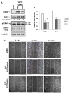
Effect of Y477F ezrin on cell motility in AC2M2 cells: Panel A) AC2M2 cells transfected with an empty pCB6 vector, or expressing Y477F ezrin, clones (A43 and C13) were lysed in Laemmli buffer, and equal protein amounts were subjected to SDS-PAGE and western blotting with antibodies against VSVG, ezrin, pY477 ezrin, pTERM and γ-tubulin. In the ezrin blot, the ~81 kDa band in pCB6 represents endogenous ezrin, while the bands in A43 and C13 represent both Y477F mutant and endogenous ezrin. Optical density (OD) ratios of ezrin vs γ-tubulin bands show a 2.6-fold and 3.5-fold increase in total ezrin expression of the A43 and C13 clones, respectively, normalized to the pCB6 clone. Ezr, ezrin; moe, moesin. Panel B) AC2M2 clones expressing either pCB6 empty vector or Y477F ezrin were grown to confluence in 10% FBS/DMEM. Confluent cells were wounded by scoring and medium was immediately replaced. Spontaneous wound closure at each of four marked wound sites for each cell clone was monitored for up to 24 h by phase contrast microscopy. The histogram shows relative cell motility for each clone calculated as the area of wound closure at 18 h and 24 h compared to T = 0 h. Values represent the mean +/- SD of 4 wound sites per clone in each of two experiments. There was significant reduction in cell motility in Y477F ezrin expressing clones (A43 and C13) at both 18 h and 24 h, as determined by a one-way ANOVA (p < 0.001). Panel C) Representative fields photographed using phase contrast microscopy (10× objective) at 0 h, 18 h and 24 h are shown. Boxed areas indicate wound area measured at each time point.
Figure 2
Effect of Y477F ezrin on morphology of AC2M2 cells in 3D Matrigel cultures: Panel A: AC2M2 cell clones expressing pCB6 empty vector or Y477F ezrin (clones A43 and C13) were cultured in 3D Matrigel. Colony morphology was assessed at the times indicated, using phase contrast microscopy. Objective magnification is shown in each panel. Panel B: Table indicates percentages of invasive colonies from each cell clone with branching cellular extensions and protrusions, and of cohesive round colonies with no extensions, respectively. The presence of 5 or more cellular extensions or protrusions per colony was considered positive. Mean percentage of colonies +/- SD (3 wells per group) in each category is shown. N = total number of colonies counted. pCB6 cells formed predominantly invasive colonies with cellular extensions, whereas the majority of Y477F ezrin cells formed round colonies with no extensions. The above differences in colony morphology were significant as determined by one way ANOVA (p < 0.001).
Figure 3
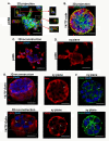
Cellular localization of ezrin and Y477F ezrin (VSVG tagged) in colonies growing in 3D Matrigel culture: Colonies formed from AC2M2 cells expressing pCB6 control vector or Y477F ezrin (clone C13) in day 12 embedded Matrigel cultures (see Figure 2) were fixed, and stained for ezrin (red), actin (green, phalloidin), and nuclei (blue, DAPI). Images of stained colonies were acquired using a Quorum Wave FX spinning disk confocal microscope. Panels A & B) 3D projection images of pCB6 control and Y477F ezrin expressing clones are shown (three colours merged). Inserts show single XY plane of actin-rich cellular protrusions with ezrin localized to the cytoplasmic side in pCB6 colonies. No actin-rich protrusions were evident in Y477F ezrin colonies. Panel C) Images of pCB6 colonies were deconvoluted and subjected to 3D reconstruction using Metamorph software. A middle section showed strong localization of ezrin in cellular extensions, compared to weak expression in the central core of colonies. Panel D) A middle xy plane of the pCB6 colony shown in C confirmed the absence of a hollow lumen. Panel E) Deconvoluted images of Y477F ezrin expressing colonies stained for ezrin or VSVG were subjected to 3D reconstruction as above. A middle xy plane showed a partial membranous staining which was strongest in the outer surface of colonies of Y477F ezrin-expressing cells, though some diffuse cytoplasmic staining was also evident. No hollow lumens were present. Panel F) XY planes corresponding to ezrin and VSVG staining in E show actin structures associated with the plasma membrane particularly at cell junctions. Images are representative of at least 20 colonies examined in each group. Scale bar indicates 50 μm.
Figure 4
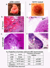
Effect of the expression of Y477F ezrin on local invasion of AC2M2 breast carcinoma cells: Two clones of AC2M2 cells expressing Y477F ezrin (A43 and C13) or empty pCB6 vector were engrafted into the mammary fat pad of nude mice. Primary tumors from the mice were excised after 21 or 23 days, and photographs of gross pathology (Panels A, D) were taken. Scale bar = 0.5 cm. 5 μm sections of FFPE processed tissues were stained with hematoxylin and eosin for histopathological analysis. Arrows indicate invasion of pCB6 tumor into underlying abdominal muscle wall (Panels A, B) or draining lymph node (Panel C). In contrast, Y477F ezrin tumors showed no adhesion to abdominal wall (Panels D, E) and rarely penetrated into the circumscribed tumor margin (Panel E) or adjacent lymph node (Panel F). Invasion was assessed categorically (presence or absence), based on combined observations of gross and histopathology. Data were expressed as the proportion of mice with local invasion relative to the total number of mice per group. Significant difference between pCB6 control and Y477F ezrin-expressing clones (A43 and C13) in two pooled experiments is shown (p = 0.003, two-sided Fisher Exact test). The overall p value for A43 + C13 clones combined is 0.0002 (Panel G). Label with "T" indicates tumor, "Mus" indicates muscle and "LN" indicates lymph node. Image magnification is shown in each panel.
Figure 5
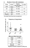
Effect of the expression of Y477F ezrin on lung metastasis formation. Mice from which primary tumors were removed (corresponding to Additional file 2: Figure S2) were allowed to survive for 20 additional days to allow overt metastases to form. Serial sections (5 μm) of FFPE processed lung tissues were stained with hematoxylin and eosin for histopathological assessment. At least two sections from the superficial and middle planes, respectively, of each mouse lung were examined and the number of lung nodules per mouse was determined. Panel A) The table shows the proportion of mice with metastasis in each group. The incidence of mice with lung metastases from Y477F ezrin tumors (A43 and C13 clones) was reduced, but this difference did not achieve significance, as determined by a two-sided Fisher Exact test. Panel B) The number of lung nodules per mouse per group is represented as a dot plot. Statistical significance was determined by a Wilcoxon Rank Sum test. Mean +/- 95% confidence intervals (bars) is shown for each group. A significant reduction in the number of nodules per mouse with clone A43 tumors was observed (p = 0.0129). The number of lung nodules in C13 tumors was also reduced, but this value was not significant (p = 0.0853), due to one outlier. Analysis of pooled data showed a significant reduction in lung nodules in mice with A43 and C13 tumors combined, compared to mice with pCB6 tumors (p = 0.0131).
Similar articles
- A novel role for ezrin in breast cancer angio/lymphangiogenesis.
Ghaffari A, Hoskin V, Szeto A, Hum M, Liaghati N, Nakatsu K, LeBrun D, Madarnas Y, Sengupta S, Elliott BE. Ghaffari A, et al. Breast Cancer Res. 2014 Sep 18;16(5):438. doi: 10.1186/s13058-014-0438-2. Breast Cancer Res. 2014. PMID: 25231728 Free PMC article. - The membrane cytoskeletal crosslinker ezrin is required for metastasis of breast carcinoma cells.
Elliott BE, Meens JA, SenGupta SK, Louvard D, Arpin M. Elliott BE, et al. Breast Cancer Res. 2005;7(3):R365-73. doi: 10.1186/bcr1006. Epub 2005 Mar 21. Breast Cancer Res. 2005. PMID: 15987432 Free PMC article. - Ezrin is key regulator of Src-induced malignant phenotype in three-dimensional environment.
Heiska L, Melikova M, Zhao F, Saotome I, McClatchey AI, Carpén O. Heiska L, et al. Oncogene. 2011 Dec 15;30(50):4953-62. doi: 10.1038/onc.2011.207. Epub 2011 Jun 13. Oncogene. 2011. PMID: 21666723 - [Advances of the Role of Ezrin in Migration and Invasion of Breast Cancer Cells].
Long ZY, Wang TH. Long ZY, et al. Sheng Li Ke Xue Jin Zhan. 2016 Feb;47(1):21-6. Sheng Li Ke Xue Jin Zhan. 2016. PMID: 27424401 Review. Chinese. - Ezrin gone rogue in cancer progression and metastasis: An enticing therapeutic target.
Barik GK, Sahay O, Paul D, Santra MK. Barik GK, et al. Biochim Biophys Acta Rev Cancer. 2022 Jul;1877(4):188753. doi: 10.1016/j.bbcan.2022.188753. Epub 2022 Jun 22. Biochim Biophys Acta Rev Cancer. 2022. PMID: 35752404 Review.
Cited by
- Targeting the Ezrin Adaptor Protein Sensitizes Metastatic Breast Cancer Cells to Chemotherapy and Reduces Neoadjuvant Therapy-induced Metastasis.
Hoskin V, Ghaffari A, Laight BJ, SenGupta S, Madarnas Y, Nicol CJB, Elliott BE, Varma S, Greer PA. Hoskin V, et al. Cancer Res Commun. 2022 Jun 17;2(6):456-470. doi: 10.1158/2767-9764.CRC-21-0117. eCollection 2022 Jun. Cancer Res Commun. 2022. PMID: 36923551 Free PMC article. - Role of Ezrin in Asthma-Related Airway Inflammation and Remodeling.
Zhao S, Luo J, Hu J, Wang H, Zhao N, Cao M, Zhang C, Hu R, Liu L. Zhao S, et al. Mediators Inflamm. 2022 Dec 7;2022:6255012. doi: 10.1155/2022/6255012. eCollection 2022. Mediators Inflamm. 2022. PMID: 36530558 Free PMC article. Review. - [Investigation of the influence of mechanical signals on the structure of CD44/FERM complex via molecular dynamics simulation].
Li Y, Fang Y, Wu J. Li Y, et al. Sheng Wu Yi Xue Gong Cheng Xue Za Zhi. 2018 Aug 25;35(4):501-508. doi: 10.7507/1001-5515.201801051. Sheng Wu Yi Xue Gong Cheng Xue Za Zhi. 2018. PMID: 30124011 Free PMC article. Chinese. - Expression of ezrin and moesin in primary breast carcinoma and matched lymph node metastases.
Bartova M, Hlavaty J, Tan Y, Singer C, Pohlodek K, Luha J, Walter I. Bartova M, et al. Clin Exp Metastasis. 2017 Jun;34(5):333-344. doi: 10.1007/s10585-017-9853-y. Epub 2017 Jun 17. Clin Exp Metastasis. 2017. PMID: 28624994 - Regulation of ErbB2 localization and function in breast cancer cells by ERM proteins.
Asp N, Kvalvaag A, Sandvig K, Pust S. Asp N, et al. Oncotarget. 2016 May 3;7(18):25443-60. doi: 10.18632/oncotarget.8327. Oncotarget. 2016. PMID: 27029001 Free PMC article.
References
Publication types
MeSH terms
Substances
LinkOut - more resources
Full Text Sources
Medical
Molecular Biology Databases
Miscellaneous


