ExoU activates NF-κB and increases IL-8/KC secretion during Pseudomonas aeruginosa infection - PubMed (original) (raw)
ExoU activates NF-κB and increases IL-8/KC secretion during Pseudomonas aeruginosa infection
Carolina Diettrich Mallet de Lima et al. PLoS One. 2012.
Abstract
ExoU, a Pseudomonas aeruginosa cytotoxin injected into host cytosol by type III secretion system, exhibits a potent proinflammatory activity that leads to a marked recruitment of neutrophils to infected tissues. To evaluate the mechanisms that account for neutrophil infiltration, we investigated the effect of ExoU on IL-8 secretion and NF-κB activation. We demonstrate that ExoU increases IL-8 mRNA and protein levels in P. aeruginosa-infected epithelial and endothelial cell lines. Also, ExoU induces the nuclear translocation of p65/p50 NF-κB transactivator heterodimer as well as NF-κB-dependent transcriptional activity. ChIP assays clearly revealed that ExoU promotes p65 binding to NF-κB site in IL-8 promoter and the treatment of cultures with the NF-κB inhibitor Bay 11-7082 led to a significant reduction in IL-8 mRNA levels and protein secretion induced by ExoU. These results were corroborated in a murine model of pneumonia that revealed a significant reduction in KC secretion and neutrophil infiltration in bronchoalveolar lavage when mice were treated with Bay 11-7082 before infection with an ExoU-producing strain. In conclusion, our data demonstrate that ExoU activates NF-κB, stimulating IL-8 expression and secretion during P. aeruginosa infection, and unveils a new mechanism triggered by this important virulence factor to interfere in host signaling pathways.
Conflict of interest statement
Competing Interests: The authors have declared that no competing interests exist.
Figures
Figure 1. ExoU increases IL-8 mRNA in A549 cells.
In (A), representative agarose gels obtained from three different semi-quantitative RT-PCR assays carried out in duplicate. In (B) and (C), graph represents the means ± SEM of values obtained by three different Real Time qRT-PCR assays performed in triplicate. **p<0.01 or ***p<0.001 when the values obtained from cultures infected with the ExoU-producing PA103 strain were compared with those obtained from the other cultures.
Figure 2. ExoU PLA2 activity stimulates IL-8 secretion by _P. aeruginosa_-infected A549 cultures.
In (A) and (B), cells were infected with the different P. aeruginosa strains for 21 hours and the concentrations of IL-8 in supernatants were assessed by ELISA. In (B), Brefeldin A was added or not to the gentamicin-containing culture medium. The graphs show the means ± SEM of three assays performed in quadruplicate. ***p<0.001 when the values obtained from untreated PA103-infected cultures were compared with those from the other cultures.
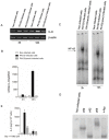
Figure 3. ExoU promotes p65/p50 nuclear translocation and NF-κB-dependent transcriptional activity in _P. aeruginosa_-infected A549 cells.
In (A), representative EMSAs showing NF-κB nuclear translocation after 2 and 12 hours of P. aeruginosa infection or treatment with culture medium. To evaluate NF-κB inhibition by Bay 11-7082, some cultures were treated with 10 µM Bay 11-7082, 1 hour prior PA103 infection. As a control for non-specific interactions, nuclear extracts from PA103-infected cells were also incubated with a probe mutated in a single nucleotide (Mut). In (B), extracts obtained from PA103-infected cells were supershifted with specific antibodies against NF-κB subunits, p50 and p65. In “Non-infected cells” and “PA103-infected cells” lanes, antibodies were not added. EMSA and supershift assays were performed in triplicate. In (C), the graph shows the luciferase activity (in arbitrary values) of whole-cell lysates obtained from A549 cultures transfected with p6κB-LUC and pRL-CMV plasmids and then infected with P. aeruginosa strains or treated with cultured medium for 24 hours. Data represent means ± SEM of three different assays carried out in quadruplicate. **p<0.01 and ***p<0.001 when the values obtained from PA103Δ_exoU_-infected cultures and non-infected cultures, respectively, were compared with those obtained from PA103-infected cultures.
Figure 4. ExoU induces NF-κB binding in IL-8 promoter of A549 cells.
In (A), representative agarose gel showing PCR products of DNA obtained after chromatin immunoprecipitation with anti-p65 antibody (IP:anti-p65) or non-immunoprecipitated DNA (input) amplified with primers specific for NF-κB site in IL-8 promoter. In (B), graph shows the means ± SEM of values obtained by Real Time PCR of three different ChIP assays. ***p<0.001 when the values obtained in non-infected cultures or cultures infected with the ExoU deficient strain were compared with those obtained in cultures infected with the ExoU-producing strain for 14 hours.
Figure 5. Inhibition of NF-κB reduces IL-8 expression and secretion induced by ExoU in _P. aeruginosa_-infected A549 cultures.
In (A), representative agarose gels of RT-PCR assays in which A549 cells were treated or not with 10 µM Bay 11-7082, 1 hour prior infection with PA103 or PA103Δ_exoU_ strains or treatment with culture medium (non-infected cells) for 18 hours. In (B), graph shows the means ± SEM of values obtained by three Real Time qRT-PCR assays. In (C), IL-8 concentrations detected in supernatants of cultures pretreated or not with 10 µM Bay 11-7082 for 1 hour and then exposed for 21 hours to PA103, PA103Δ_exoU_ or culture medium, as assessed by ELISA. In (D), IL-8 concentrations, as detected by ELISA, in supernatants of cultures pretreated or not with 10 µM Bay 11-7082 and/or 10 µM SP600125 for 1 hour and then exposed for 21 hours to bacterial strains or culture medium. In (C) and (D), data represent means ± SEM of three assays performed in quadruplicate. ***p<0.001 when the values obtained from untreated PA103-infected cultures were compared with those from the other cultures.
Figure 6. ExoU activates p65/p50 NF-κB and increases IL-8 expression and secretion in HMEC-1 capillary endothelial cells.
In (A), representative agarose gels of three different semi-quantitative RT-PCR assays carried out in duplicate. In (B), graph shows the means ± SEM of values obtained in three Real Time qRT-PCR assays. **p<0.01 and ***p<0.001 when the values obtained from PA103Δ_exoU_-infected cultures and non-infected cultures, respectively, were compared with those obtained from cultures infected with the ExoU-producing strain. In (C), representative EMSAs showing NF-κB nuclear translocation after 2 and 12 hours of P. aeruginosa infection or treatment with culture medium. In (D), extracts obtained from PA103-infected cells were incubated with specific antibodies for NF-κB subunits, p50, p52, p65 or cRel, but only supershifted with p50 and p65. In non-infected cells, antibodies were not added. EMSA and supershift assays were performed in triplicate. In (E), IL-8 concentrations, detected by ELISA, in supernatants of cultures pretreated or not with 5 µM Bay 11-7082, for 1 hour, and exposed to PA103, PA103Δ_exoU_ or culture medium, for 21 hours. The graph represents the means ± SEM of two assays performed in quadruplicate and shows ***p<0.001 when the values obtained from untreated PA103-infected cultures were compared with those from the other untreated cultures or with those from Bay 11-7082-treated cultures infected with the ExoU-producing strain.
Figure 7. ExoU induces NF-κB-dependent KC secretion and neutrophil infiltration in mice airways at 24 hours post-infection.
In (A), concentrations of KC assessed by ELISA, in (B) total leukocytes and in (C) neutrophils (PMN), both assessed by light microscopy, in BALF of mice pretreated or not with Bay 11-7082 at 20 mg/Kg, for 1 hour, and inoculated intratracheally with PA103, PA103Δ_exoU_ or saline. The graphs show the mean values ± SEM of three independent assays (n = 16 for each group). *p<0,05 or **p<0.01 when the values obtained in untreated mice infected with PA103 strain were compared with the values obtained in the other groups.
Similar articles
- Central role of PAFR signalling in ExoU-induced NF-κB activation.
Mallet de Lima CD, da Conceição Costa J, de Oliveira Lima Santos SA, Carvalho S, de Carvalho L, Albano RM, Teixeira MM, Plotkowski MC, Saliba AM. Mallet de Lima CD, et al. Cell Microbiol. 2014 Aug;16(8):1244-54. doi: 10.1111/cmi.12280. Epub 2014 Mar 21. Cell Microbiol. 2014. PMID: 24612488 - NF-kappaB activation and sustained IL-8 gene expression in primary cultures of cystic fibrosis airway epithelial cells stimulated with Pseudomonas aeruginosa.
Joseph T, Look D, Ferkol T. Joseph T, et al. Am J Physiol Lung Cell Mol Physiol. 2005 Mar;288(3):L471-9. doi: 10.1152/ajplung.00066.2004. Epub 2004 Oct 29. Am J Physiol Lung Cell Mol Physiol. 2005. PMID: 15516493 - Yersinia enterocolitica invasin protein triggers IL-8 production in epithelial cells via activation of Rel p65-p65 homodimers.
Schulte R, Grassl GA, Preger S, Fessele S, Jacobi CA, Schaller M, Nelson PJ, Autenrieth IB. Schulte R, et al. FASEB J. 2000 Aug;14(11):1471-84. doi: 10.1096/fj.14.11.1471. FASEB J. 2000. PMID: 10928981 - Association between Pseudomonas aeruginosa type III secretion, antibiotic resistance, and clinical outcome: a review.
Sawa T, Shimizu M, Moriyama K, Wiener-Kronish JP. Sawa T, et al. Crit Care. 2014 Dec 13;18(6):668. doi: 10.1186/s13054-014-0668-9. Crit Care. 2014. PMID: 25672496 Free PMC article. Review.
Cited by
- Interesting Cytokine Profile Caused by Clinical Strains of Pseudomonas aeruginosa MDR Carrying the exoU Gene.
Badillo-Larios NS, Turrubiartes-Martínez EA, Layseca-Espinosa E, González-Amaro R, Pérez-González LF, Niño-Moreno P. Badillo-Larios NS, et al. Int J Microbiol. 2024 Jun 30;2024:2748842. doi: 10.1155/2024/2748842. eCollection 2024. Int J Microbiol. 2024. PMID: 38974708 Free PMC article. - Contribution of the platelet activating factor signaling pathway to cerebral microcirculatory dysfunction during experimental sepsis by ExoU producing Pseudomonas aeruginosa.
Plotkowski MC, Estato V, Santos SA, da Silva MC, Miranda AS, de Miranda PE, Pinho V, Tibiriça E, Morandi V, Teixeira MM, Vianna A, Saliba AM. Plotkowski MC, et al. Pathog Dis. 2015 Oct;73(7):ftv046. doi: 10.1093/femspd/ftv046. Epub 2015 Jul 17. Pathog Dis. 2015. PMID: 26187894 Free PMC article. - Impact of Bacterial Toxins in the Lungs.
Lucas R, Hadizamani Y, Gonzales J, Gorshkov B, Bodmer T, Berthiaume Y, Moehrlen U, Lode H, Huwer H, Hudel M, Mraheil MA, Toque HAF, Chakraborty T, Hamacher J. Lucas R, et al. Toxins (Basel). 2020 Apr 2;12(4):223. doi: 10.3390/toxins12040223. Toxins (Basel). 2020. PMID: 32252376 Free PMC article. Review. - Pseudomonas aeruginosa: pathogenesis, virulence factors, antibiotic resistance, interaction with host, technology advances and emerging therapeutics.
Qin S, Xiao W, Zhou C, Pu Q, Deng X, Lan L, Liang H, Song X, Wu M. Qin S, et al. Signal Transduct Target Ther. 2022 Jun 25;7(1):199. doi: 10.1038/s41392-022-01056-1. Signal Transduct Target Ther. 2022. PMID: 35752612 Free PMC article. Review. - ExoU-induced redox imbalance and oxidative stress in airway epithelial cells during Pseudomonas aeruginosa pneumosepsis.
da Cunha LG Jr, Ferreira MF, de Moraes JA, Reis PA, Castro-Faria-Neto HC, Barja-Fidalgo C, Plotkowski MC, Saliba AM. da Cunha LG Jr, et al. Med Microbiol Immunol. 2015 Dec;204(6):673-80. doi: 10.1007/s00430-015-0418-x. Epub 2015 Apr 23. Med Microbiol Immunol. 2015. PMID: 25904542 Free PMC article.
References
- Schulert GS, Feltman H, Rabin SDP, Martin CG, Battle SE, et al. Secretion of the toxin ExoU is a marker for highly virulent Pseudomonas aeruginosa isolates obtained from patients with hospital-acquired pneumonia. J Infect Dis. 2003;188:1695–1706. - PubMed
- Wareham DW, Curtis MA. A genotypic and phenotypic comparison of type III secretion profiles of Pseudomonas aeruginosa cystic fibrosis and bacteremia isolates. Int J Med Microbiol. 2007;297:227–234. - PubMed
- Engel J, Balachandran P. Role of Pseudomonas aeruginosa type III effectors in disease. Curr Opin Microbiol. 2009;12:61–66. - PubMed
- Saliba AM, Nascimento DO, Silva MCA, Assis MC, Gayer CRM, et al. Eicosanoid-mediated proinflammatory activity of Pseudomonas aeruginosa ExoU. Cell.Microbiol. 2005;7:1811–1822. - PubMed
- Hoffmann E, Dittrich-Breiholz O, Holtmann H, Kracht M. Multiple control of interleukin-8 gene expression. J Leukoc Biol. 2002;72:847–855. - PubMed
Publication types
MeSH terms
Substances
LinkOut - more resources
Full Text Sources
Research Materials


