hnRNP U enhances caspase-9 splicing and is modulated by AKT-dependent phosphorylation of hnRNP L - PubMed (original) (raw)
hnRNP U enhances caspase-9 splicing and is modulated by AKT-dependent phosphorylation of hnRNP L
Ngoc T Vu et al. J Biol Chem. 2013.
Abstract
Caspase-9 has two splice variants, pro-apoptotic caspase-9a and anti-apoptotic caspase-9b, which are regulated by RNA trans-factors associated with exon 3 of caspase-9 pre-mRNA (C9/E3). In this study, we identified hnRNP U as an RNA trans-factor associated with C9/E3. Down-regulation of hnRNP U led to a decrease in the caspase-9a/9b mRNA ratio, demonstrating a novel enhancing function. Importantly, hnRNP U bound specifically to C9/E3 at an RNA cis-element previously reported as the binding site for the splicing repressor, hnRNP L. Phosphorylated hnRNP L interfered with hnRNP U binding to C9/E3, and our results demonstrate the importance of the phosphoinositide 3-kinase/AKT pathway in modulating the association of hnRNP U to C9/E3. Taken together, these findings show that hnRNP U competes with hnRNP L for binding to C9/E3 to enhance the inclusion of the four-exon cassette, and this splice-enhancing function is blocked by the AKT pathway via phosphorylation of hnRNP L.
Figures
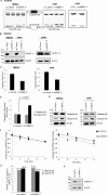
FIGURE 1.
hnRNP U, but not hnRNP R, represses the formation of caspase-9b via RNA splicing. A549 or HBEpC cells were transfected with control siRNA (100 n
m
), hnRNP U SMARTpool siRNA (100 n
m
), or hnRNP R siRNA (100 n
m
) for 48 h. A, total RNA was isolated and analyzed by competitive/quantitative RT-PCR for caspase-9 splice variant ratio using ratio-based quantitation. Ctr, control. B, total protein extracts were subjected to SDS/PAGE/immunoblotting for hnRNP U, hnRNP R, or β-actin. C, graphs were created from the results in A. D, total RNA isolated from A549 cells transfected with control or hnRNP U siRNA was analyzed for relative amount of caspase-9a or 9b mRNA by quantitative RT-PCR using the standard curve method and 18 S rRNA as a normalizing control. E, total protein extracts were subjected to SDS/PAGE/immunoblotting for caspase-9 or β-actin. The data are shown as the means ± S.D. (n ≥ 4 on at least two independent occasions). F, A549 cells were transfected with siRNA as in A. After 48 h, the cells were treated with actinomycin D (5 μg/ml) for 0, 2, 4, or 8 h. RNA was isolated, and the relative amount of caspase-9a or 9b mRNA (fold change compared with the si-control or si-hnRNP U samples at 0 h) was individually determined by quantitative RT-PCR. G, half-life of caspase-9a or caspase-9b mRNA was calculated based on the data in F. H, total protein from siRNA transfected cells (48 h) was subjected to SDS/PAGE/immunoblotting for hnRNP U or β-actin. Statistical significance was evaluated by Student's t test (p < 0.0001, n = 5 on two independent occasions).
FIGURE 2.
hnRNP U binds to exon 3 of caspase-9. A, schematic illustration of caspase-9 pre-mRNA structure. 18-mer C9/E3 ROs used in RNA binding assays are corresponding to the purine-rich sequence in C9/E3. oligos, oligonucleotides. B and C, EMSAs were performed in the absence of protein phosphatase inhibitors using 5′-FITC-tagged C9/E3 ROs and in the presence of IgG (control), A549 cell lysates (B), or lysates from A549 cells transfected with control siRNA or hnRNP U SMARTpool siRNA (C). Ab, antibody. D, cell lysate input in C was subjected to SDS-PAGE and immunoblotted for hnRNP U and β-actin. E, SBAP was performed using 5′-biotinylated nonspecific control (NSC), 5′-biotinylated wild-type C9/E3 ROs (E3/C9 WT), or 5′-biotinylated mutant C9/E3 ROs (Mut1–Mut4) in the presence of IgG (control) or A549 cell lysates. F, EMSAs were performed as in C, but using additional ROs (5′-FITC-tagged C9/E3 Mut1–Mut4). Arrows indicate the supershifts obtained by the addition of the hnRNP U antibody. The graphs shown in B, C, E, and F are representative of n = 4 from three independent experiments.
FIGURE 3.
Phosphorylation regulates the association of hnRNP U with exon 3 of caspase-9. A, hnRNP U is expressed at the same level in both transformed and nontransformed cells. A549 (transformed) and HBEpC (nontransformed) cells were cultured under the same condition (DMEM with/without FBS) and confluency. Total protein extracts were subjected to SDS-PAGE/immunoblotting for hnRNP U and β-actin. B and C, hnRNP U binds to C9/E3 in higher amounts in HBEpCs than in A549 cells. SBAP assays (B) or EMSAs in the presence of protein phosphatase inhibitors (C) were performed with either IgG (control) or lysates from A549 or HBEpC cells using a 5′-biotinylated or 5-FITC-tagged wild-type C9/E3 ROs. Ab, antibody. D and E, dephosphorylation increases the binding affinity of hnRNP U to C9/E3. A549 cell lysates (fresh) were preincubated with denatured calf intestinal alkaline phosphatase (De-CIP) or active protein phosphatase (CIP) followed by EMSA (D) in the absence of protein phosphatase inhibitors or SBAP (E) to assay the binding with 5′-FITC tagged (D) or 5′-biotinylated (E) C9/E3 ROs. IgG was used as a control. F, dephosphorylation abolishes the difference between A549 versus HBEpC regarding the binding of hnRNP U to E3/C9. A549 (transformed) and HBEpC (nontransformed) cells were cultured under the same conditions and confluency, and cell lysates were produced. Lysates (fresh) were again treated and assayed as in E. G, hypothetical model for regulation of hnRNP U/L-C9/E3 interaction by phosphorylation. Whereas the phosphorylation event augments the association between hnRNP L and C9/E3, phosphorylation of hnRNP U attenuates its binding to C9/E3. The graphs shown in A–F are representative of n = 4 from three independent experiments.
FIGURE 4.
AKT pathway regulates the association of hnRNP U to exon 3 of caspase-9. A, inhibition of AKT1/2 activity increases the caspase-9a/9b ratio and enhances the binding of hnRNP U to C9/E3 in a dose-response manner. A549 cells were treated with 0.1% DMSO control or increasing concentrations of the AKT1/2 inhibitor (AKT VIII). Total RNA was isolated and analyzed for caspase-9 splice variants by competitive/quantitative RT-PCR. Cell lysates from a concomitant experiment were utilized in SBAP to assay the binding of hnRNP U to 5′-biotinylated C9/E3 ROs. Cell lysate input for the SBAP was subjected to SDS-PAGE and immunoblotted for hnRNP U and β-actin. B, overexpression of AKT decreases both caspase-9a/9b ratio and hnRNP U-E3/C9 association. HBEpC cells were transfected with the control adenovirus or the adenovirus expressing constitutive active AKT2 (caAKT2; multiplicity of infection of 25) for 48 h. Total protein or RNA extracts from the transfected cells were then subjected to SDS-PAGE/immunoblotting or competitive/quantitative RT-PCR. Cell lysates were also generated for SBAP to assay the binding of hnRNP U to E3/C9 ROs. The graphs shown in A and B are representative of n = 4 from three independent experiments.
FIGURE 5.
AKT inhibition does not alter the phospho-state of hnRNP U. A, A549 cells were treated with 0.1% dimethyl sulfoxide (DMSO) control or AKT1/2 inhibitor (20 μ
m
). Endogenous hnRNP U was then immunoprecipitated (IP) and resolved by SDS-PAGE/immunoblotting for phospho-serine/threonine or hnRNP U. B, A549 protein extracts were incubated with denatured or active CIP. Endogenous hnRNP U in the resulted protein extracts was immunoprecipitated and resolved by SDS-PAGE/immunoblotting for phosphoserine/phosphothreonine or hnRNP U. The graphs shown in A and B are representative of n = 3 from two independent experiments.
FIGURE 6.
AKT-dependent phosphorylation of hnRNP L leads to the competition with hnRNP U for C9/E3 binding. A, phosphorylation of hnRNP L by AKT enhances binding to E3/C9. Recombinant FLAG-tagged hnRNP L was phosphorylated in vitro using active AKT2 (denatured AKT2 was utilized as a negative control). Products from the kinase assay were then subjected to SDS-PAGE/immunoblotting for phosphoserine/phosphothreonine, or the FLAG tag and also utilized in the EMSA to examine the interaction with E3/C9 ROs. B, AKT or EGFR inhibition in NSCLC cells induces a reduction in the phospho-status of hnRNP L. A549 cells were treated with either 0.1% dimethyl sulfoxide (DMSO) control, AKT inhibitor (20 μ
m
), or erlotinib (EGFR inhibitor, 1 μ
m
). Endogenous hnRNP L was immunoprecipitated (IP) from the protein extracts. Immunoprecipitated hnRNP L was resolved by SDS-PAGE and immunoblotted with Ser(P)52-hnRNP L or hnRNP L antibodies. C, exogenous hnRNP L phosphorylated by AKT competes with endogenous hnRNP U for binding to E3/C9. Recombinant hnRNP L was incubated with denatured or active AKT2 in the in vitro kinase assay and then utilized in the SBAP-based competitive binding assay using HBEpC cell lysates and E3/C9 ROs. D, model for controlling hnRNPU-hnRNP L-E3/C9 interaction by AKT pathway. In the nontransformed cells, the recruitment of hnRNP L to E3/C9 is interfered with by hnRNP U binding to the same position. Through activation of AKT pathway in transformed cells, hnRNP L is phosphorylated, which results in the enhanced binding of this splicing repressor to E3/C9. Consequently, the elevated levels of hnRNP L associated with E3/C9 prevent the access of hnRNP U to E3/C9 for splice-enhancing function. The graphs shown in A–C are representative of n = 4 from two independent experiments.
Similar articles
- hnRNP L regulates the tumorigenic capacity of lung cancer xenografts in mice via caspase-9 pre-mRNA processing.
Goehe RW, Shultz JC, Murudkar C, Usanovic S, Lamour NF, Massey DH, Zhang L, Camidge DR, Shay JW, Minna JD, Chalfant CE. Goehe RW, et al. J Clin Invest. 2010 Nov;120(11):3923-39. doi: 10.1172/JCI43552. Epub 2010 Oct 25. J Clin Invest. 2010. PMID: 20972334 Free PMC article. - Alternative splicing of caspase 9 is modulated by the phosphoinositide 3-kinase/Akt pathway via phosphorylation of SRp30a.
Shultz JC, Goehe RW, Wijesinghe DS, Murudkar C, Hawkins AJ, Shay JW, Minna JD, Chalfant CE. Shultz JC, et al. Cancer Res. 2010 Nov 15;70(22):9185-96. doi: 10.1158/0008-5472.CAN-10-1545. Epub 2010 Nov 2. Cancer Res. 2010. PMID: 21045158 Free PMC article. - hnRNP L controls HPV16 RNA polyadenylation and splicing in an Akt kinase-dependent manner.
Kajitani N, Glahder J, Wu C, Yu H, Nilsson K, Schwartz S. Kajitani N, et al. Nucleic Acids Res. 2017 Sep 19;45(16):9654-9678. doi: 10.1093/nar/gkx606. Nucleic Acids Res. 2017. PMID: 28934469 Free PMC article. - hnRNP proteins and splicing control.
Martinez-Contreras R, Cloutier P, Shkreta L, Fisette JF, Revil T, Chabot B. Martinez-Contreras R, et al. Adv Exp Med Biol. 2007;623:123-47. doi: 10.1007/978-0-387-77374-2_8. Adv Exp Med Biol. 2007. PMID: 18380344 Review. - The role of SAF-A/hnRNP U in regulating chromatin structure.
Marenda M, Lazarova E, Gilbert N. Marenda M, et al. Curr Opin Genet Dev. 2022 Feb;72:38-44. doi: 10.1016/j.gde.2021.10.008. Epub 2021 Nov 22. Curr Opin Genet Dev. 2022. PMID: 34823151 Review.
Cited by
- Regulation of HNRNP family by post-translational modifications in cancer.
Li B, Wen M, Gao F, Wang Y, Wei G, Duan Y. Li B, et al. Cell Death Discov. 2024 Oct 4;10(1):427. doi: 10.1038/s41420-024-02198-7. Cell Death Discov. 2024. PMID: 39366930 Free PMC article. Review. - B Cell Receptor Activation Predominantly Regulates AKT-mTORC1/2 Substrates Functionally Related to RNA Processing.
Mohammad DK, Ali RH, Turunen JJ, Nore BF, Smith CI. Mohammad DK, et al. PLoS One. 2016 Aug 3;11(8):e0160255. doi: 10.1371/journal.pone.0160255. eCollection 2016. PLoS One. 2016. PMID: 27487157 Free PMC article. - RNA Binding Proteins that Mediate LPS-induced Alternative Splicing of the MyD88 Innate Immune Regulator.
Lee FFY, Harris C, Alper S. Lee FFY, et al. J Mol Biol. 2024 Apr 15;436(8):168497. doi: 10.1016/j.jmb.2024.168497. Epub 2024 Feb 17. J Mol Biol. 2024. PMID: 38369277 - Circular RNA CircSLC22A23 Promotes Gastric Cancer Progression by Activating HNRNPU Expression.
Wu X, Cao C, Li Z, Xie Y, Zhang S, Sun W, Guo J. Wu X, et al. Dig Dis Sci. 2024 Apr;69(4):1200-1213. doi: 10.1007/s10620-024-08291-2. Epub 2024 Feb 24. Dig Dis Sci. 2024. PMID: 38400886 - Phosphorylation-mediated regulation of alternative splicing in cancer.
Naro C, Sette C. Naro C, et al. Int J Cell Biol. 2013;2013:151839. doi: 10.1155/2013/151839. Epub 2013 Aug 28. Int J Cell Biol. 2013. PMID: 24069033 Free PMC article. Review.
References
- Seol D.-W., Billiar T. R. (1999) A caspase-9 variant missing the catalytic site is an endogenous inhibitor of apoptosis. J. Biol. Chem. 274, 2072–2076 - PubMed
- Srinivasula S. M., Ahmad M., Guo Y., Zhan Y., Lazebnik Y., Fernandes-Alnemri T., Alnemri E. S. (1999) Identification of an endogenous dominant-negative short isoform of caspase-9 that can regulate apoptosis. Cancer Res. 59, 999–1002 - PubMed
- Goehe R. W., Shultz J. C., Murudkar C., Usanovic S., Lamour N. F., Massey D. H., Zhang L., Camidge D. R., Shay J. W., Minna J. D., Chalfant C. E. (2010) hnRNP L regulates the tumorigenic capacity of lung cancer xenografts in mice via caspase-9 pre-mRNA processing. J. Clin. Invest. 120, 3923–3939 - PMC - PubMed
Publication types
MeSH terms
Substances
LinkOut - more resources
Full Text Sources
Other Literature Sources
Research Materials
Miscellaneous


