CD40 ligand as a potential biomarker for atherosclerotic instability - PubMed (original) (raw)
CD40 ligand as a potential biomarker for atherosclerotic instability
Jing-Hua Wang et al. Neurol Res. 2013 Sep.
Abstract
Objective: The signaling protein CD40 and its ligand, CD40L, are thought to contribute to atherosclerotic plaque formation and rupture. We sought to determine their utility as markers of cerebral atherosclerosis and neurological dysfunction.
Methods: We recruited 82 patients with acute cerebral infarction (ACI) and classified each as having large-artery atherosclerosis (LAA, 30), small-artery occlusion (36), or cardioaortic embolism (16). We also recruited 17 patients who had carotid artery stenosis (CAS) without stroke and 20 healthy individuals as controls. CD40L expression on peripheral blood monocytes (PBMCs) was detected by direct immunofluorescence with flow cytometry, and serum soluble CD40L (sCD40L) was measured by ELISA.
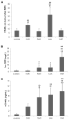
Results: CD40L expression on PBMCs was highest in the LAA group, followed by that in the CAS group (both P < 0·01 versus control). It was also higher in patients with atherosclerotic infarction than in those without atherosclerosis (P < 0·05). PBMC CD40L expression was sensitive and specific for detecting atherosclerosis and LAA cerebral infarction and was superior to serum C-reactive protein for predicting cerebral atherosclerosis (P < 0·01). Serum sCD40L was higher in patients with ACI than in healthy controls (P < 0·01) or patients with CAS (P < 0·01) and correlated with increased disability on three scales (all P < 0·01).
Conclusions: We conclude that patients with ACI have an upregulated CD40-CD40L system, which could be used as a clinical biomarker for assessing atherosclerotic instability and severity of neurological dysfunction.
Figures
Figure 1
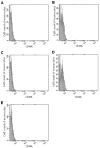
Detection of CD40L-expressing peripheral blood monocytes by flow cytometry. Monocytes gated by flow cytometry and expression of CD40L were assessed by the mean fluorescence intensity. Green area represents isotype control and red area represents CD40L detection. The _x_-axis represents CD40L fluorescence intensity and the _y_-axis represents the number of blood monocytes. (A) Normal control. (B) Carotid artery stenosis only. (C) Small-artery occlusion infarct. (D) Large-artery atherosclerotic infarct. (E) Cardioaortic cerebral embolism infarct.
Figure 2
Comparisons of CD40L and high-sensitivity C-reactive protein (hs-CRP) between healthy controls and patients with carotid artery stenosis (CAS), small-artery occlusion (SAO), large-artery atherosclerosis (LAA), and cardioaortic embolism (CAE). (A) Mean fluorescence intensity (MFI) of CD40L on peripheral blood monocytes as assessed by flow cytometry. **P<0.01 versus control; ††P<0.01 versus CAS; ##P<0.01 versus SAO or CAE. (B) Serum hs-CRP levels. *P<0.05, **P<0.01 versus control; †P<0.05, ††P<0.01 versus CAS; ##P<0.01 versus SAO or LAA. (C) Serum sCD40L levels. **P<0.01 versus control; †† P<0.01 versus CAS; ##P<0.01 versus SAO.
Figure 3
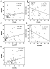
Correlations between CD40L expression on PBMCs, serum hp-CRP, serum sCD40L, and neurological dysfunction. CD40L expression on PBMCs was positively correlated with NIHSS score (Spearman rho=0.282, P<0.05; A) and negatively correlated with Barthel Index score (Spearman rho=−0.253, P<0.05; B). Serum hs-CRP level was positively correlated with NIHSS score (Spearman rho=0.433, P<0.01; C) and negatively correlated with Barthel Index score (Spearman rho=0.569, P<0.01; D). There was a positive linear correlation between serum sCD40L and NIHSS score (Spearman rho=0.237, P<0.05, E).
Similar articles
- The CD40/CD40L system regulates rat cerebral microvasculature after focal ischemia/reperfusion via the mTOR/S6K signaling pathway.
Jiang RH, Xu XQ, Wu CJ, Lu SS, Zu QQ, Zhao LB, Liu S, Shi HB. Jiang RH, et al. Neurol Res. 2018 Sep;40(9):717-723. doi: 10.1080/01616412.2018.1473075. Epub 2018 May 29. Neurol Res. 2018. PMID: 29843579 - CD40 ligand: a novel target in the fight against cardiovascular disease.
Vishnevetsky D, Kiyanista VA, Gandhi PJ. Vishnevetsky D, et al. Ann Pharmacother. 2004 Sep;38(9):1500-8. doi: 10.1345/aph.1D611. Epub 2004 Jul 27. Ann Pharmacother. 2004. PMID: 15280513 Review. - The CD40-CD40L system in cardiovascular disease.
Pamukcu B, Lip GY, Snezhitskiy V, Shantsila E. Pamukcu B, et al. Ann Med. 2011 Aug;43(5):331-40. doi: 10.3109/07853890.2010.546362. Epub 2011 Jan 18. Ann Med. 2011. PMID: 21244217 Review.
Cited by
- Analysis of Carotid color ultrasonography and high sensitive C-reactive protein in patients with atherosclerotic cerebral infarction.
Zhao L, Zhai Z, Hou W. Zhao L, et al. Pak J Med Sci. 2016 Jul-Aug;32(4):931-4. doi: 10.12669/pjms.324.9731. Pak J Med Sci. 2016. PMID: 27648042 Free PMC article. - Biomarker Profiling of Neurovascular Diseases in Patients with Stage 5 Chronic Kidney Disease.
Lee J, Bontekoe J, Trac B, Bansal V, Biller J, Hoppensteadt D, Maia P, Walborn A, Fareed J. Lee J, et al. Clin Appl Thromb Hemost. 2018 Dec;24(9_suppl):248S-254S. doi: 10.1177/1076029618807565. Epub 2018 Oct 22. Clin Appl Thromb Hemost. 2018. PMID: 30348002 Free PMC article. - Evaluation of plasma levels of neopterin and soluble CD40 ligand in patients with acute ischemic stroke in upper Egypt: can they surrogate the severity and functional outcome?
Tony AA, Tony EA, Mohammed WS, Kholef EF. Tony AA, et al. Neuropsychiatr Dis Treat. 2019 Feb 22;15:575-586. doi: 10.2147/NDT.S177726. eCollection 2019. Neuropsychiatr Dis Treat. 2019. PMID: 30863079 Free PMC article. - Changes in the cellular immune system and circulating inflammatory markers of stroke patients.
Jiang C, Kong W, Wang Y, Ziai W, Yang Q, Zuo F, Li F, Wang Y, Xu H, Li Q, Yang J, Lu H, Zhang J, Wang J. Jiang C, et al. Oncotarget. 2017 Jan 10;8(2):3553-3567. doi: 10.18632/oncotarget.12201. Oncotarget. 2017. PMID: 27682880 Free PMC article. - Soluble CD40L is associated with increased oxidative burst and neutrophil extracellular trap release in Behçet's disease.
Perazzio SF, Soeiro-Pereira PV, Dos Santos VC, de Brito MV, Salu B, Oliva MLV, Stevens AM, de Souza AWS, Ochs HD, Torgerson TR, Condino-Neto A, Andrade LEC. Perazzio SF, et al. Arthritis Res Ther. 2017 Oct 19;19(1):235. doi: 10.1186/s13075-017-1443-5. Arthritis Res Ther. 2017. PMID: 29052524 Free PMC article.
References
- Ross R. Atherosclerosis — an inflammatory disease. N Engl J Med. 1999;340:115–26. - PubMed
- Hassan GS, Merhi Y, Mourad W. CD40 ligand: a neo-inflammatory molecule in vascular diseases. Immunobiology. 2012;217:521–32. - PubMed
- Schönbeck U, Libby P. CD40 signaling and plaque instability. Circ Res. 2001;89:1092–103. - PubMed
- Mach F, Schonbeck U, Sukhova GK, Bourcier T, Bonnefoy JY, Pober JS, et al. Functional CD40 ligand is expressed on human vascular endothelial cells, smooth muscle cells, and macrophages: implications for CD40–CD40 ligand signaling in atherosclerosis. Proc Natl Acad Sci USA. 1997;94:1931–6. - PMC - PubMed
- Hansson GK, Robertson AK, Soderberg-Naucler C. Inflammation and atherosclerosis. Annu Rev Pathol. 2006;1:297–329. - PubMed
Publication types
MeSH terms
Substances
LinkOut - more resources
Full Text Sources
Other Literature Sources
Research Materials


