Structure of the mammalian ribosomal 43S preinitiation complex bound to the scanning factor DHX29 - PubMed (original) (raw)
Structure of the mammalian ribosomal 43S preinitiation complex bound to the scanning factor DHX29
Yaser Hashem et al. Cell. 2013.
Abstract
Eukaryotic translation initiation begins with assembly of a 43S preinitiation complex. First, methionylated initiator methionine transfer RNA (Met-tRNAi(Met)), eukaryotic initiation factor (eIF) 2, and guanosine triphosphate form a ternary complex (TC). The TC, eIF3, eIF1, and eIF1A cooperatively bind to the 40S subunit, yielding the 43S preinitiation complex, which is ready to attach to messenger RNA (mRNA) and start scanning to the initiation codon. Scanning on structured mRNAs additionally requires DHX29, a DExH-box protein that also binds directly to the 40S subunit. Here, we present a cryo-electron microscopy structure of the mammalian DHX29-bound 43S complex at 11.6 Å resolution. It reveals that eIF2 interacts with the 40S subunit via its α subunit and supports Met-tRNAi(Met) in an unexpected P/I orientation (eP/I). The structural core of eIF3 resides on the back of the 40S subunit, establishing two principal points of contact, whereas DHX29 binds around helix 16. The structure provides insights into eukaryote-specific aspects of translation, including the mechanism of action of DHX29.
Copyright © 2013 Elsevier Inc. All rights reserved.
Figures
Fig.1. Cryo-EM structure of the DHX29-bound 43S preinitiation complex
The map was segmented and colored variably. In all panels: 40S subunit is displayed in yellow, eIF2-tRNAiMet-GDPNP TC in orange, DHX29 in green, eIF3 structural core in red and its peripheral domains in light red. (A) The preinitiation complex viewed from the intersubunit face, (B) back, (C) solvent face and (D) top. Green arrows indicate the spatial relationship between views. See also figures S1, S2 and S3.
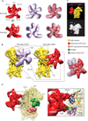
Fig.2. Structure and ribosomal interactions of eIF3
The map was segmented and colored variably. In all panels: 40S subunit, DHX29, eIF3 structural core and its peripheral domains are colored as in Fig. 1. (A) Comparison of the 30Å structure of individual eIF3 from HeLa cells (light purple) (Siridechadilok et al., 2005) with the 11.6Å structure of ribosome-bound eIF3 from RRL (red; present study). A small area of potential difference between structures is highlighted by a dashed circle. (B) Side-by side comparison of eIF3-40S subunit interactions as modeled previously (left) (Siridechadilok et al., 2005) and reconstructed in the present study (middle). Right panel shows an overlay of eIF3. (C) Comparison of eIF3-40S subunit interactions determined in the present study (top) and in a 48Å negative stain reconstruction of a ‘native’ 40S subunit (bottom) (Srivastava et al., 1992), in which eIF3 connections with the 40S that are called ls and us correspond to eIF3’s left arm and head, respectively. (D) Ribosomal proteins involved in interaction of eIF3 with the 40S subunit. Left, global view showing the T. thermophila 40S subunit crystal structure (light yellow ribbon) (Rabl et al., 2011) rigid-body fitted into our segmented 40S subunit Cryo-EM map (yellow mesh). Right, close-up view on the left arm and head regions of eIF3 showing the ribosomal proteins contacting eIF3 (colored ribbons). See also figure S4.
Fig.3. Compatibility of the ribosomal position of eIF3 with the 60S subunit, HCV IRES and expansion segments ES6S and ES7S of the _T. brucei_40S subunit
(A) Structural basis for the ribosomal anti-association activity of eIF3. eIF3 from our reconstruction (red mesh) was fitted on the 40S subunit in the crystal structure of yeast 80S ribosome (left) (Ben-Shem et al., 2011). Close-up view (right) showing the potential clash (red arrow) between eIF3’s head, covering rpS13e, and rpL30e of the 60S subunit. (B) Compatibility of eIF3 and the HCV IRES on the 40S subunit. eIF3 from our reconstruction (red mesh) was fitted on the 19.8Å cryo-EM structure of the 40S/HCV IRES complex (purple) (Spahn et al., 2001). A small clash between eIF3’s left arm and the pseudoknot/domain IIIf area of the IRES is indicated by the black arrow. (C) Compatibility of eIF3 with the T. brucei 40S subunit. Fitting of eIF3 from our reconstruction onto the 5Å cryo-EM structure of the T. brucei 40S subunit (Hashem et al., 2013), showing shape complementarity between eIF3 (red mesh) and the T. brucei expansion segments ES6S (hot pink) and ES7S (cyan).
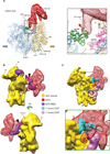
Fig.4. Peripheral domains of eIF3
(A) eIF3 peripheral domain (light red) located on the head of the 40S subunit, behind RACK1 (left). Close-up views (middle, right) showing interactions of this domain with ribosomal proteins (colored ribbons). (B) eIF3 peripheral doughnut-shaped domain (light red) located next to DHX29 (green) (left). Close-up views (middle, right) showing shape and size complementarity of the cryo-EM density of this domain with the crystal structure of eIF3i/TIF34 (Herrmannová et al. 2012). In all panels, the crystal structure of the T. thermophila 40S subunit (light yellow ribbon) (Rabl et al., 2011) was rigid-body fitted into our segmented 40S cryo-EM map (yellow mesh). See also figure S5.
Fig.5. The ribosomal position of the eIF2-ternary complex
(A) The location of the TC (orange) at the 40S subunit interface in the area of the P-site (left). The structures of eIF1 (cyan ribbon) and eIF1A (dark blue ribbon) were docked onto our map based on (Rabl et al., 2011) and (Battiste et al., 2000; Carter et al., 2001), respectively. Close-up views (middle, right) showing rigid body fitting of the crystal structure of the archaeal aIF2/Met-tRNAiMet/GDPNP complex (Schmitt et al., 2012) on the density assigned to the TC. (B) (Left) Poor accommodation of eIF2α D1–D2 domains after rigid-body fitting of the crystal structure of the archaeal aIF2-ternary complex (Schmitt et al., 2012) on the cryo-EM density of the DHX29-bound 43S complex. (Right) A different orientation of the aIF2αD1–D2 domains relative to Met-tRNAiMet when better fitted into density after rotation by 45 degrees and displacement further towards the 40S subunit neck by ~15 Å. The thick black lines in both panels highlight the main axis of the D1–D2 domain. See also figures S5 and S6.
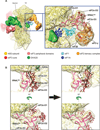
Fig.6. DHX29 binding on the 40S subunit
(A) Density assigned to DHX29 (green) showing that it mainly binds around h16, with a small domain residing at the subunit interface near the A-site (left). eIF1 (cyan ribbon) and eIF1A (dark blue ribbon) were docked as in Fig. 5. (Right) DHX29 density at a lower threshold, displaying several contacts with the 40S subunit beak (highlighted by black arrows), formed by interaction with ribosomal proteins and rRNA (colored ribbons). The magenta arrow indicates the weak linker between the main DHX29 density and the small domain. (B) Atomic model of DHX29 aa 551–1302 (colored to show its different domains, as in (Dhote et al., 2012)) rigid-body fitted into the DHX29 density. The brown ribbon shows the path of mRNA through DHX29, modeled on the basis of the crystal structure of single-stranded DNA bound to the structurally related DExH-box DNA helicase Hel308 (Büttner et al., 2005), and the thick cyan dashed line shows the conventional mRNA path. See also figure S5 and S6.
Similar articles
- Structure of mammalian eIF3 in the context of the 43S preinitiation complex.
des Georges A, Dhote V, Kuhn L, Hellen CU, Pestova TV, Frank J, Hashem Y. des Georges A, et al. Nature. 2015 Sep 24;525(7570):491-5. doi: 10.1038/nature14891. Epub 2015 Sep 7. Nature. 2015. PMID: 26344199 Free PMC article. - Functional role and ribosomal position of the unique N-terminal region of DHX29, a factor required for initiation on structured mammalian mRNAs.
Sweeney TR, Dhote V, Guca E, Hellen CUT, Hashem Y, Pestova TV. Sweeney TR, et al. Nucleic Acids Res. 2021 Dec 16;49(22):12955-12969. doi: 10.1093/nar/gkab1192. Nucleic Acids Res. 2021. PMID: 34883515 Free PMC article. - DHX29 reduces leaky scanning through an upstream AUG codon regardless of its nucleotide context.
Pisareva VP, Pisarev AV. Pisareva VP, et al. Nucleic Acids Res. 2016 May 19;44(9):4252-65. doi: 10.1093/nar/gkw240. Epub 2016 Apr 11. Nucleic Acids Res. 2016. PMID: 27067542 Free PMC article. - The scanning mechanism of eukaryotic translation initiation.
Hinnebusch AG. Hinnebusch AG. Annu Rev Biochem. 2014;83:779-812. doi: 10.1146/annurev-biochem-060713-035802. Epub 2014 Jan 29. Annu Rev Biochem. 2014. PMID: 24499181 Review. - Structural Insights into the Mechanism of Scanning and Start Codon Recognition in Eukaryotic Translation Initiation.
Hinnebusch AG. Hinnebusch AG. Trends Biochem Sci. 2017 Aug;42(8):589-611. doi: 10.1016/j.tibs.2017.03.004. Epub 2017 Apr 22. Trends Biochem Sci. 2017. PMID: 28442192 Review.
Cited by
- Conformational rearrangements upon start codon recognition in human 48S translation initiation complex.
Yi SH, Petrychenko V, Schliep JE, Goyal A, Linden A, Chari A, Urlaub H, Stark H, Rodnina MV, Adio S, Fischer N. Yi SH, et al. Nucleic Acids Res. 2022 May 20;50(9):5282-5298. doi: 10.1093/nar/gkac283. Nucleic Acids Res. 2022. PMID: 35489072 Free PMC article. - Suppression of MEHMO Syndrome Mutation in eIF2 by Small Molecule ISRIB.
Young-Baird SK, Lourenço MB, Elder MK, Klann E, Liebau S, Dever TE. Young-Baird SK, et al. Mol Cell. 2020 Feb 20;77(4):875-886.e7. doi: 10.1016/j.molcel.2019.11.008. Epub 2019 Dec 10. Mol Cell. 2020. PMID: 31836389 Free PMC article. - Ribosomal Protein L13 Promotes IRES-Driven Translation of Foot-and-Mouth Disease Virus in a Helicase DDX3-Dependent Manner.
Han S, Sun S, Li P, Liu Q, Zhang Z, Dong H, Sun M, Wu W, Wang X, Guo H. Han S, et al. J Virol. 2020 Jan 6;94(2):e01679-19. doi: 10.1128/JVI.01679-19. Print 2020 Jan 6. J Virol. 2020. PMID: 31619563 Free PMC article. - Role of aIF1 in Pyrococcus abyssi translation initiation.
Monestier A, Lazennec-Schurdevin C, Coureux PD, Mechulam Y, Schmitt E. Monestier A, et al. Nucleic Acids Res. 2018 Nov 16;46(20):11061-11074. doi: 10.1093/nar/gky850. Nucleic Acids Res. 2018. PMID: 30239976 Free PMC article. - Cellular functions of eukaryotic RNA helicases and their links to human diseases.
Bohnsack KE, Yi S, Venus S, Jankowsky E, Bohnsack MT. Bohnsack KE, et al. Nat Rev Mol Cell Biol. 2023 Oct;24(10):749-769. doi: 10.1038/s41580-023-00628-5. Epub 2023 Jul 20. Nat Rev Mol Cell Biol. 2023. PMID: 37474727 Review.
References
- Allen GS, Zavialov A, Gursky R, Ehrenberg M, Frank J. The cryo-EM structure of a translation initiation complex from Escherichia coli. Cell. 2005;121:703–712. - PubMed
- Battiste JL, Pestova TV, Hellen CU, Wagner G. The eIF1A solution structure reveals a large RNA-binding surface important for scanning function. Mol. Cell. 2000;5:109–119. - PubMed
Publication types
MeSH terms
Substances
Grants and funding
- R01 GM059660/GM/NIGMS NIH HHS/United States
- R01 GM029169/GM/NIGMS NIH HHS/United States
- R01 GM59660/GM/NIGMS NIH HHS/United States
- R01 GM29169/GM/NIGMS NIH HHS/United States
- HHMI/Howard Hughes Medical Institute/United States
LinkOut - more resources
Full Text Sources
Other Literature Sources
Molecular Biology Databases
Research Materials
Miscellaneous


