The stem cell organisation, and the proliferative and gene expression profile of Barrett's epithelium, replicates pyloric-type gastric glands - PubMed (original) (raw)
. 2014 Dec;63(12):1854-63.
doi: 10.1136/gutjnl-2013-306508. Epub 2014 Feb 18.
Anna M Nicholson 2, Richard Poulsom 3, Rosemary Jeffery 3, Alia Hussain 1, Laura J Gay 1, Janusz A Jankowski 3, Sebastian S Zeki 1, Hugh Barr 4, Rebecca Harrison 5, James Going 6, Sritharan Kadirkamanathan 7, Peter Davis 7, Timothy Underwood 8, Marco R Novelli 9, Manuel Rodriguez-Justo 9, Neil Shepherd 10, Marnix Jansen 11, Nicholas A Wright 1, Stuart A C McDonald 1
Affiliations
- PMID: 24550372
- PMCID: PMC4251192
- DOI: 10.1136/gutjnl-2013-306508
The stem cell organisation, and the proliferative and gene expression profile of Barrett's epithelium, replicates pyloric-type gastric glands
Danielle L Lavery et al. Gut. 2014 Dec.
Abstract
Objective: Barrett's oesophagus shows appearances described as 'intestinal metaplasia', in structures called 'crypts' but do not typically display crypt architecture. Here, we investigate their relationship to gastric glands.
Methods: Cell proliferation and migration within Barrett's glands was assessed by Ki67 and iododeoxyuridine (IdU) labelling. Expression of mucin core proteins (MUC), trefoil family factor (TFF) peptides and LGR5 mRNA was determined by immunohistochemistry or by in situ hybridisation, and clonality was elucidated using mitochondrial DNA (mtDNA) mutations combined with mucin histochemistry.
Results: Proliferation predominantly occurs in the middle of Barrett's glands, diminishing towards the surface and the base: IdU dynamics demonstrate bidirectional migration, similar to gastric glands. Distribution of MUC5AC, TFF1, MUC6 and TFF2 in Barrett's mirrors pyloric glands and is preserved in Barrett's dysplasia. MUC2-positive goblet cells are localised above the neck in Barrett's glands, and TFF3 is concentrated in the same region. LGR5 mRNA is detected in the middle of Barrett's glands suggesting a stem cell niche in this locale, similar to that in the gastric pylorus, and distinct from gastric intestinal metaplasia. Gastric and intestinal cell lineages within Barrett's glands are clonal, indicating derivation from a single stem cell.
Conclusions: Barrett's shows the proliferative and stem cell architecture, and pattern of gene expression of pyloric gastric glands, maintained by stem cells showing gastric and intestinal differentiation: neutral drift may suggest that intestinal differentiation advances with time, a concept critical for the understanding of the origin and development of Barrett's oesophagus.
Keywords: Barrett's Oesophagus; Gene Expression; Mucins; Stem Cells; Trefoil Factors.
Published by the BMJ Publishing Group Limited. For permission to use (where not already granted under a licence) please go to http://group.bmj.com/group/rights-licensing/permissions.
Figures
Figure 1
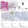
(A) (i) H&E (highlighted with s(surface), m(middle) and b(base)) and (ii) showing Ki67 expression in Barrett's glands; (iii) The number of Ki67+ cells in each region of Barrett's glands; (B) (i) IdU+ cells in the base, middle and surface of Barrett's glands 7 days. Inserts show high-power images of IdU+ cells; (ii) IdU+ cells at 11 days (arrowed). Inserts show a high-power image of IdU+ cells. (C) The changes in the distribution of IdU+ cells Barrett's glands with time after IdU injection. (i) IdU+ cells within the foveolus of the gland rapidly disappear and cannot be identified after 11 days; (ii) IdU+ cells identified within the middle of the gland decrease more rapidly after 11 days; (iii) the incidence of IdU+ cells in the base of Barrett's glands falls slowly up to 67 days after infusion.
Figure 2
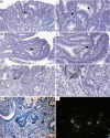
LGR5 mRNA expression using in situ hybridisation. (A, B) A bright field image and accompanying dark field image of LGR5 mRNA in Barrett's glands; (C and D) A bright field image and accompanying dark field image of LGR5 mRNA of pyloric gastric glands; (E and F) A bright field image and accompanying dark field image of LGR5 mRNA in gastric intestinal metaplasia.
Figure 3
A well-orientated Barrett's gland. (A) An H&E; (B and C) stained with MUC5AC and MUC2 (figure 3B prelaser capture microdissection (LCM), figure 3C post-LCM). (D) Cells microdissected from the gland all contain the same heteroplasmic m.825 G>T mutation in the MT-RNR1 gene. MUC2 cells (i) wild-type cells; (ii) MUC2 cells, (iii) MUC5AC cells, (iv) basal mucous-secreting cells (note: a lower level of heteroplasmy was detected), (iv) all share this mutation, but cells from the neighbouring wild-type gland do not. Online
supplementary figure
S4 shows high power views of the cells dissected.
Figure 4
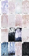
Gene expression in Barrett's glands compared with pyloric glands. Well-orientated glands displaying a contiguous surface, middle and base were analysed. (A) (i) Barrett's stained with D/PAS/Alcian Blue; (ii) Ki67 protein expression in Barrett's glands; (iii) in pyloric glands; (iv) MUC5AC protein expression in Barrett's glands. Figure 4B (i) MUC5AC protein expression in pyloric glands; (ii) MUC6 protein expression in Barrett's glands; (iii) in pyloric glands; (iv) MUC2 expression in Barrett's glands (see online
supplementary figure
S5A shows MUC2 to be absent from pyloric glands); figure 4C (i) TTF1 protein and (ii) mRNA expression in Barrett's glands; (iii) trefoil family factor 1 (TFF1) mRNA expression in pyloric glands. Supplementary figure 5B shows MUC5AC protein also in the upper part of pyloric glands; (iv) TTF2 protein in Barrett's glands. Figure 4D (i) mRNA expression in Barrett's glands; (ii) TFF2 mRNA in pyloric glands; (iii) TFF3 protein and (iv) mRNA in Barrett's glands.
Figure 5
Protein expression in low-grade Barrett's dysplasia. (A) An H&E; (B) Ki67 expression: (C) MUC2 expression; (D) MUC5AC expression; trefoil family factor 2 (TFF2); (E) and MUC6 (F) colocalise in the mucous cell bases of the gland, which remain in dysplasia.
Figure 6
LGR5 mRNA expression in Barrett's dysplasia and carcinoma. (A-C) low- (i) and high-power (ii) images showing non-isotopic ISH for LGR5 mRNA localisation in Barrett's dysplasia; (D) (i) bright field image and accompanying dark field image (ii) of isotopic ISH for LGR5 mRNA localisation in invasive Barrett's carcinoma glands.
Figure 7
(A) An H&E of a well-orientated Barrett's glands with diagrammatic representation of a model of organisation in Barrett's glands; the stem cell zone, here visualised as a ring of 6–7 cells, occupies the centre of the gland immediately above the point of branching. The trefoil family factor 1 (TFF1)+/MUC5AC+/MUC2+ cells migrate upwards from this zone while the TFF2+/MUC6+ cells migrate towards the base. (B, C) Possible models for stem/committed progenitor lineage relationships in Barrett's glands. Two possibilities are shown: (B) where a single stem cell gives rise to committed progenitors for the TFF1+/MUC5AC+ cells, the TFF3+/MUC2+ cells and the TFF2+/MUC6+ cells. (C) A neutral drift model where there are stem cells which produce TFF2+/MUC6+ cells, and stem cells which produce TFF1+/MUC5AC+ cells: following an event such as activation of CDX2, this stem cell(s) commit to produce TFF3+/MUC2+ cells, and stochastic niche succession will eventually, in some glands, move entirely to a niche containing stem cells committed to the TFF3+/MUC2+ lineage. We propose a conversion from non-goblet containing columnar to a specialised epithelium and finally to intestinal metaplasia.
Comment in
- Barrett oesophagus: origin of Barrett oesophagus.
Smith K. Smith K. Nat Rev Gastroenterol Hepatol. 2014 Apr;11(4):203. doi: 10.1038/nrgastro.2014.34. Epub 2014 Mar 11. Nat Rev Gastroenterol Hepatol. 2014. PMID: 24614345 No abstract available.
Similar articles
- Gastric-type mucin and TFF-peptide expression in Barrett's oesophagus is disturbed during increased expression of MUC2.
Van De Bovenkamp JH, Korteland-Van Male AM, Warson C, Büller HA, Einerhand AW, Ectors NL, Dekker J. Van De Bovenkamp JH, et al. Histopathology. 2003 Jun;42(6):555-65. doi: 10.1046/j.1365-2559.2003.01619.x. Histopathology. 2003. PMID: 12786891 - Barrett's esophagus is characterized by expression of gastric-type mucins (MUC5AC, MUC6) and TFF peptides (TFF1 and TFF2), but the risk of carcinoma development may be indicated by the intestinal-type mucin, MUC2.
Warson C, Van De Bovenkamp JH, Korteland-Van Male AM, Büller HA, Einerhand AW, Ectors NL, Dekker J. Warson C, et al. Hum Pathol. 2002 Jun;33(6):660-8. doi: 10.1053/hupa.2002.124907. Hum Pathol. 2002. PMID: 12152167 - Differential expression of mucins and trefoil peptides in native epithelium, Barrett's metaplasia and squamous cell carcinoma of the oesophagus.
Labouvie C, Machado JC, Carneiro F, Sarbia M, Vieth M, Porschen R, Seitz G, Blin N. Labouvie C, et al. J Cancer Res Clin Oncol. 1999;125(2):71-6. doi: 10.1007/s004320050244. J Cancer Res Clin Oncol. 1999. PMID: 10190312 - [Histochemical diagnosis of short segment Barrett's esophagus].
Fujiyama Y, Ishizuka I, Koyama S. Fujiyama Y, et al. Nihon Rinsho. 2005 Aug;63(8):1420-6. Nihon Rinsho. 2005. PMID: 16101233 Review. Japanese. - Origins of Metaplasia in Barrett's Esophagus: Is this an Esophageal Stem or Progenitor Cell Disease?
Zhang W, Wang DH. Zhang W, et al. Dig Dis Sci. 2018 Aug;63(8):2005-2012. doi: 10.1007/s10620-018-5069-5. Dig Dis Sci. 2018. PMID: 29675663 Free PMC article. Review.
Cited by
- Hybrid Stomach-Intestinal Chromatin States Underlie Human Barrett's Metaplasia.
Singh H, Ha K, Hornick JL, Madha S, Cejas P, Jajoo K, Singh P, Polak P, Lee H, Shivdasani RA. Singh H, et al. Gastroenterology. 2021 Sep;161(3):924-939.e11. doi: 10.1053/j.gastro.2021.05.057. Epub 2021 Jun 4. Gastroenterology. 2021. PMID: 34090884 Free PMC article. - Oesophageal adenocarcinoma and gastric cancer: should we mind the gap?
Hayakawa Y, Sethi N, Sepulveda AR, Bass AJ, Wang TC. Hayakawa Y, et al. Nat Rev Cancer. 2016 Apr 26;16(5):305-18. doi: 10.1038/nrc.2016.24. Nat Rev Cancer. 2016. PMID: 27112208 Review. - Clonal Transitions and Phenotypic Evolution in Barrett's Esophagus.
Evans JA, Carlotti E, Lin ML, Hackett RJ, Haughey MJ, Passman AM, Dunn L, Elia G, Porter RJ, McLean MH, Hughes F, ChinAleong J, Woodland P, Preston SL, Griffin SM, Lovat L, Rodriguez-Justo M, Huang W, Wright NA, Jansen M, McDonald SAC. Evans JA, et al. Gastroenterology. 2022 Apr;162(4):1197-1209.e13. doi: 10.1053/j.gastro.2021.12.271. Epub 2021 Dec 29. Gastroenterology. 2022. PMID: 34973296 Free PMC article. - Pathogenesis and Cells of Origin of Barrett's Esophagus.
Que J, Garman KS, Souza RF, Spechler SJ. Que J, et al. Gastroenterology. 2019 Aug;157(2):349-364.e1. doi: 10.1053/j.gastro.2019.03.072. Epub 2019 May 10. Gastroenterology. 2019. PMID: 31082367 Free PMC article. Review. - The cellular origins of cancer with particular reference to the gastrointestinal tract.
Alison MR. Alison MR. Int J Exp Pathol. 2020 Oct;101(5):132-151. doi: 10.1111/iep.12364. Epub 2020 Aug 14. Int J Exp Pathol. 2020. PMID: 32794627 Free PMC article. Review.
References
- Souza RF, Krishnan K, Spechler SJ. Acid, bile, and CDX: the ABCs of making Barrett's metaplasia. Am J Physiol Gastrointest Liver Physiol 2008;295:G211–8. - PubMed
- Chandrasoma PT, Der R, Dalton P, et al. Distribution and significance of epithelial types in columnar-lined esophagus. Am J Surg Pathol 2001;25:1188–93. - PubMed
Publication types
MeSH terms
Substances
LinkOut - more resources
Full Text Sources
Other Literature Sources
Medical
Miscellaneous


