The journey from stem cell to macrophage - PubMed (original) (raw)
Review
The journey from stem cell to macrophage
Mikael J Pittet et al. Ann N Y Acad Sci. 2014 Jun.
Abstract
Essential protectors against infection and injury, macrophages can also contribute to many common and fatal diseases. Here, we discuss the mechanisms that control different types of macrophage activities in mice. We follow the cells' maturational pathways over time and space and elaborate on events that influence the type of macrophage eventually settling a particular destination. The nature of the precursor cells, developmental niches, tissues, environmental cues, and other connecting processes appear to contribute to the identity of macrophage type. Together, the spatial and developmental relationships of macrophages compose a topo-ontogenic map that can guide our understanding of their biology.
Keywords: hematopoesis; macrophage; monocyte.
© 2014 New York Academy of Sciences.
Figures
Fig. 1
Topo-ontogenic map of macrophages and their progenitors in steady state. HSCs in bone marrow produce discrete intermediate progenitor populations, which increasingly lose self-renewing capacity as they commit to a given lineage. HSC-derived monocytes are found in circulation and in a splenic reservoir and can renew macrophages upon tissue extravasation. Some tissue macrophages develop in the embryo (yolk sac) before the appearance of HSCs and are maintained independently from the bone marrow.
Fig. 2
Monocyte production in bone marrow. Working model describing how long-term HSCs may produce monocytes in vivo. Several exogenous cells and soluble factors promote the retention and self-renewal/survival of HSCs (presumably in a quiescent niche) but also instruct commitment toward the myeloid lineage (presumably in an instructive/proliferative niche). The cartoon describes how monocyte production is controlled in steady state (white inserts) and in response to inflammatory cues (orange insert), which can induce HSCs to proliferate and skew them to the myeloid lineage.
Fig. 3
Monocyte production in extramedullary tissue. (A) A small fraction of HSPCs constitutively exit the bone marrow. Under inflammatory conditions, extramedullary HSPCs can be retained and amplified in the spleen, where they produce and replenish reservoir monocytes. Bottom left inserts provide additional information on how bone marrow and splenic activities differ in steady-state and inflammation. (B) Intravital micrographs depict the formation of splenic proliferative niches upon adoptive transfer of green GMPs. Cell clusters are highlighted in circles, venous sinuses in red. Bottom right: co-transfer of green and red GMPs identifies the formation of distinct green and red colonies. Images reproduced from refs, .
Fig. 4
Distinct mechanisms of monocyte mobilization from medullary and extramedullary tissue. The mobilization of monocytes from the bone marrow or spleen is a controlled process and is reviewed in detail elsewhere. In order to leave the bone marrow, Ly-6Chi monocytes require the chemokine receptor CCR2, which recognizes MCP-1 and MCP-3, two chemokines produced by medullary MSCs and CAR cells and secreted close to (and perhaps into) the lumen, and increase monocyte movement and intravasation. This process can be activated through sensing of bacterial products by TLRs expressed on MSCs and CAR cells. Panel A shows bone marrow sections illustrating emigration of monocytes in response to LPS, a process that depends on CCR2 signaling. Mobilization of monocytes from extramedullary sites such as the spleen, however, is independent of CCR2 and instead can be triggered by the peptide hormone angiotensin II (panel B). Whether accessory cells are involved in this process remains unknown. Increased concentrations of angiotensin II induce AGTR1A receptor dimerization on splenic monocytes, an event that increases monocyte motility and promotes intravasation (tracking of a departing monocyte shows time in min:sec). Images are reproduced from Refs and .
Fig. 5
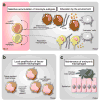
Expanding macrophage diversity in tissue. The cartoon illustrates three different origins of tissue macrophages (A) Selective accumulation of monocyte subsets committed with separate functions contributes different types of macrophages. For instance, CCR2-dependent recruitment of Ly-6Chi monocytes mediates tissue inflammation and proteolysis. This response is triggered upon intracellular pathogen infection or tissue damage. Ly-6Chi monocytes made both in medullary and extramedullary sites express CCR2 and response to MCP-1 for recruitment to tissues; however these cells might be equipped with divergent effector functions. Also, CX3CR1-dependent recruitment of Ly-6Clo monocytes may be associated with resolution of inflammation and mediation of tissue remodeling and healing. The recruited cells are also educated and polarized by their environment through cell-cell communication, sensing of soluble factors and interactions with extracellular matrix. (B) Local proliferation of tissue-resident macrophages. Some macrophages may proliferate at very low levels in steady-state conditions to maintain cell repertoires locally. Abnormal conditions, such as helminth infection and cardiovascular disease can induce potent macrophage proliferation in pleural cavity and atherosclerotic lesions, respectively. Such amplification can an occur in presence or absence of Ly-6Chi monocytes, and be promoted by locally produced factors including IL-4. (C) Maintenance of primitive macrophages. A lineage independent of HSCs is seeded during embryonic life and generates a repertoire of macrophages in various tissues, that is maintained after birth. These cells are often associated with epithelial structures, are thought to be unable to exit their tissue of residence, and may be poor antigen-presenting cells. These cells might serve in part as structural components that protect the tissue by sequestering foreign material and/or be involved in specific (non-immune) tissue functions.
Fig. 6
New targets in macrophage pathways? Drugs can attack cells at different developmental stages, from HSC to macrophage. The cartoon illustrates drugs that can interfere with macrophage differentiation survival and proliferation, monocyte recruitment to tissue, monocyte release from bone marrow or spleen, and production, amplification and release of bone marrow HSPCs.
Similar articles
- TGF-beta 1 and IFN-gamma direct macrophage activation by TNF-alpha to osteoclastic or cytocidal phenotype.
Fox SW, Fuller K, Bayley KE, Lean JM, Chambers TJ. Fox SW, et al. J Immunol. 2000 Nov 1;165(9):4957-63. doi: 10.4049/jimmunol.165.9.4957. J Immunol. 2000. PMID: 11046022 - New vistas on macrophage differentiation and activation.
Mantovani A, Sica A, Locati M. Mantovani A, et al. Eur J Immunol. 2007 Jan;37(1):14-6. doi: 10.1002/eji.200636910. Eur J Immunol. 2007. PMID: 17183610 Review. - Macrophage plasticity and polarization in tissue repair and remodelling.
Mantovani A, Biswas SK, Galdiero MR, Sica A, Locati M. Mantovani A, et al. J Pathol. 2013 Jan;229(2):176-85. doi: 10.1002/path.4133. Epub 2012 Nov 29. J Pathol. 2013. PMID: 23096265 Review. - Niche signals and transcription factors involved in tissue-resident macrophage development.
T'Jonck W, Guilliams M, Bonnardel J. T'Jonck W, et al. Cell Immunol. 2018 Aug;330:43-53. doi: 10.1016/j.cellimm.2018.02.005. Epub 2018 Feb 13. Cell Immunol. 2018. PMID: 29463401 Free PMC article. Review. - Surface antigens as markers of mouse macrophage differentiation.
Hirsch S, Gordon S. Hirsch S, et al. Int Rev Exp Pathol. 1983;25:51-75. Int Rev Exp Pathol. 1983. PMID: 6365822 Review. No abstract available.
Cited by
- Lifestyle effects on hematopoiesis and atherosclerosis.
Nahrendorf M, Swirski FK. Nahrendorf M, et al. Circ Res. 2015 Feb 27;116(5):884-94. doi: 10.1161/CIRCRESAHA.116.303550. Circ Res. 2015. PMID: 25722442 Free PMC article. Review. - Macrophage polarization and its role in the pathogenesis of acute lung injury/acute respiratory distress syndrome.
Chen X, Tang J, Shuai W, Meng J, Feng J, Han Z. Chen X, et al. Inflamm Res. 2020 Sep;69(9):883-895. doi: 10.1007/s00011-020-01378-2. Epub 2020 Jul 10. Inflamm Res. 2020. PMID: 32647933 Free PMC article. Review. - Clinical analysis and artificial intelligence survival prediction of serous ovarian cancer based on preoperative circulating leukocytes.
Feng Y, Wang Z, Cui R, Xiao M, Gao H, Bai H, Delvoux B, Zhang Z, Dekker A, Romano A, Wang S, Traverso A, Liu C, Zhang Z. Feng Y, et al. J Ovarian Res. 2022 May 24;15(1):64. doi: 10.1186/s13048-022-00994-2. J Ovarian Res. 2022. PMID: 35610701 Free PMC article. - Hematopoiesis and Cardiovascular Disease.
Poller WC, Nahrendorf M, Swirski FK. Poller WC, et al. Circ Res. 2020 Apr 10;126(8):1061-1085. doi: 10.1161/CIRCRESAHA.120.315895. Epub 2020 Apr 9. Circ Res. 2020. PMID: 32271679 Free PMC article. Review. - Spatio-Temporal Dynamics of M1 and M2 Macrophages in a Multiphase Model of Tumor Growth.
Lampropoulos I, Kevrekidis PG, Zois CE, Byrne H, Kavousanakis M. Lampropoulos I, et al. Bull Math Biol. 2025 Jun 4;87(7):92. doi: 10.1007/s11538-025-01466-6. Bull Math Biol. 2025. PMID: 40464993 Free PMC article.
References
- Metschnikoff E. Der Kampf der Phagocyten gegen Krankeitserreger. Virchows Archiv. 1884;96
- Chow A, Brown BD, Merad M. Studying the mononuclear phagocyte system in the molecular age. Nat Rev Immunol. 2011;11:788–798. - PubMed
- Weissleder R, Nahrendorf M, Pittet MJ. Imaging macrophages with nanoparticles. Nat Mater. 2014;13:125–138. - PubMed
- Janeway CAJ, Medzhitov R. Innate immune recognition. Annu Rev Immunol. 2002;20:197–216. - PubMed
- Stuart LM, Ezekowitz RA. Phagocytosis and comparative innate immunity: learning on the fly. Nat Rev Immunol. 2008;8:131–141. - PubMed
Publication types
MeSH terms
Grants and funding
- R01 HL095612/HL/NHLBI NIH HHS/United States
- R56 AI104695/AI/NIAID NIH HHS/United States
- R01 AI084880/AI/NIAID NIH HHS/United States
- R56-AI104695/AI/NIAID NIH HHS/United States
- P50 CA086355/CA/NCI NIH HHS/United States
- P50-CA086355/CA/NCI NIH HHS/United States
- R01-AI084880/AI/NIAID NIH HHS/United States
- R01 HL096576/HL/NHLBI NIH HHS/United States
- R01-HL095612/HL/NHLBI NIH HHS/United States
- U54 CA126515/CA/NCI NIH HHS/United States
- U54-CA126515/CA/NCI NIH HHS/United States
- R01-HL096576/HL/NHLBI NIH HHS/United States
- R01 HL095629/HL/NHLBI NIH HHS/United States
- R01-HL095629/HL/NHLBI NIH HHS/United States
LinkOut - more resources
Full Text Sources
Other Literature Sources


