Regulatory T cell reprogramming toward a Th2-cell-like lineage impairs oral tolerance and promotes food allergy - PubMed (original) (raw)
Regulatory T cell reprogramming toward a Th2-cell-like lineage impairs oral tolerance and promotes food allergy
Magali Noval Rivas et al. Immunity. 2015.
Abstract
Oral immunotherapy has had limited success in establishing tolerance in food allergy, reflecting failure to elicit an effective regulatory T (Treg) cell response. We show that disease-susceptible (Il4ra(F709)) mice with enhanced interleukin-4 receptor (IL-4R) signaling exhibited STAT6-dependent impaired generation and function of mucosal allergen-specific Treg cells. This failure was associated with the acquisition by Treg cells of a T helper 2 (Th2)-cell-like phenotype, also found in peripheral-blood allergen-specific Treg cells of food-allergic children. Selective augmentation of IL-4R signaling in Treg cells induced their reprogramming into Th2-like cells and disease susceptibility, whereas Treg-cell-lineage-specific deletion of Il4 and Il13 was protective. IL-4R signaling impaired the capacity of Treg cells to suppress mast cell activation and expansion, which in turn drove Th2 cell reprogramming of Treg cells. Interruption of Th2 cell reprogramming of Treg cells might thus provide candidate therapeutic strategies in food allergy.
Copyright © 2015 Elsevier Inc. All rights reserved.
Figures
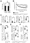
Figure 1. Deficiency of allergen-specific Treg cells in OVA-allergic Il4raF709 mice
(A) Core body temperature changes in PBS or OVA-SEB-sensitized WT and Il4raF709 mice after oral OVA challenge. (B, C) Total and OVA-specific serum IgE concentrations (B), intestinal mast cell number per low power field (LPF) and serum MMCP-1 concentrations post anaphylaxis (C) in mouse groups shown in (A). (D) Flow cytometric analysis and enumeration of CD4+IL-4+ and CD4+IFN-γ+ T cells in the MLN of PBS or OVA-SEB-sensitized WT and Il4raF709 mice. (E, F) Frequency (E) and numbers (F) of CD4+Foxp3+ Treg cells in different tissues of PBS, SEB and OVA-SEB-sensitized WT and Il4raF709 mice. (G) Foxp3 mRNA in the SI, MLN and spleens of PBS or OVA-SEB-sensitized WT and Il4raF709 mice. (H) Flow cytometric analysis of CD4+Foxp3+ Treg cell frequencies in cultures of MLN cells isolated from OVA-SEB-sensitized WT and Il4raF709 mice and stimulated with OVA323-339 peptide-pulsed DCs. (I) Frequencies of proliferating cells in gated CD4+Foxp3+ Treg cells shown in (H) and identified by proliferative dye (CellTrace Violet) dilution. Results are representative of 3 independent experiments. *p<0.05; **p<0.01; ***p<0.001 by Student’s unpaired two tailed t test and 2-way ANOVA with post-test analysis. N=5–10 mice/group.
Figure 2. Impaired allergen-specific iTreg cell formation in OVA-allergic Il4raF709 mice
(A) TGF-β induction of iTreg cells from naïve CD4+ T cells isolated from WT, Il4raF709, _Stat6_−/− and Il4raF709Stat6−/− mice in the absence or presence of IL-4 (10 ng/ml). (B) Effect of different IL-4 concentrations on TGF-β induction of iTreg cells from naïve T cells of groups shown in (A). (C) Absolute numbers of proliferating WT and Il4raF709 CD4+Foxp3+ iTreg cells and CD4+Foxp3− effector cells. (D) Schema of the experimental design of in vivo iTreg cell induction studies. Naïve DO11.10+CD4+ Tconv cells from WT or Il4raF709 DO11.10Rag2−/−_Foxp3_EGFP mice were loaded with CellTrace Violet dye and injected into OVA-SEB-sensitized Il4raF709 recipients. (E) Core body temperature change following OVA challenge. (F) Total and OVA-specific serum IgE concentrations and MMCP-1 release post anaphylaxis. (G) Flow cytometric analysis of iTreg cell conversion of adoptively transferred DO11.10+CD4+ Tconv cells in MLN of recipient OVA-SEB-sensitized Il4raF709 mice. (H) Percentages of donor CD4+_Foxp3_EGFP+ (left panel) and CD4+Foxp3EGFP− (right panel) in the MLN of recipient mice of panel (G). (I) Frequencies of proliferating cells in gated CD4+_Foxp3_EGFP+ (left panel) and CD4+Foxp3EGFP− (right panel) from panel (G). Results are representative of 3 independent experiments. *p<0.05; **p<0.01; ***p<0.001 by Student’s unpaired two tailed t test and 2-way ANOVA with post-test analysis. N=5–10 mice/group.
Figure 3. OVA-specific Il4raF709 Treg cells fail to suppress food allergy
(A) Core body temperature changes following OVA challenge of OVA-SEB-sensitized Il4raF709 mice that had received in vitro generated WT- or Il4raF709 DO11.10_+Foxp3EGFP_ iTreg, as described in Figure S3A. (B) Total and OVA-specific serum IgE concentrations, MMCP-1 release and small intestinal mast cell counts in mouse groups shown in panel (A). (C) Core body temperature changes following OVA challenge of OVA-SEB-sensitized Il4raF709 mice that were either left untreated or given either WT DO11.10+_Foxp3_EGFP+ Treg cells or _Il4raF709_DO11.10+_Foxp3_EGFP+ STAT6-sufficient or deficient Treg cells. (D) Total and OVA-specific serum IgE and serum MMCP-1 concentrations post anaphylaxis of the mouse groups from panel (C). N=5–17 mice per group, pooled from two different experiments. *p<0.05; **p<0.01; ***p<0.001, 1- and 2-way ANOVA with post-test analysis.
Figure 4. Treg cells of food allergic Il4raF709 mice undergo pathogenic Th2 cell-like reprogramming
(A) IL-4 expression by MLN CD4+Foxp3+ Treg cells from OVA-SEB-sensitized WT and Il4raF709 mice. (B) Frequencies (upper panel) and absolute numbers (bottom panel) of IL-4 expressing CD4+Foxp3+ Treg cells isolated from the MLN of PBS and OVA-SEB-sensitized WT and Il4raF709 mice. (C) Flow cytometric analysis of EGFP and YFP expression in CD4+ T cells isolated from the MLN of OVA-SEB-sensitized Foxp3EGFPCreR26YFP/YFP and Il4raF709Foxp3EGFPCreR26YFP/YFP mice. (D) Percentages (top panel) and numbers of Treg (EGFP+YFP+) and ex-Treg cells (EGFP−YFP+) in the MLN of the mouse groups described in panel (C). (E) Numbers of IL-4 secreting Tconv (EGFP−YFP−), ex-Treg (EGFP−YFP+) and Treg (EGFP+YFP+) CD4+ T cells isolated from the MLN of OVA-SEB sensitized Foxp3EGFPCreR26YFP/YFP and Il4raF709Foxp3EGFPCreR26YFP/YFP mice. (F–H) Flow cytometric analysis (F), frequencies (G) and numbers (H) of GATA-3+ or IRF-4+ Treg cells isolated from the MLN of PBS and OVA-SEB-sensitized WT and Il4raF709 mice. Results are representative of 2–3 independent experiments. N=3–10 mice/group; *p<0.05, **p<0.01 and ***p<0.001 by 1- and 2-way ANOVA with post-test analysis and Student’s unpaired two tailed t test.
Figure 5. Th2 cell cytokine production by Th2-reprogrammed Treg cells is critical to the food allergic response
(A) Real time PCR analysis of Il4 mRNA transcripts in Tconv (CD4+Foxp3−) and Treg cells (CD4+Foxp3+) sorted from spleens of Il4raF709 and Il4raF709 Foxp3EGFPCreIl4-Il13Δ/Δ. (B) Core body temperature changes following OVA challenge of OVA-SEB sensitized Il4raF709 and Il4raF709Foxp3EGFPCreIl4-Il13_Δ/_Δ mice. (C) Serum total IgE, OVA-specific IgE and MMCP-1 concentrations post anaphylaxis of mice from panel (B). (D) Percentages and numbers of CD4+Foxp3+ Treg cells in the MLN of OVA-SEB-sensitized Il4raF709 and Il4raF709 Foxp3EGFPCre Il4-Il13Δ/Δ mice. (E) Frequencies of GATA-3+ or IRF-4+ Treg cells isolated from the MLN of OVA-SEB-sensitized Il4raF709 and Il4raF709 Foxp3EGFPCre Il4-Il13−/− mice. (F) Percentages (top panel) and numbers (bottom panel) of CD4+ Tconv cells producing IL-4 in the MLN of OVA-SEB-sensitized Il4raF709 and Il4raF709 Foxp3EGFPCre Il4-Il13Δ/Δ mice. Results are representative of 2 independent experiments. N=3–18 mice/group; *p<0.05, **p<0.01 and ***p<0.001 by 1- and 2-way ANOVA with post-test analysis and Student’s unpaired two tailed t test.
Figure 6. Allergen-specific Treg cells of food allergic human subjects acquire a Th2-like phenotype
(A,B) Flow cytometric analysis (A) and frequencies (B) of circulating CD4+CD25+Foxp3+ Treg cells in the peripheral blood of food allergic children and age-matched healthy controls. Cells were gated on live CD3+CD4+ then analyzed for Foxp3 and CD25 expression. (C, D) Flow cytometric analysis (C) and frequencies (D) of GATA-3+ and IRF-4+ Treg cells in food allergic and control subjects. (E, F) Flow cytometric analysis (E) and frequencies (F) of IL-4+ and IL-13+ allergen-specific Treg cells in milk-stimulated PBMC cultures from healthy control, food allergic and milk allergic subjects. (G, H) Flow cytrometric analysis (G) and frequencies (H) of IL-4 and IL-13 expressing CD4+Foxp3− Tconv cells in PBMC cultures of the respective groups shown in (E, F). Numbers in the flow plots represents means ± SEM of positive cells. N=5–17; *p<0.05, **p<0.01 and ***p<0.001 by Student’s unpaired two tailed t test and 1-way ANOVA with post-test analysis.
Figure 7. Lineage specific upregulation of IL-4R signaling in Treg cells confers susceptibility to food allergy
(A). Flow cytometric analysis of MLN congenic Treg cells in Foxp3K276X mice rescued at birth with either WT or Il4raF709 Thy1.1+CD4+ T cells from Foxp3EGFP mice then sham or OVA-SEB-sensitized and challenged with OVA. (B). Flow cytometric analysis of MLN cells of WT Foxp3 competent mice injected at birth with either WT or Il4raF709 Thy1.1+CD4+ T cells isolated from Foxp3EGFP mice then sham or OVA-SEB-sensitized and challenged with OVA. (C) Core body temperature changes in PBS and OVA-SEB-sensitized rescued Foxp3K276X mice following OVA challenge. (D) Total and OVA-specific serum IgE concentrations, intestinal mast cell count and MMCP-1 release post-anaphylaxis in PBS and OVA-SEB-sensitized rescued Foxp3K276X mice. (E) Frequencies of IL-4, IL-13, IFN-γ and IL-9-expressing CD4+Foxp3+ Treg cells in the MLN of PBS and OVA-SEB-sensitized Foxp3K276X rescued mice. (F) Core body temperature changes in Foxp3EGFPCre and _Foxp3EGFPCrePtpn6_Δ/Δ mice sensitized and challenged with OVA. (G–I) Serum total and OVA-specific IgE concentrations (G), chloroacetate esterase staining of jejunal mast cells (revealed in red) (H) and mast cells counts (I) of OVA-SEB-sensitized Foxp3EGFPCre and _Foxp3EGFPCrePtpn6_Δ/Δ mice. Results representative of two independent experiments. N=3–10 mice per group. *p<0.05; **p<0.01; ***p<0.001 by 1-and 2-way ANOVA with post-test analysis and Student’s unpaired two tailed t test.
Similar articles
- Allergen-specific IgG antibody signaling through FcγRIIb promotes food tolerance.
Burton OT, Tamayo JM, Stranks AJ, Koleoglou KJ, Oettgen HC. Burton OT, et al. J Allergy Clin Immunol. 2018 Jan;141(1):189-201.e3. doi: 10.1016/j.jaci.2017.03.045. Epub 2017 May 4. J Allergy Clin Immunol. 2018. PMID: 28479335 Free PMC article. - IL-4 production by group 2 innate lymphoid cells promotes food allergy by blocking regulatory T-cell function.
Noval Rivas M, Burton OT, Oettgen HC, Chatila T. Noval Rivas M, et al. J Allergy Clin Immunol. 2016 Sep;138(3):801-811.e9. doi: 10.1016/j.jaci.2016.02.030. Epub 2016 Apr 18. J Allergy Clin Immunol. 2016. PMID: 27177780 Free PMC article. - Control and regulation of peripheral tolerance in allergic inflammatory disease: therapeutic consequences.
Li L, Boussiotis V. Li L, et al. Chem Immunol Allergy. 2008;94:178-188. doi: 10.1159/000155086. Chem Immunol Allergy. 2008. PMID: 18802347 Free PMC article. Review. - T regulatory cells and their counterparts: masters of immune regulation.
Ozdemir C, Akdis M, Akdis CA. Ozdemir C, et al. Clin Exp Allergy. 2009 May;39(5):626-39. doi: 10.1111/j.1365-2222.2009.03242.x. Clin Exp Allergy. 2009. PMID: 19422105 Review.
Cited by
- Microbiome and its impact on gastrointestinal atopy.
Muir AB, Benitez AJ, Dods K, Spergel JM, Fillon SA. Muir AB, et al. Allergy. 2016 Sep;71(9):1256-63. doi: 10.1111/all.12943. Epub 2016 Jun 23. Allergy. 2016. PMID: 27240281 Free PMC article. Review. - IL-4 inhibits regulatory T cells differentiation by HDAC9-mediated epigenetic regulation.
Cui J, Xu H, Yu J, Li Y, Chen Z, Zou Y, Zhang X, Du Y, Xia J, Wu J. Cui J, et al. Cell Death Dis. 2021 May 18;12(6):501. doi: 10.1038/s41419-021-03769-7. Cell Death Dis. 2021. PMID: 34006836 Free PMC article. - Pathogenic mechanisms in the evolution of food allergy.
Martinez-Blanco M, Mukhatayev Z, Chatila TA. Martinez-Blanco M, et al. Immunol Rev. 2024 Sep;326(1):219-226. doi: 10.1111/imr.13398. Epub 2024 Sep 17. Immunol Rev. 2024. PMID: 39285835 Free PMC article. Review. - The rise of food allergy: Environmental factors and emerging treatments.
Benedé S, Blázquez AB, Chiang D, Tordesillas L, Berin MC. Benedé S, et al. EBioMedicine. 2016 May;7:27-34. doi: 10.1016/j.ebiom.2016.04.012. Epub 2016 Apr 16. EBioMedicine. 2016. PMID: 27322456 Free PMC article. Review. - Primary atopic disorders.
Lyons JJ, Milner JD. Lyons JJ, et al. J Exp Med. 2018 Apr 2;215(4):1009-1022. doi: 10.1084/jem.20172306. Epub 2018 Mar 16. J Exp Med. 2018. PMID: 29549114 Free PMC article. Review.
References
- Amoli MM, Hand S, Hajeer AH, Jones KP, Rolf S, Sting C, Davies BH, Ollier WER. Polymorphism in the STAT6 gene encodes risk for nut allergy. Genes Immun. 2002;3:220–224. - PubMed
- Bilate AM, Lafaille JJ. Induced CD4+Foxp3+ regulatory T cells in immune tolerance. Annu Rev Immunol. 2012;30:733–758. - PubMed
Publication types
MeSH terms
Substances
Grants and funding
- R01 AI065617/AI/NIAID NIH HHS/United States
- R01 AI085090/AI/NIAID NIH HHS/United States
- R56 AI100889/AI/NIAID NIH HHS/United States
- T32 AI007512/AI/NIAID NIH HHS/United States
LinkOut - more resources
Full Text Sources
Other Literature Sources
Medical
Molecular Biology Databases
Research Materials
Miscellaneous


