CGRP as a neuropeptide in migraine: lessons from mice - PubMed (original) (raw)
Review
CGRP as a neuropeptide in migraine: lessons from mice
Andrew F Russo. Br J Clin Pharmacol. 2015 Sep.
Abstract
Migraine is a neurological disorder that is far more than just a bad headache. A hallmark of migraine is altered sensory perception. A likely contributor to this altered perception is the neuropeptide calcitonin gene-related peptide (CGRP). Over the past decade, CGRP has become firmly established as a key player in migraine. Although the mechanisms and sites of action by which CGRP might trigger migraine remain speculative, recent advances with mouse models provide some hints. This brief review focuses on how CGRP might act as both a central and peripheral neuromodulator to contribute to the migraine-like symptom of light aversive behaviour in mice.
Keywords: CGRP; calcitonin gene-related peptide; migraine; mouse model; neuropeptide; photophobia.
© 2015 The British Pharmacological Society.
Figures
Figure 1
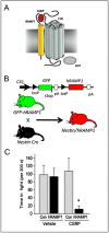
Calcitonin gene-related peptide (CGRP)-induced light-aversive behaviour in nestin/human receptor activity-modifying protein 1 (hRAMP1) mice. (A) A schematic of the CGRP receptor complex consisting of calcitonin-like receptor (CLR), receptor activity-modifying protein 1 (RAMP1), and receptor component protein (RCP) is shown (reproduced from Russo [8]). (B) The conditional hRAMP1 expression strategy is outlined (modified from Zhang et al. [60]). The parental green fluorescent protein (GFP)–hRAMP1 mouse transgene contains a GFP stop sequence that prevents the expression of hRAMP1 in the absence of Cre recombinase action at loxP sites. After crossing GFP–hRAMP1 mice with nestin–cre mice, the GFP stop sequence is removed and hRAMP1 is expressed in the nervous system of double transgenic nestin/hRAMP1 mice. (C) CGRP administration by intracerebroventricular injection caused nestin/hRAMP1 mice to spend less time in the light compared with control mice or mice injected with vehicle. *P < 0.05). Data obtained from Recober et al. [34]
Figure 2
Potential sites of CGRP action in light-aversive behaviour. The rodent brain is shown schematically, with arrows indicating pathways between relevant nuclei and input from the trigeminovascular system and light detected by the eye. These nuclei have calcitonin gene-related peptide (CGRP) receptors and respond to CGRP to modulate either affective behaviour or spinal trigeminal nucleus activity. Not all pathways are shown in this simplified presentation. Abbreviations are as follows: Amg, amygdala; Hyp, hypothalamus (which refers to the A11 nucleus and paraventricular nucleus); TG, trigeminal ganglion (which includes neurones and satellite glia); PAG, periaqueductal gray; Po, posterior thalamic nuclei (which include the posterior, ventroposteromedial and lateral posterior thalamus); Rmg, raphe magnus nucleus; SpV, spinal trigeminal nucleus; S1, S2, somatosensory cortex; V1, V2, visual cortex. Dural mast cells and blood vessels with associated trigeminal fibres and Schwann cells (not shown) are also indicated (brown line represents the dura)
Similar articles
- Calcitonin gene-related peptide (CGRP): a new target for migraine.
Russo AF. Russo AF. Annu Rev Pharmacol Toxicol. 2015;55:533-52. doi: 10.1146/annurev-pharmtox-010814-124701. Epub 2014 Oct 8. Annu Rev Pharmacol Toxicol. 2015. PMID: 25340934 Free PMC article. Review. - Induction of Migraine-Like Photophobic Behavior in Mice by Both Peripheral and Central CGRP Mechanisms.
Mason BN, Kaiser EA, Kuburas A, Loomis MM, Latham JA, Garcia-Martinez LF, Russo AF. Mason BN, et al. J Neurosci. 2017 Jan 4;37(1):204-216. doi: 10.1523/JNEUROSCI.2967-16.2016. J Neurosci. 2017. PMID: 28053042 Free PMC article. - Targeting CGRP: A New Era for Migraine Treatment.
Wrobel Goldberg S, Silberstein SD. Wrobel Goldberg S, et al. CNS Drugs. 2015 Jun;29(6):443-52. doi: 10.1007/s40263-015-0253-z. CNS Drugs. 2015. PMID: 26138383 Review. - Calcitonin gene-related peptide (CGRP) and migraine current understanding and state of development.
Bigal ME, Walter S, Rapoport AM. Bigal ME, et al. Headache. 2013 Sep;53(8):1230-44. doi: 10.1111/head.12179. Epub 2013 Jul 12. Headache. 2013. PMID: 23848260 Review. - [Calcitonin gene-related peptide: a key player neuropeptide in migraine].
Ramos-Romero ML, Sobrino-Mejia FE. Ramos-Romero ML, et al. Rev Neurol. 2016 Nov 16;63(10):460-468. Rev Neurol. 2016. PMID: 27819404 Review. Spanish.
Cited by
- Calcitonin gene-related peptide potentiated the excitatory transmission and network propagation in the anterior cingulate cortex of adult mice.
Li XH, Matsuura T, Liu RH, Xue M, Zhuo M. Li XH, et al. Mol Pain. 2019 Jan-Dec;15:1744806919832718. doi: 10.1177/1744806919832718. Mol Pain. 2019. PMID: 30717631 Free PMC article. - Navigating the Neurobiology of Migraine: From Pathways to Potential Therapies.
Tanaka M, Tuka B, Vécsei L. Tanaka M, et al. Cells. 2024 Jun 25;13(13):1098. doi: 10.3390/cells13131098. Cells. 2024. PMID: 38994951 Free PMC article. - Analysis of the DNA methylation pattern of the promoter region of calcitonin gene-related peptide 1 gene in patients with episodic migraine: An exploratory case-control study.
Rubino E, Boschi S, Giorgio E, Pozzi E, Marcinnò A, Gallo E, Roveta F, Grassini A, Brusco A, Rainero I. Rubino E, et al. Neurobiol Pain. 2022 Apr 2;11:100089. doi: 10.1016/j.ynpai.2022.100089. eCollection 2022 Jan-Jul. Neurobiol Pain. 2022. PMID: 35445161 Free PMC article. - Calcitonin gene-related peptide (receptor) antibodies: an exciting avenue for migraine treatment.
MaassenVanDenBrink A, Terwindt GM, van den Maagdenberg AMJM. MaassenVanDenBrink A, et al. Genome Med. 2018 Feb 22;10(1):10. doi: 10.1186/s13073-018-0524-7. Genome Med. 2018. PMID: 29471874 Free PMC article. - The Epigenetics of Migraine.
Zobdeh F, Eremenko II, Akan MA, Tarasov VV, Chubarev VN, Schiöth HB, Mwinyi J. Zobdeh F, et al. Int J Mol Sci. 2023 May 23;24(11):9127. doi: 10.3390/ijms24119127. Int J Mol Sci. 2023. PMID: 37298078 Free PMC article. Review.
References
- de Tommaso M, Ambrosini A, Brighina F, Coppola G, Perrotta A, Pierelli F, Sandrini G, Valeriani M, Marinazzo D, Stramaglia S, Schoenen J. Altered processing of sensory stimuli in patients with migraine. Nat Rev Neurol. 2014;10:144–55. - PubMed
- Charles A. Migraine: a brain state. Curr Opin Neurol. 2013;26:235–9. - PubMed
- Goadsby PJ, Lipton RB, Ferrari MD. Migraine – current understanding and treatment. N Engl J Med. 2002;346:257–70. - PubMed
- Pietrobon D, Moskowitz MA. Pathophysiology of migraine. Annu Rev Physiol. 2013;75:365–91. - PubMed
- Noseda R, Burstein R. Migraine pathophysiology: anatomy of the trigeminovascular pathway and associated neurological symptoms, cortical spreading depression, sensitization, and modulation of pain. Pain. 2013;154:S44–53. - PubMed
Publication types
MeSH terms
Substances
LinkOut - more resources
Full Text Sources
Other Literature Sources
Medical
Research Materials
Miscellaneous

