Focal enhancement of the skeleton to exercise correlates with responsivity of bone marrow mesenchymal stem cells rather than peak external forces - PubMed (original) (raw)
. 2015 Oct;218(Pt 19):3002-9.
doi: 10.1242/jeb.118729. Epub 2015 Jul 31.
Affiliations
- PMID: 26232415
- PMCID: PMC4631774
- DOI: 10.1242/jeb.118729
Focal enhancement of the skeleton to exercise correlates with responsivity of bone marrow mesenchymal stem cells rather than peak external forces
Ian J Wallace et al. J Exp Biol. 2015 Oct.
Abstract
Force magnitudes have been suggested to drive the structural response of bone to exercise. As importantly, the degree to which any given bone can adapt to functional challenges may be enabled, or constrained, by regional variation in the capacity of marrow progenitors to differentiate into bone-forming cells. Here, we investigate the relationship between bone adaptation and mesenchymal stem cell (MSC) responsivity in growing mice subject to exercise. First, using a force plate, we show that peak external forces generated by forelimbs during quadrupedal locomotion are significantly higher than hindlimb forces. Second, by subjecting mice to treadmill running and then measuring bone structure with μCT, we show that skeletal effects of exercise are site-specific but not defined by load magnitudes. Specifically, in the forelimb, where external forces generated by running were highest, exercise failed to augment diaphyseal structure in either the humerus or radius, nor did it affect humeral trabecular structure. In contrast, in the ulna, femur and tibia, exercise led to significant enhancements of diaphyseal bone areas and moments of area. Trabecular structure was also enhanced by running in the femur and tibia. Finally, using flow cytometry, we show that marrow-derived MSCs in the femur are more responsive to exercise-induced loads than humeral cells, such that running significantly lowered MSC populations only in the femur. Together, these data suggest that the ability of the progenitor population to differentiate toward osteoblastogenesis may correlate better with bone structural adaptation than peak external forces caused by exercise.
Keywords: Bone adaptation; Cortical bone; Ground reaction forces; Mechanical loading; Osteoprogenitor cells; Physical activity; Trabecular bone.
© 2015. Published by The Company of Biologists Ltd.
Conflict of interest statement
Competing interests
The authors declare no competing or financial interests.
Figures
Fig. 1.
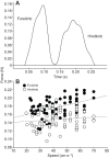
Mouse forelimbs generated higher peak vertical ground reaction forces than hindlimbs during quadrupedal locomotion. (A) Representative vertical ground reaction force trace from a 22.3 g mouse running at 39.4 cm s−1. (B) Bivariate plot of peak vertical ground reaction forces sustained by forelimbs and hindlimbs versus subject speed. Plotted lines were determined by least-squares regression. P<0.0001 for ANCOVA comparison between forelimb and hindlimb forces with speed as a covariate.
Fig. 2.
Treadmill exercise did not alter mouse body mass, muscle mass or home-cage activity level. (A) Change in mean body mass (±s.d.) among controls and treadmill runners during the experiment. (B) Mean limb muscle mass (+s.d.) among controls and runners at the end of the experiment. (C) Mean hourly ambulatory counts (±s.d.) among controls and runners throughout a 24 h period on a non-running day. (D) Total number of ambulatory counts among controls and runners during the 24 h. Boxes, median±first and third quartile; whiskers, median±range of non-outliers; circles, outliers. _P_>0.05 for all comparisons between controls and treadmill runners.
Fig. 3.
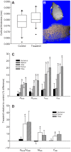
Skeletal adaptation to treadmill exercise was site-specific. (A) Interparietal cortical bone thickness among controls and treadmill runners. Boxes are median±first and third quartile; whiskers, median±range of non-outliers; circles, outliers. (B) μCT images of a mouse cranium showing the location of the interparietal (top, arrow) and the separation of cortical bone from diploë (bottom). Relative differences in (C) limb bone cortical properties and (D) trabecular properties between controls and runners. _A_total, total area; _A_cortex, cortical area; _I_max and _I_min, maximum and minimum second moments of area; _V_bone, bone volume; _V_total, total volume; _N_trab, trabecular number; _T_trab, trabecular thickness. Differences between group means are plotted as percentage difference+s.d. of the sampling distribution of the relative difference. *P<0.05 for comparisons between controls and treadmill runners. _P_>0.05 for all other differences.
Fig. 4.
Responsiveness of bone marrow mesenchymal stem cells to treadmill exercise. (A) Flow cytometric analysis illustrating the marrow cell population of a representative femur, with cells that positively stained with fluorescent antibody surface markers within the gates shown: (i) 10.3% SCA-1+; (ii) 3.65% CD105+; (iii) 4.63% c-Kit+; (iv) 60.9% CD44+; (v) 6.09% CD90.2+. (B) Subgating of surface markers selecting for discrete MSC populations: (i) 99.4% SCA-1+, c-Kit+; (ii) 99.3% SCA-1+, c-Kit+, CD90.2+; (iii) 100% SCA-1+, c-Kit+, CD90.2+, CD105+; (iv) 100% SCA-1+, c-Kit+, CD90.2+, CD105+, CD44+. (C) Percentage of total gated cells identified as MSCs in the humerus and femur of controls and treadmill runners. *P<0.05 for between-group comparison. (D) The number of MSCs per unit of marrow cavity volume in the humerus and femur of controls (_P_>0.05). Data are means+s.d.
Similar articles
- Effects of load-bearing exercise on skeletal structure and mechanics differ between outbred populations of mice.
Wallace IJ, Judex S, Demes B. Wallace IJ, et al. Bone. 2015 Mar;72:1-8. doi: 10.1016/j.bone.2014.11.013. Epub 2014 Nov 22. Bone. 2015. PMID: 25460574 - Mechanical, morphological and biochemical adaptations of bone and muscle to hindlimb suspension and exercise.
Shaw SR, Zernicke RF, Vailas AC, DeLuna D, Thomason DB, Baldwin KM. Shaw SR, et al. J Biomech. 1987;20(3):225-34. doi: 10.1016/0021-9290(87)90289-2. J Biomech. 1987. PMID: 3584148 - [Morphological analysis of bone dynamics and metabolic bone disease. Effect of loading on bone tissue].
Sakai A. Sakai A. Clin Calcium. 2011 Apr;21(4):569-74. Clin Calcium. 2011. PMID: 21447924 Review. Japanese. - Acute exercise mobilizes hematopoietic stem and progenitor cells and alters the mesenchymal stromal cell secretome.
Emmons R, Niemiro GM, Owolabi O, De Lisio M. Emmons R, et al. J Appl Physiol (1985). 2016 Mar 15;120(6):624-32. doi: 10.1152/japplphysiol.00925.2015. Epub 2016 Jan 7. J Appl Physiol (1985). 2016. PMID: 26744505 - The skeleton in a physical world.
Rubin J, Styner M. Rubin J, et al. Exp Biol Med (Maywood). 2022 Dec;247(24):2213-2222. doi: 10.1177/15353702221113861. Epub 2022 Aug 19. Exp Biol Med (Maywood). 2022. PMID: 35983849 Free PMC article. Review.
Cited by
- Exercise affects biological characteristics of mesenchymal stromal cells derived from bone marrow and adipose tissue.
Liu SY, He YB, Deng SY, Zhu WT, Xu SY, Ni GX. Liu SY, et al. Int Orthop. 2017 Jun;41(6):1199-1209. doi: 10.1007/s00264-017-3441-2. Epub 2017 Mar 31. Int Orthop. 2017. PMID: 28364139 - Swimming as Treatment for Osteoporosis: A Systematic Review and Meta-analysis.
Su Y, Chen Z, Xie W. Su Y, et al. Biomed Res Int. 2020 May 15;2020:6210201. doi: 10.1155/2020/6210201. eCollection 2020. Biomed Res Int. 2020. PMID: 32509864 Free PMC article. - Sprint Interval Training Induces A Sexual Dimorphism but does not Improve Peak Bone Mass in Young and Healthy Mice.
Koenen K, Knepper I, Klodt M, Osterberg A, Stratos I, Mittlmeier T, Histing T, Menger MD, Vollmar B, Bruhn S, Müller-Hilke B. Koenen K, et al. Sci Rep. 2017 Mar 17;7:44047. doi: 10.1038/srep44047. Sci Rep. 2017. PMID: 28303909 Free PMC article. - Use of adult mesenchymal stromal cells in tissue repair: impact of physical exercise.
Bourzac C, Bensidhoum M, Pallu S, Portier H. Bourzac C, et al. Am J Physiol Cell Physiol. 2019 Oct 1;317(4):C642-C654. doi: 10.1152/ajpcell.00530.2018. Epub 2019 Jun 26. Am J Physiol Cell Physiol. 2019. PMID: 31241985 Free PMC article. Review. - Quantitative Assessment of Optimal Bone Marrow Site for the Isolation of Porcine Mesenchymal Stem Cells.
McDaniel JS, Antebi B, Pilia M, Hurtgen BJ, Belenkiy S, Necsoiu C, Cancio LC, Rathbone CR, Batchinsky AI. McDaniel JS, et al. Stem Cells Int. 2017;2017:1836960. doi: 10.1155/2017/1836960. Epub 2017 Apr 30. Stem Cells Int. 2017. PMID: 28539939 Free PMC article.
References
- Bab I. A., Hajbi-Yonissi C., Gabet Y. and Müller R. (2007). Micro-tomographic Atlas of the Mouse Skeleton. New York: Springer.
- Biewener A. A. (1983). Locomotory stresses in the limb bones of two small mammals: the ground squirrel and chipmunk. J. Exp. Biol. 103, 131-154. - PubMed
- Biewener A. A. and Bertram J. E. A. (1994). Structural response of growing bone to exercise and disuse. J. Appl. Physiol. 72, 946-955. - PubMed
Publication types
MeSH terms
LinkOut - more resources
Full Text Sources
Other Literature Sources
Research Materials


