A long noncoding RNA AB073614 promotes tumorigenesis and predicts poor prognosis in ovarian cancer - PubMed (original) (raw)
A long noncoding RNA AB073614 promotes tumorigenesis and predicts poor prognosis in ovarian cancer
Zhongping Cheng et al. Oncotarget. 2015.
Abstract
Long noncoding RNA (lncRNA) profiles in ovarian cancer (OC) remain largely unknown. In the present study, we screened AB073614 as a new candidate lncRNA which promotes development of OC, in two independent datasets (GSE18521 and GSE38666) from the Gene Expression Omnibus (GEO). The level of AB073614 was then detected in 75 paired OC tissues and adjacent normal tissues by qRT-PCR. Results showed that AB073614 expression was significantly up-regulated in 85.3% (64/75) cancerous tissues compared with normal counterparts (P < 0.01). Further, the 5-year overall survival (OS) in OC patients with high expression of AB073614 was inferior to that with low expression (17.2 months vs 30.0 months, P = 0.0025). To investigate the functional role of AB073614, AB073614 siRNA was transfected into OC cell lines. Knockdown of AB073614 expression significantly inhibited cell proliferation and invasion, resulted in cell arrest in G1 phase of cell cycle and a dramatic increase of apoptosis, both in HO-8910 and OVCAR3 cells. In vivo experiment also revealed that knockdown AB073614 inhibited OVCAR3 cells proliferation. Finally, western blot assays indicated that lncRNA AB073614 may exert its function by targeting ERK1/2 and AKT-mediated signaling pathway. In conclusion, our study suggests that lncRNA AB073614 acts as a functional oncogene in OC development.
Keywords: AB073614; RNAi; lincRNA; ovarian cancer.
Conflict of interest statement
CONFLICTS OF INTEREST
The authors declare no competing financial interests.
Figures
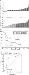
Figure 1. Screen of OC specific LncRNA in GEO database
LncRNA AB073614 was consistently over-expressed in OC tissue compared to the normal tissue in both of GSE18521 (A) and GSE38666 (B).
Figure 2. LncRNA AB073614 expression in human ovarian cancer tissues
A. Relative expression of AB073614 in OC tissues (n = 75) compared with corresponding non-tumor tissues (n = 75). AB073614 expression was examined by qPCR and normalized to GAPDH expression. Results are presented as the fold-change in tumor tissues relative to normal tissues. B. LncRNA AB073614 expression was classified into two groups, according the expression level in OC tissues. C. The correlation between AB073614 expression and prognosis. 5 year overall survival was analyzed by Kaplan-Meier survival curve. D. The receiver operating characteristic (ROC) curve for prognosis prediction of patients using AB073614 level. The area under curve (AUC) was shown in the plots.
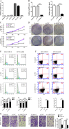
Figure 3. Knockdown AB073614 inhibits OC cells proliferation and invasion in vitro
A. Expression of lncRNA AB073614 in human ovarian epithelial cell line (HOEpiC) and five OC cell lines as determined by qRT-PCR. B. Knockdown efficiency was determined by qRT-PCR in HO-8910 and OVCAR3 cells. Knockdown AB073614 in HO-8910 and OVCAR3 cells significantly reduced their proliferative capacities, as determined by cell number counting assay C. and colony formation assay D. Knockdown AB073614 in HO-8910 and OVCAR3 cells resulted in cell arrest in G1 phase of cell cycle E. and a dramatic increase of apoptosis F. Invasion and metastasis capacities determined by Transwell assays G.
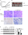
Figure 4. Knockdown AB073614 inhibits OC cells proliferation in vivo
OVCAR3 cells transfected with Si-AB073614 or Si-NC were subcutaneously inoculated into nude mice (6 per group). A. The tumor size was monitored every three days. B. Mice were sacrificed and the tumors were isolated after 33 days. C. Transplanted tumors with H&E staining and PCNA immunostaining. D. The expression of MMP-2 and MMP-9 in xenograft from the nude mice was determined by western blot.
Figure 5. Mechanisms of LncRNA AB073614 exerts its function
Signal pathway and key moderators in tumor progression were determined by western blotting in HO-8910 A, C. and OVCAR3 cells B, D. Left panel, representative results of western blot; right panal, protein levels relative to GAPDH. Data were presented as the mean value from three independent experiments ± S.D. **P < 0.01, **P < 0.001.
Similar articles
- Upregulation of lncRNA AB073614 functions as a predictor of epithelial ovarian cancer prognosis and promotes tumor growth in vitro and in vivo.
Zeng S, Liu S, Feng J, Gao J, Xue F. Zeng S, et al. Cancer Biomark. 2019;24(4):421-428. doi: 10.3233/CBM-182160. Cancer Biomark. 2019. PMID: 30909184 - LncRNA AB073614 regulates proliferation and metastasis of colorectal cancer cells via the PI3K/AKT signaling pathway.
Wang Y, Kuang H, Xue J, Liao L, Yin F, Zhou X. Wang Y, et al. Biomed Pharmacother. 2017 Sep;93:1230-1237. doi: 10.1016/j.biopha.2017.07.024. Epub 2017 Jul 20. Biomed Pharmacother. 2017. PMID: 28738539 - Up-Regulation of Long Non-Coding RNA AB073614 Predicts a Poor Prognosis in Patients with Glioma.
Hu L, Lv QL, Chen SH, Sun B, Qu Q, Cheng L, Guo Y, Zhou HH, Fan L. Hu L, et al. Int J Environ Res Public Health. 2016 Apr 19;13(4):433. doi: 10.3390/ijerph13040433. Int J Environ Res Public Health. 2016. PMID: 27104549 Free PMC article. - The Role of Long Non-Coding RNAs in Ovarian Cancer.
Nikpayam E, Tasharrofi B, Sarrafzadeh S, Ghafouri-Fard S. Nikpayam E, et al. Iran Biomed J. 2017 Jan;21(1):3-15. doi: 10.6091/.21.1.24. Epub 2016 Sep 24. Iran Biomed J. 2017. PMID: 27664137 Free PMC article. Review. - Role of lncRNAs in ovarian cancer: defining new biomarkers for therapeutic purposes.
Tripathi MK, Doxtater K, Keramatnia F, Zacheaus C, Yallapu MM, Jaggi M, Chauhan SC. Tripathi MK, et al. Drug Discov Today. 2018 Sep;23(9):1635-1643. doi: 10.1016/j.drudis.2018.04.010. Epub 2018 Apr 23. Drug Discov Today. 2018. PMID: 29698834 Free PMC article. Review.
Cited by
- Screening and Identification of an Immune-Associated lncRNA Prognostic Signature in Ovarian Carcinoma: Evidence from Bioinformatic Analysis.
Li Y, Huo FF, Wen YY, Jiang M. Li Y, et al. Biomed Res Int. 2021 Apr 30;2021:6680036. doi: 10.1155/2021/6680036. eCollection 2021. Biomed Res Int. 2021. PMID: 33997040 Free PMC article. - Next generation deep sequencing identified a novel lncRNA n375709 associated with paclitaxel resistance in nasopharyngeal carcinoma.
Ren S, Li G, Liu C, Cai T, Su Z, Wei M, She L, Tian Y, Qiu Y, Zhang X, Liu Y, Wang Y. Ren S, et al. Oncol Rep. 2016 Oct;36(4):1861-7. doi: 10.3892/or.2016.4981. Epub 2016 Jul 28. Oncol Rep. 2016. PMID: 27498905 Free PMC article. - Long noncoding RNA DATOC-1 that associate with DICER promotes development in epithelial ovarian cancer by upregulating miR-7 expression.
Qin W, Miao Y, Sun G, Chen S, Zang YS, Dong C. Qin W, et al. Transl Cancer Res. 2021 May;10(5):2379-2388. doi: 10.21037/tcr-20-189. Transl Cancer Res. 2021. PMID: 35116553 Free PMC article. - Long non-coding RNA H19 promotes tumorigenesis of ovarian cancer by sponging miR-675.
Ji F, Chen B, Du R, Zhang M, Liu Y, Ding Y. Ji F, et al. Int J Clin Exp Pathol. 2019 Jan 1;12(1):113-122. eCollection 2019. Int J Clin Exp Pathol. 2019. PMID: 31933725 Free PMC article. Retracted. - Role of STAT3 in cancer cell epithelial‑mesenchymal transition (Review).
Zhang G, Hou S, Li S, Wang Y, Cui W. Zhang G, et al. Int J Oncol. 2024 May;64(5):48. doi: 10.3892/ijo.2024.5636. Epub 2024 Mar 15. Int J Oncol. 2024. PMID: 38488027 Free PMC article. Review.
References
- Lalwani N, Prasad SR, Vikram R, Shanbhogue AK, Huettner PC, Fasih N. Histologic, molecular, and cytogenetic features of ovarian cancers: implications for diagnosis and treatment. Radiographics. 2011;31:625–646. - PubMed
- Bowtell DD. The genesis and evolution of high-grade serous ovarian cancer. Nat Rev Cancer. 2010;10:803–808. - PubMed
- Rustin G, van der Burg M, Griffin C, Qian W, Swart AM. Early versus delayed treatment of relapsed ovarian cancer. Lancet. 2011;377:380–381. - PubMed
- Yang G, Lu X, Yuan L. LncRNA: a link between RNA and cancer. Biochim Biophys Acta. 2014;1839:1097–1109. - PubMed
MeSH terms
Substances
LinkOut - more resources
Full Text Sources
Other Literature Sources
Medical
Miscellaneous


