Rare type of pancreatitis as the first presentation of anti-neutrophil cytoplasmic antibody-related vasculitis - PubMed (original) (raw)
Review
Rare type of pancreatitis as the first presentation of anti-neutrophil cytoplasmic antibody-related vasculitis
Tomoya Iida et al. World J Gastroenterol. 2016.
Abstract
A pancreatic tumor was suspected on the abdominal ultrasound of a 72-year-old man. Abdominal computed tomography showed pancreatic enlargement as well as a diffuse, poorly enhanced area in the pancreas; endoscopic ultrasound-guided fine needle aspiration biopsy and endoscopic retrograde cholangiopancreatography failed to provide a definitive diagnosis. Based on the trend of improvement of the pancreatic enlargement, the treatment plan involved follow-up examinations. Later, he was hospitalized with an alveolar hemorrhage and rapidly progressive glomerulonephritis; he tested positive for myeloperoxidase-anti-neutrophil cytoplasmic antibody (ANCA) and was diagnosed with ANCA-related vasculitis, specifically microscopic polyangiitis. It appears that factors such as thrombus formation caused by the vasculitis in the early stages of ANCA-related vasculitis cause abnormal distribution of the pancreatic blood flow, resulting in non-uniform pancreatitis. Pancreatic lesions in ANCA-related vasculitis are very rare. Only a few cases have been reported previously. Therefore, we report our case and a review of the literature.
Keywords: Anti-neutrophil cytoplasmic; Antibodies; Microscopic polyangiitis; Pancreas; Pancreatitis; Vasculitis.
Figures
Figure 1
Contrast-enhanced abdominal/pelvic computed tomography showing pancreatic enlargement with a diffuse, poorly enhanced area in the uncinate process (arrow) and pancreatic body tail (A-C).
Figure 2
On endoscopic ultrasound, a hypoechoic mass in a form in which the pancreatic lobe structure was maintained in the uncinate process and from the pancreatic body to the tail.
Figure 3
Endoscopic retrograde pancreatography. A: A slight disparity of the opening diameter in the main pancreatic duct, but no localized stenosis or diffuse narrowing observed; B: No abnormality in the papilla of Vater (arrow); C: The biopsy reveals thrombus formation (arrow).
Figure 4
Contrast-enhanced abdominal/pelvic computed tomography performed in May 2014 showing a trend of improvement in the pancreatic enlargement.
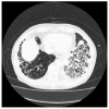
Figure 5
Chest computed tomography demonstrating an alveolar hemorrhage and pneumonia.
Similar articles
- [Myeloperoxidase antineutrophil cytoplasmic antibody (MPO-ANCA) -associated glomerulonephritis with acute pancreatitis: a case report].
Iida T, Amari Y, Yurugi T, Nakajima F. Iida T, et al. Nihon Jinzo Gakkai Shi. 2015;57(4):783-8. Nihon Jinzo Gakkai Shi. 2015. PMID: 26126336 Japanese. - Pancreatic mass as an initial manifestation of polyarteritis nodosa: a case report and review of the literature.
Yokoi Y, Nakamura I, Kaneko T, Sawayanagi T, Watahiki Y, Kuroda M. Yokoi Y, et al. World J Gastroenterol. 2015 Jan 21;21(3):1014-9. doi: 10.3748/wjg.v21.i3.1014. World J Gastroenterol. 2015. PMID: 25624739 Free PMC article. Review. - A case of microscopic polyangiitis with skin manifestations in a seven-year-old girl.
Yamada Y, Kitagawa C, Kamioka I, Chen KR, Oka M. Yamada Y, et al. Dermatol Online J. 2013 Sep 14;19(9):19624. Dermatol Online J. 2013. PMID: 24050297 - [Case of microscopic polyangiitis presenting initially as prostatic vasculitis].
Ashikaga E, Iyoda M, Suzuki H, Nagai H, Shibata T, Akizawa T. Ashikaga E, et al. Nihon Jinzo Gakkai Shi. 2009;51(8):1075-9. Nihon Jinzo Gakkai Shi. 2009. PMID: 19999587 Japanese. - From fibrosis to diagnosis: a paediatric case of microscopic polyangiitis and review of the literature.
Roszkiewicz J, Smolewska E. Roszkiewicz J, et al. Rheumatol Int. 2018 Apr;38(4):683-687. doi: 10.1007/s00296-017-3923-y. Epub 2018 Jan 2. Rheumatol Int. 2018. PMID: 29294176 Review.
Cited by
- Unique case of granulomatous arteritis in a grey mouse lemur (Microcebus murinus) - first case description.
Cichon N, Lampe K, Bremmer F, Becker T, Mätz-Rensing K. Cichon N, et al. Primate Biol. 2017 Apr 3;4(1):71-75. doi: 10.5194/pb-4-71-2017. eCollection 2017. Primate Biol. 2017. PMID: 32110694 Free PMC article. - Idiopathic acute pancreatitis: a review on etiology and diagnostic work-up.
Del Vecchio Blanco G, Gesuale C, Varanese M, Monteleone G, Paoluzi OA. Del Vecchio Blanco G, et al. Clin J Gastroenterol. 2019 Dec;12(6):511-524. doi: 10.1007/s12328-019-00987-7. Epub 2019 Apr 30. Clin J Gastroenterol. 2019. PMID: 31041651 Review. - Causal relationship between acute pancreatitis and methylprednisolone pulse therapy for fulminant autoimmune hepatitis: a case report and review of literature.
Nango D, Nakashima H, Hirose Y, Shiina M, Echizen H. Nango D, et al. J Pharm Health Care Sci. 2018 May 31;4:14. doi: 10.1186/s40780-018-0111-5. eCollection 2018. J Pharm Health Care Sci. 2018. PMID: 29881634 Free PMC article.
References
- Jennette JC, Falk RJ, Andrassy K, Bacon PA, Churg J, Gross WL, Hagen EC, Hoffman GS, Hunder GG, Kallenberg CG. Nomenclature of systemic vasculitides. Proposal of an international consensus conference. Arthritis Rheum. 1994;37:187–192. - PubMed
- Jennette JC, Falk RJ, Bacon PA, Basu N, Cid MC, Ferrario F, Flores-Suarez LF, Gross WL, Guillevin L, Hagen EC, et al. 2012 revised International Chapel Hill Consensus Conference Nomenclature of Vasculitides. Arthritis Rheum. 2013;65:1–11. - PubMed
- Villiger PM, Guillevin L. Microscopic polyangiitis: Clinical presentation. Autoimmun Rev. 2010;9:812–819. - PubMed
- Kallenberg CG, Heeringa P, Stegeman CA. Mechanisms of Disease: pathogenesis and treatment of ANCA-associated vasculitides. Nat Clin Pract Rheumatol. 2006;2:661–670. - PubMed
- Vamvakopoulos J, Savage CO, Harper L. ANCA-associated vasculitides-lessons from the adult literature. Pediatr Nephrol. 2010;25:1397–1407. - PubMed
Publication types
MeSH terms
Substances
LinkOut - more resources
Full Text Sources
Other Literature Sources
Medical
Research Materials


