Skin photoprotective and antiageing effects of a combination of rosemary (Rosmarinus officinalis) and grapefruit (Citrus paradisi) polyphenols - PubMed (original) (raw)
Skin photoprotective and antiageing effects of a combination of rosemary (Rosmarinus officinalis) and grapefruit (Citrus paradisi) polyphenols
Vincenzo Nobile et al. Food Nutr Res. 2016.
Abstract
Background: Plant polyphenols have been found to be effective in preventing ultraviolet radiation (UVR)-induced skin alterations. A dietary approach based of these compounds could be a safe and effective method to provide a continuous adjunctive photoprotection measure. In a previous study, a combination of rosemary (Rosmarinus officinalis) and grapefruit (Citrus paradisi) extracts has exhibited potential photoprotective effects both in skin cell model and in a human pilot trial.
Objective: We investigated the efficacy of a combination of rosemary (R. officinalis) and grapefruit (C. paradisi) in decreasing the individual susceptibility to UVR exposure (redness and lipoperoxides) and in improving skin wrinkledness and elasticity.
Design: A randomised, parallel group study was carried out on 90 subjects. Furthermore, a pilot, randomised, crossover study was carried out on five subjects. Female subjects having skin phototype from I to III and showing mild to moderate chrono- or photoageing clinical signs were enrolled in both studies. Skin redness (a* value of CIELab colour space) after UVB exposure to 1 minimal erythemal dose (MED) was assessed in the pilot study, while MED, lipoperoxides (malondialdehyde) skin content, wrinkle depth (image analysis), and skin elasticity (suction and elongation method) were measured in the main study.
Results: Treated subjects showed a decrease of the UVB- and UVA-induced skin alterations (decreased skin redness and lipoperoxides) and an improvement of skin wrinkledness and elasticity. No differences were found between the 100 and 250 mg extracts doses, indicating a plateau effect starting from 100 mg extracts dose. Some of the positive effects were noted as short as 2 weeks of product consumption.
Conclusions: The long-term oral intake of Nutroxsun™ can be considered to be a complementary nutrition strategy to avoid the negative effects of sun exposure. The putative mechanism for these effects is most likely to take place through the inhibition of UVR-induced reactive oxygen species and the concomitant inflammatory markers (lipoperoxides and cytokines) together with their direct action on intracellular signalling pathways.
Keywords: Citrus paradisi; Rosmarinus officinalis; antiageing; clinical study; photoprotection; plants extracts.
Figures
Fig. 1
Study flow and schedule of assessments chart. Subjects were first screened in the Farcoderm volunteers database (keywords: Sex = ‘female’, Age = ‘18’, Skin phototype = ‘I < phototype < III’, Skin type: ‘ageing or photoageing’, Testing preferences: ‘food supplements’). Eligible participants were then screened by a board certified dermatologist. During the screening visit, a physical examination was carried out in order to assess the uniformity of the test area (back) and the clinical sign of skin ageing on the face. Subjects meeting the inclusion criteria were then enrolled and randomised to participate in the short- or in the long-term study. Legend:
, physical examination; , informed consent signature; , eligibility check; , randomisation; PM*, provisional MED measurement (carried out only before study start); M, MED; L, lipoperoxides; W, wrinkle depth; E, skin elasticity; R, skin redness product intake.
Fig. 2
(a) UV exposure site and subsites. (b) Minimal erythema dose (MED).
Fig. 3
Skin elasticity curve. (a) R2 parameter calculation. (b) R5 parameter calculation.
Fig. 4
Flow chart of inclusion of subjects.
Fig. 5
Skin redness time course after 1 MED UVB exposure. Data are means (arbitrary units)±SE. *Statistically significant (p<0.05) when compared to 24 h;
Product intake.
Fig. 6
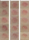
Skin redness variation after 1 MED UVB exposure. Digital pictures of (a) placebo, (b) 100 mg extracts dose, and (c) 250 mg extracts dose were taken using a Nikon D300 camera (Nikon corporate, Japan) equipped with a Nikon macro lens (AF-S Micro Nikkor 60 mm f/2.8 G ED) and parallel-polarised filters.
Fig. 7
Minimal erythemal dose (MED) before and after 0.5, 1, and 2 months treatment. Intragroup (vs. 0) statistical analysis is reported inside the bars of the histogram. Intergroup (vs. placebo) statistical analysis is reported upon the bars of the histogram. Statistical analysis is reported as follows: *p < 0.05, **p < 0.01, and ***p < 0.001. Data are means (mJ/cm2)±SE.
Fig. 8
Digital pictures of (a) placebo, (b) 100 mg extracts dose, and (c) 250 mg extracts dose were taken using a Nikon D300 camera (Nikon corporate, Japan) equipped with a Nikon macro lens (AF-S Micro Nikkor 60 mm f/2.8 G ED) and parallel-polarised filters. The a* (CIELab chromatic space) channel image is reported in order to enhance image contrast. The blue circle indicates the MED.
Fig. 9
Wrinkle depth before and after 0.5, 1, and 2 months treatment. Intragroup (vs. 0) statistical analysis is reported inside the bars of the histogram. Intergroup (vs. placebo) statistical analysis is reported upon the bars of the histogram. Statistical analysis is reported as follows: *p < 0.05, **p < 0.01, and ***p < 0.001. Data are means (µm)±SE.
Similar articles
- Protective effects of citrus and rosemary extracts on UV-induced damage in skin cell model and human volunteers.
Pérez-Sánchez A, Barrajón-Catalán E, Caturla N, Castillo J, Benavente-García O, Alcaraz M, Micol V. Pérez-Sánchez A, et al. J Photochem Photobiol B. 2014 Jul 5;136:12-8. doi: 10.1016/j.jphotobiol.2014.04.007. Epub 2014 Apr 20. J Photochem Photobiol B. 2014. PMID: 24815058 Clinical Trial. - Photoprotective and Antiaging Effects of a Standardized Red Orange (Citrus sinensis (L.) Osbeck) Extract in Asian and Caucasian Subjects: A Randomized, Double-Blind, Controlled Study.
Nobile V, Burioli A, Yu S, Zhifeng S, Cestone E, Insolia V, Zaccaria V, Malfa GA. Nobile V, et al. Nutrients. 2022 May 27;14(11):2241. doi: 10.3390/nu14112241. Nutrients. 2022. PMID: 35684041 Free PMC article. Clinical Trial. - Antioxidant and reduced skin-ageing effects of a polyphenol-enriched dietary supplement in response to air pollution: a randomized, double-blind, placebo-controlled study.
Nobile V, Schiano I, Peral A, Giardina S, Spartà E, Caturla N. Nobile V, et al. Food Nutr Res. 2021 Mar 29;65. doi: 10.29219/fnr.v65.5619. eCollection 2021. Food Nutr Res. 2021. PMID: 33889065 Free PMC article. - Rosemary (Rosmarinus officinalis L., syn Salvia rosmarinus Spenn.) and Its Topical Applications: A Review.
de Macedo LM, Santos ÉMD, Militão L, Tundisi LL, Ataide JA, Souto EB, Mazzola PG. de Macedo LM, et al. Plants (Basel). 2020 May 21;9(5):651. doi: 10.3390/plants9050651. Plants (Basel). 2020. PMID: 32455585 Free PMC article. Review. - Rosmarinus officinalis L. (rosemary) as therapeutic and prophylactic agent.
de Oliveira JR, Camargo SEA, de Oliveira LD. de Oliveira JR, et al. J Biomed Sci. 2019 Jan 9;26(1):5. doi: 10.1186/s12929-019-0499-8. J Biomed Sci. 2019. PMID: 30621719 Free PMC article. Review.
Cited by
- Evaluation of the acute toxicity, phototoxicity and embryotoxicity of a residual aqueous fraction from extract of the Antarctic moss Sanionia uncinata.
da Silva Fernandes A, Brito LB, Oliveira GAR, Ferraz ERA, Evangelista H, Mazzei JL, Felzenszwalb I. da Silva Fernandes A, et al. BMC Pharmacol Toxicol. 2019 Dec 19;20(Suppl 1):77. doi: 10.1186/s40360-019-0353-3. BMC Pharmacol Toxicol. 2019. PMID: 31852531 Free PMC article. - A Critical Review on Polyphenols and Health Benefits of Black Soybeans.
Ganesan K, Xu B. Ganesan K, et al. Nutrients. 2017 May 4;9(5):455. doi: 10.3390/nu9050455. Nutrients. 2017. PMID: 28471393 Free PMC article. Review. - Aesthetic Radiofrequency Associated with Rosmarinus officinalis Supplementation is Safe and Reduces Oxidative Stress in Women: Randomized, and Double-Blind Clinical Trial.
Tremêa GTF, Kleibert KRU, Krause LS, Fell APW, Scapini AR, Marschall KW, Baiotto CS, da Silva MHT, da Silva JAG, Colet CF. Tremêa GTF, et al. J Evid Based Integr Med. 2024 Jan-Dec;29:2515690X241246293. doi: 10.1177/2515690X241246293. J Evid Based Integr Med. 2024. PMID: 39135397 Free PMC article. Clinical Trial. - Ursodeoxycholic Acid May Inhibit Environmental Aging-Associated Hyperpigmentation.
Moon IJ, Yoo H, Paik SH, Kim HT, Kim SY, Song Y, Chang SE. Moon IJ, et al. Antioxidants (Basel). 2021 Feb 9;10(2):267. doi: 10.3390/antiox10020267. Antioxidants (Basel). 2021. PMID: 33572325 Free PMC article. - The Promising Role of Polyphenols in Skin Disorders.
Farhan M. Farhan M. Molecules. 2024 Feb 15;29(4):865. doi: 10.3390/molecules29040865. Molecules. 2024. PMID: 38398617 Free PMC article. Review.
References
- Sabziparvar AA, Shine KP, Forster PM. A model-derived global climatology of UV irradiation at the Earth's surface. Photochem Photobiol. 1999;69:193–202. - PubMed
- Tewari A, Grage MM, Harrison GI, Sarkany R, Young AR. UVA1 is skin deep: molecular and clinical implications. Photochem Photobiol Sci. 2013;12:95–103. doi: http://dx.doi.org/10.1039/c2pp25323b. - DOI - PubMed
- Cotran RS, Pathak MA. The pattern of vascular leakage induced by monochromatic UV irradiation in rats, guinea pigs and hairless mice. J Invest Dermatol. 1968;51:155–64. - PubMed
- Hruza LL, Pentland AP. Mechanisms of UV-induced inflammation. J Invest Dermatol. 1993;100:35S–41S. - PubMed
- Benrath J, Eschenfelder C, Zimmermann M, Gillardon F. Calcitonin gene-related peptide, substance P and nitric oxide are involved in cutaneous inflammation following ultraviolet irradiation. Eur J Pharmacol. 1995;293:87–96. - PubMed
LinkOut - more resources
Full Text Sources
Other Literature Sources


