Multiple transmitter systems contribute neurites to individual senile plaques - PubMed (original) (raw)
Multiple transmitter systems contribute neurites to individual senile plaques
L C Walker et al. J Neuropathol Exp Neurol. 1988 Mar.
Abstract
Senile plaques (SP), which consist largely of abnormal neuronal processes in proximity to deposits of amyloid, are a characteristic neuropathological feature of Alzheimer's disease. In lesser numbers, SP also occur in the brains of nondemented aged humans and nonhuman primates. To date, it is not known whether neurites in individual SP derive from neurons of one or several neurotransmitter systems. In aged monkeys, two strategies were used to test the hypothesis that individual SP can contain abnormal neurites arising from multiple neuronal systems. First, immunocytochemical methods were used to identify somatostatin-immunoreactive neurites in plaques, and these sections were subsequently stained with silver to visualize other neurites. Numerous plaques contained both somatostatin-positive and somatostatin-negative (i.e. argyrophilic only) neurites, suggesting that more than one transmitter system contributed neurites to each of these plaques. Second, two-color immunocytochemical techniques showed, in a small percentage of plaques, that cholinergic neurites coexist with neuropeptide Y (NPY)-containing neurites or catecholaminergic neurites. These results suggest that the formation of SP may result from events that involve abnormalities of neuronal processes arising from multiple transmitter systems.
Figures
Figure 1.
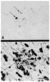
A. Abnormal somatostatin-immunoreactive neurites (thin arrows) in a senile plaque in the amygdala of the 26-year-old rhesus monkey. B. The same plaque as in Figure 1A, post-stained using a nonspecific silver stain [15]. Thin arrows designate the somatostatin-immunoreactive neurites indicated in Figure 1A. Thick arrows show two neurites that were not stained previously for somatostatin. Many surrounding circular and elliptical structures are silver-stained cell nuclei. Bar = 20 microns.
Figure 2.
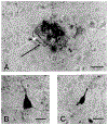
A. Neocortical senile plaque double-immunostained for choline acetyltransferase/DAB (light brown in original publication) and NPY/BDHC (granular blue in original publication). The black arrow indicates an abnormal cholinergic neurite, and the white arrow indicates an abnormal NPY-containing neurite. Numerous additional, partially overlapping neurites are also present. B. Choline acetyltransferase-immunoreactive neuron in the nucleus basalis of Meynert in the same tissue section as the plaque in Figure 2A. C. Neuropeptide Y-immunoreactive neuron in the neocortex, also from the same tissue section as the plaque in Figure 2A. This section was taken from the 28-year-old rhesus monkey. Bars = 20 microns.
Similar articles
- Neuropeptidergic systems in plaques of Alzheimer's disease.
Struble RG, Powers RE, Casanova MF, Kitt CA, Brown EC, Price DL. Struble RG, et al. J Neuropathol Exp Neurol. 1987 Sep;46(5):567-84. doi: 10.1097/00005072-198709000-00006. J Neuropathol Exp Neurol. 1987. PMID: 2442313 Review. - Current advances on different kinases involved in tau phosphorylation, and implications in Alzheimer's disease and tauopathies.
Ferrer I, Gomez-Isla T, Puig B, Freixes M, Ribé E, Dalfó E, Avila J. Ferrer I, et al. Curr Alzheimer Res. 2005 Jan;2(1):3-18. doi: 10.2174/1567205052772713. Curr Alzheimer Res. 2005. PMID: 15977985 Review.
Cited by
- Experimental microembolism induces localized neuritic pathology in guinea pig cerebrum.
Li JM, Cai Y, Liu F, Yang L, Hu X, Patrylo PR, Cai H, Luo XG, Xiao D, Yan XX. Li JM, et al. Oncotarget. 2015 May 10;6(13):10772-85. doi: 10.18632/oncotarget.3599. Oncotarget. 2015. PMID: 25871402 Free PMC article. - Development of senile plaques. Relationships of neuronal abnormalities and amyloid deposits.
Cork LC, Masters C, Beyreuther K, Price DL. Cork LC, et al. Am J Pathol. 1990 Dec;137(6):1383-92. Am J Pathol. 1990. PMID: 1701963 Free PMC article. - Comparative neuropathology in aging primates: A perspective.
Freire-Cobo C, Edler MK, Varghese M, Munger E, Laffey J, Raia S, In SS, Wicinski B, Medalla M, Perez SE, Mufson EJ, Erwin JM, Guevara EE, Sherwood CC, Luebke JI, Lacreuse A, Raghanti MA, Hof PR. Freire-Cobo C, et al. Am J Primatol. 2021 Nov;83(11):e23299. doi: 10.1002/ajp.23299. Epub 2021 Jul 13. Am J Primatol. 2021. PMID: 34255875 Free PMC article. Review. - Functional deprivation promotes amyloid plaque pathogenesis in Tg2576 mouse olfactory bulb and piriform cortex.
Zhang XM, Xiong K, Cai Y, Cai H, Luo XG, Feng JC, Clough RW, Patrylo PR, Struble RG, Yan XX. Zhang XM, et al. Eur J Neurosci. 2010 Feb;31(4):710-21. doi: 10.1111/j.1460-9568.2010.07103.x. Eur J Neurosci. 2010. PMID: 20384814 Free PMC article. - Aged rhesus monkeys: Cognitive performance categorizations and preclinical drug testing.
Plagenhoef MR, Callahan PM, Beck WD, Blake DT, Terry AV Jr. Plagenhoef MR, et al. Neuropharmacology. 2021 Apr 1;187:108489. doi: 10.1016/j.neuropharm.2021.108489. Epub 2021 Feb 6. Neuropharmacology. 2021. PMID: 33561449 Free PMC article.
References
- Armstrong DM, et al., Somatostatin-like immunoreactivity within neuritic plaques. Brain Res, 1985. 338(1): p. 71–9. - PubMed
- Berger B, et al., Catecholaminergic innervation of the human cerebral cortex in presenile and senile dementia: Histochemical and biochemical studies., in Enzymes and neurotransmitters in mental disease, Usdin E, Sourkes TL, and Youdim MBH, Editors. 1980, Wiley: Chichester. p. 317–328.
- Chan-Palay V, et al., Cortical neurons immunoreactive with antisera against neuropeptide Y are altered in Alzheimer’s-type dementia. J Comp Neurol, 1985. 238(4): p. 390–400. - PubMed
- Chan-Palay V, et al., Distribution of altered hippocampal neurons and axons immunoreactive with antisera against neuropeptide Y in Alzheimer’s-type dementia. J Comp Neurol, 1986. 248(3): p. 376–94. - PubMed
- Kitt CA, et al., Evidence for cholinergic neurites in senile plaques. Science, 1984. 226(4681): p. 1443–5. - PubMed
Publication types
MeSH terms
LinkOut - more resources
Full Text Sources
Medical
Miscellaneous

