Membrane Protein of Human Coronavirus NL63 Is Responsible for Interaction with the Adhesion Receptor - PubMed (original) (raw)
Membrane Protein of Human Coronavirus NL63 Is Responsible for Interaction with the Adhesion Receptor
Antonina Naskalska et al. J Virol. 2019.
Abstract
Human coronavirus NL63 (HCoV-NL63) is a common respiratory virus that causes moderately severe infections. We have previously shown that the virus uses heparan sulfate proteoglycans (HSPGs) as the initial attachment factors, facilitating viral entry into the cell. In the present study, we show that the membrane protein (M) of HCoV-NL63 mediates this attachment. Using viruslike particles lacking the spike (S) protein, we demonstrate that binding to the cell is not S protein dependent. Furthermore, we mapped the M protein site responsible for the interaction with HSPG and confirmed its relevance using a viable virus. Importantly, in silico analysis of the region responsible for HSPG binding in different clinical isolates and the Amsterdam I strain did not exhibit any signs of cell culture adaptation.IMPORTANCE It is generally accepted that the coronaviral S protein is responsible for viral interaction with a cellular receptor. Here we show that the M protein is also an important player during early stages of HCoV-NL63 infection and that the concerted action of the two proteins (M and S) is a prerequisite for effective infection. We believe that this study broadens the understanding of HCoV-NL63 biology and may also alter the way in which we perceive the first steps of cell infection with the virus. The data presented here may also be important for future research into vaccine or drug development.
Keywords: HCoV-NL63; attachment; heparan sulfate proteoglycans; membrane protein; viruslike particles.
Copyright © 2019 American Society for Microbiology.
Figures
FIG 1
Production of HCoV-NL63 VLPs. (A) Fluorescence microscope images of insect cells expressing N and M proteins. N protein was detected with mouse monoclonal antibody and is immunostained in green, whereas M protein was detected with rabbit polyclonal serum and is immunostained in red. Cell nuclei are immunostained with DAPI. Bar = 10 μm. (B) As anti-S antibody could not be used for immunostaining, S protein incorporation was demonstrated by Western blotting. Culture medium was harvested from insect cells infected with (M+E) rBV, N rBV, and S rBV. M, N, and S are lanes from blots detected with anti-M, anti-N, and anti-S antibodies, respectively. (C) Purification profiles of MEN and MENS VLPs on a heparin column. Collected fractions were analyzed by dot blotting with anti-M antibody. Numbers correspond to eluted fractions numbered on the chromatogram. “0,” sample prior purification; FT, flowthrough fraction; W, wash fraction. (D) TEM image of purified and positively stained MENS VLPs.
FIG 2
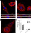
MEN VLP and MENS VLP adhesion to and internalization in susceptible cells. (A to C) Confocal microscopy images of LLC-Mk2 cells incubated with purified MEN VLPs (A) and MENS VLPs (B) and mock-incubated control cells (C). The bottom lanes of panels A and B show the xz plane in orthogonal views. VLPs were detected with anti-M polyclonal serum and are immunostained in green, actin fibers were immunostained with phalloidin and are shown in red, and nuclei were immunostained with DAPI and are shown in blue. Bars = 10 μm. (D) Graph showing quantification of the adsorbed and internalized VLPs. Data are presented as means ± standard deviations (SD). Each bar shows data from a minimum of 24 106- by 106-μm fields of view, registered from three different samples.
FIG 3
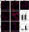
Adhesion and internalization of MEN and MENS VLPs in the presence of anti-ACE2 antibodies. (A) Cells preincubated with anti-ACE antibody (denoted αACE) or isotype antibody control (denoted Izo) were incubated with purified MEN or MENS VLPs. VLPs were detected with anti-M polyclonal serum and are immunostained in green, actin fibers were stained with phalloidin and are shown in red, and nuclei were stained with DAPI and are shown in blue. The bottom lanes show the xz plane in orthogonal views. Bars = 10 μm. (B) Graph showing the total number of VLPs per cell. Data were normalized to the values for untreated control samples (MEN and MENS. (C) Graph showing the proportion of VLPs inside the cell compared to the total number of VLPs observed on the cell. Each bar on graphs shows data from a minimum of 60 212- by 212-μm fields of view, registered from seven different samples, collected in three independent experiments. Data are presented as means ± 95% confidence intervals (CI). The total number of particles and the number of cells were quantified using the ImageJ Fiji tool 3D Objects Counter, and internalized particles were counted manually (*, P < 0.05; ****, P < 0.0001; ns, not significant). Ab, antibody.
FIG 4
Adhesion of MEN VLPs and MENS VLPs to LLC-Mk2 cells in the presence of heparan sulfate. Flow cytometry analysis was performed on cells incubated with VLPs or HCoV-NL63 in the presence of control PBS (denoted as Ø) or heparan sulfate (denoted as HS). Data are presented as means ± SD. Each bar shows data from a minimum of 5 replicates (****, P < 0.0001).
FIG 5
Peptide array. Synthetic peptides covering the M protein sequence were immobilized on a cellulose membrane and incubated with biotinylated heparin. The interaction was detected as a chemiluminescence signal with HRP-conjugated streptavidin. Numbers on the cellulose membrane (bottom) correspond to peptide numbers listed at the top.
FIG 6
Theoretical topology prediction of selected coronaviruses. Graphs were generated with the TMHMM server online tool (
). GenBank accession numbers of analyzed sequences are
AY567487
for human coronavirus NL63;
DQ010921
for feline coronavirus strain FIPV 79-1146;
AF353511
for porcine epidemic diarrhea virus strain CV777;
DQ811789
for virulent TGEV Purdue;
DQ811786
for TGEV Miller M60;
NC_001451
for avian infectious bronchitis virus;
NC_004718
,
NC_028845
,
NC_028858
,
NC_028866
,
NC_028873
, and
NC_028884
for SARS coronavirus;
KJ556336
for Middle East respiratory syndrome coronavirus isolate Jeddah_1_2013; and
FJ647225
for murine coronavirus inf-MHV-A59.
FIG 7
In situ ELISA. The 6×His-M153–226 protein interacts with LLC-Mk2 cells. Cells were incubated with 6×His-M153–226 protein and 6×His-N HKU1 protein, as a control (A), or with HS-preincubated 6×His-M153–226 protein (B). Protein adhesion to cells was detected with anti-His-HRP antibody and measured in a spectrophotometer. All measurements were done in triplicates, and the background signal was subtracted from the calculated mean values. The results shown are representative of data from three independent experiments.
FIG 8
VLP and HCoV-NL63 pseudoneutralization assay. (A and B) LLC-Mk2 cells were inoculated with MEN or MENS VLPs or HCoV-NL63 preincubated with anti-M serum, denoted “Ab” (A), or the respective preimmune serum (B). (C) The specificity of anti-M serum was examined by testing its effect on HCoV-OC43 adhesion to HRT-18G cells. Cells fixed and stained with anti-N monoclonal antibody were visualized using confocal microscopy. The number of particles and number of cells were quantified using the ImageJ Fiji 3D Objects Counter tool. The number of particles per cell is presented as a minimum-maximum graph with a line set at the median value. Each bar shows data from a minimum of 24 212- by 212-μm fields of view, registered from three different samples (**, P < 0.01; ****, P < 0.0001).
Similar articles
- Human coronavirus NL63 utilizes heparan sulfate proteoglycans for attachment to target cells.
Milewska A, Zarebski M, Nowak P, Stozek K, Potempa J, Pyrc K. Milewska A, et al. J Virol. 2014 Nov;88(22):13221-30. doi: 10.1128/JVI.02078-14. Epub 2014 Sep 3. J Virol. 2014. PMID: 25187545 Free PMC article. - Entry of Human Coronavirus NL63 into the Cell.
Milewska A, Nowak P, Owczarek K, Szczepanski A, Zarebski M, Hoang A, Berniak K, Wojarski J, Zeglen S, Baster Z, Rajfur Z, Pyrc K. Milewska A, et al. J Virol. 2018 Jan 17;92(3):e01933-17. doi: 10.1128/JVI.01933-17. Print 2018 Feb 1. J Virol. 2018. PMID: 29142129 Free PMC article. - Replication-dependent downregulation of cellular angiotensin-converting enzyme 2 protein expression by human coronavirus NL63.
Dijkman R, Jebbink MF, Deijs M, Milewska A, Pyrc K, Buelow E, van der Bijl A, van der Hoek L. Dijkman R, et al. J Gen Virol. 2012 Sep;93(Pt 9):1924-1929. doi: 10.1099/vir.0.043919-0. Epub 2012 Jun 20. J Gen Virol. 2012. PMID: 22718567 - Antiviral Action of Tryptanthrin Isolated from Strobilanthes cusia Leaf against Human Coronavirus NL63.
Tsai YC, Lee CL, Yen HR, Chang YS, Lin YP, Huang SH, Lin CW. Tsai YC, et al. Biomolecules. 2020 Feb 27;10(3):366. doi: 10.3390/biom10030366. Biomolecules. 2020. PMID: 32120929 Free PMC article. - Heparan Sulfate Proteoglycans and Viral Attachment: True Receptors or Adaptation Bias?
Cagno V, Tseligka ED, Jones ST, Tapparel C. Cagno V, et al. Viruses. 2019 Jul 1;11(7):596. doi: 10.3390/v11070596. Viruses. 2019. PMID: 31266258 Free PMC article. Review.
Cited by
- Endogenous IFITMs boost SARS-coronavirus 1 and 2 replication whereas overexpression inhibits infection by relocalizing ACE2.
Xie Q, Bozzo CP, Eiben L, Noettger S, Kmiec D, Nchioua R, Niemeyer D, Volcic M, Lee JH, Zech F, Sparrer KMJ, Drosten C, Kirchhoff F. Xie Q, et al. iScience. 2023 Apr 21;26(4):106395. doi: 10.1016/j.isci.2023.106395. Epub 2023 Mar 13. iScience. 2023. PMID: 36968088 Free PMC article. - Reversible rearrangement of the cellular cytoskeleton: A key to the broad-spectrum antiviral activity of novel amphiphilic polymers.
Dabrowska A, Botwina P, Barreto-Duran E, Kubisiak A, Obloza M, Synowiec A, Szczepanski A, Targosz-Korecka M, Szczubialka K, Nowakowska M, Pyrc K. Dabrowska A, et al. Mater Today Bio. 2023 Aug 7;22:100763. doi: 10.1016/j.mtbio.2023.100763. eCollection 2023 Oct. Mater Today Bio. 2023. PMID: 37600352 Free PMC article. - Cell surface nucleocapsid protein expression: A betacoronavirus immunomodulatory strategy.
López-Muñoz AD, Santos JJS, Yewdell JW. López-Muñoz AD, et al. Proc Natl Acad Sci U S A. 2023 Jul 11;120(28):e2304087120. doi: 10.1073/pnas.2304087120. Epub 2023 Jul 3. Proc Natl Acad Sci U S A. 2023. PMID: 37399385 Free PMC article. - Molecular mechanisms of human coronavirus NL63 infection and replication.
Castillo G, Mora-Díaz JC, Breuer M, Singh P, Nelli RK, Giménez-Lirola LG. Castillo G, et al. Virus Res. 2023 Apr 2;327:199078. doi: 10.1016/j.virusres.2023.199078. Epub 2023 Feb 22. Virus Res. 2023. PMID: 36813239 Free PMC article. Review. - The method utilized to purify the SARS-CoV-2 N protein can affect its molecular properties.
Tarczewska A, Kolonko-Adamska M, Zarębski M, Dobrucki J, Ożyhar A, Greb-Markiewicz B. Tarczewska A, et al. Int J Biol Macromol. 2021 Oct 1;188:391-403. doi: 10.1016/j.ijbiomac.2021.08.026. Epub 2021 Aug 6. Int J Biol Macromol. 2021. PMID: 34371045 Free PMC article.
References
Publication types
MeSH terms
Substances
LinkOut - more resources
Full Text Sources
Molecular Biology Databases
Research Materials


