Yap suppresses T-cell function and infiltration in the tumor microenvironment - PubMed (original) (raw)
Yap suppresses T-cell function and infiltration in the tumor microenvironment
Eleni Stampouloglou et al. PLoS Biol. 2020.
Abstract
A major challenge for cancer immunotherapy is sustaining T-cell activation and recruitment in immunosuppressive solid tumors. Here, we report that the levels of the Hippo pathway effector Yes-associated protein (Yap) are sharply induced upon the activation of cluster of differentiation 4 (CD4)-positive and cluster of differentiation 8 (CD8)-positive T cells and that Yap functions as an immunosuppressive factor and inhibitor of effector differentiation. Loss of Yap in T cells results in enhanced T-cell activation, differentiation, and function, which translates in vivo to an improved ability for T cells to infiltrate and repress tumors. Gene expression analyses of tumor-infiltrating T cells following Yap deletion implicates Yap as a mediator of global T-cell responses in the tumor microenvironment and as a negative regulator of T-cell tumor infiltration and patient survival in diverse human cancers. Collectively, our results indicate that Yap plays critical roles in T-cell biology and suggest that Yap inhibition improves T-cell responses in cancer.
Conflict of interest statement
The authors have declared that no competing interests exist.
Figures
Fig 1. Yap expression is induced upon T-cell activation resulting in suppression of T-cell activation.
WT and Yap-cKO CD4+ and CD8+ T cells were isolated from mouse spleens and stimulated with anti-CD3 and anti-CD28 coated magnetic beads to test expression of Yap protein and TEAD1–4 mRNA. T-cell activation was tested using increasing concentrations of plate-bound anti-CD3 with soluble anti-CD28 in Yap-cKO and WT T cells. Activation marker expression was also tested in WT CD4+ and CD8+ T cells treated with increasing concentrations of verteporfin under IL-2, and CD3/CD28 stimulation. Statistical differences were determined by using an F test to identify differences between nonlinear curve fits (C–F), unpaired two-sample t test (G–H), or one-way repeated-measures ANOVA with post hoc test for linear trend with increasing verteporfin (I–J). The underlying data for the graphs in this figure can be found in S1 Data and for the immunoblots in S1 Raw Images. (A) Yap protein levels in CD4+ T cells isolated from WT mouse spleens at various time points following CD3/CD28 stimulation. (B) Yap protein levels in CD8+ T cells isolated from WT mouse spleens at various time points following CD3/CD28 stimulation. (C) CD44 expression by flow cytometry on WT and Yap-cKO CD4+ T cells 72 hours post CD3/CD28 stimulation (n = 2–3 per dose/group). (D) CD44 expression by flow cytometry on WT and Yap-cKO CD8+ T cells 72 hours post CD3/CD28 stimulation (n = 2–3 per dose/group). (E) CD25 expression by flow cytometry on WT and Yap-cKO CD4+ T cells 72 hours post CD3/CD28 stimulation (n = 2–3 per dose/group). (F) CD25 expression by flow cytometry on WT and Yap-cKO CD8+ T cells 72 hours post CD3/CD28 stimulation (n = 2–3 per dose/group). (G) TEAD1–4 mRNA expression in CD4+ T cells 24 hours post CD3/CD28 stimulation (n = 3/group). (H) TEAD1–4 mRNA expression in CD8+ T cells 24 hours post CD3/CD28 stimulation (n = 3/group). (I) CD71 expression on WT CD4+ T cells 72 hours post IL-2 and CD3/CD28 stimulation and increasing concentration of verteporfin (n = 4/group). (J) CD71 expression on WT CD8+ T cells 72 hours post IL-2 and CD3/CD28 stimulation and increasing concentration of verteporfin (n = 4/group). CD, cluster of differentiation; cKO, conditional knockout; GAPDH, glyceraldehyde 3-phosphate dehydrogenase; IL-2, interleukin 2; MFI, median fluorescence intensity; No Stim, no stimulation; Stim, stimulation with IL-2 and anti-CD3/CD28; TEAD, TEA domain family member; WT, wild type; Yap, Yes-associated protein.
Fig 2. Deletion of Yap in CD4+ T cells results in increased IFNγ, IL-17, GATA3, and Foxp3 expression under Th1-, Th17-, Th2-, and Treg-polarizing conditions, respectively.
Naïve CD4+ T cells from WT and Yap-cKO mice were isolated using magnetic beads. WT and Yap-cKO T cells were cultured under Th1-, Th17-, Th2-, or Treg-polarizing conditions for 5 days in the presence of CD3 and CD28 antibodies. On day 5, IFNγ, IL-17, GATA3, and Foxp3 expression were measured using flow cytometry. Statistical differences were determined by using a Student t test (A–C) or an F test to identify differences between nonlinear curve fits (D). The underlying data for the graphs in this figure can be found in S2 Data. (A) IFNγ and IL-17 expression in WT and Yap-cKO CD4+ T cells under Th1-polarizing conditions (n = 3/group). (B) IFNγ and IL-17 expression in WT and Yap-cKO CD4+ T cells under Th17-polarizing conditions (n = 3/group). (C) IFNγ and GATA3 expression in WT and Yap-cKO CD4+ T cells under Th2-polarizing conditions (n = 3/group). (D) CD25 and Foxp3 expression in WT and Yap-cKO CD4+ T cells under Treg-polarizing conditions (n = 3–6 per dose/group). CD, cluster of differentiation; cKO, conditional knockout; Foxp3, forkhead box protein 3; GATA3, GATA binding protein 3; IFNγ, interferon gamma; Iono, ionomycin; IL-17, interleukin 17; No Stim, no stimulation; PMA, phorbol 12-myristate 13-acetate; TGFβ, transforming growth factor beta; Th, T helper cell type; Treg, regulatory T cell; WT, wild type; Yap, Yes-associated protein.
Fig 3. Thymocyte development is similar between WT and Yap-cKO T cells.
(A) Total thymocytes were isolated from thymuses of WT and Yap-cKO mice. Cells were stained and analyzed by flow cytometry for coreceptor maturation (A), positive selection (B), medullary maturation (C–E), and negative selection (F). Statistical differences were determined using two-way ANOVA followed by Sidak’s pairwise multiple comparisons tests. The underlying data for the graphs in this figure can be found in S3 Data. (A) Absolute numbers of thymocytes in different maturation stages: DN, DP, and SP for CD4 and CD8 coreceptor expression (n = 6–7 mice per group). (B) Frequency of thymocytes progressing through positive selection determined by TCRβ and CD69 expression (n = 6–7 mice per group). (C) Frequency of TCRβ+CCR7+ SP thymocytes in SM stage (CD69+MHCI−) (n = 6–7 mice per group). (D) Frequency of TCRβ+CCR7+ SP thymocytes in M1 stage (CD69+ MHCI+) (n = 6–7 mice per group). (E) Frequency of TCRβ+CCR7+ SP thymocytes in M2 stage (CD69−MHCI+) (n = 6–7 mice per group). (F) Frequency of Nur77+ cells among DP, CD4SP, or CD8SP thymocytes (n = 6–7 mice per group). CCR, chemokine receptor; CD, cluster of differentiation; cKO, conditional knockout; DN, double negative; DP, double positive; MHCI, major histocompatibility complex class I; M1, mature 1; M2, mature 2; N.S., not significant; Nur77, nuclear receptor 77; pos. sel., positive selection; pre-sel., pre-selection; SM, semimature; SP, single positive; TCR, T cell receptor; WT, wild type; Yap, Yes-associated protein.
Fig 4. T-cell–specific deletion of Yap results in reduced tumor growth and enhanced T-cell tumor infiltration.
Mice were challenged subcutaneously with B16F10 or LLC tumor cells on the right flank. Some mice carrying B16F10 tumors received adoptive cell transfer of WT and Yap-cKO CD8+ T cells. Tumor growth was monitored over the course of 15 days, until the maximum size of the tumors reached 500 mm3. B16F10 tumors were harvested for immunofluorescence or flow cytometric analysis. Statistical differences were determined by using a Student t test. The underlying data for the graphs in this figure can be found in S4 Data. (A) B16 tumor growth curve of WT and Yap-cKO mice (n = 9/group). (B) Tumor weight of B16 tumors derived from WT and Yap-cKO mice on day 15 post injection (n = 9/group). (C) LLC tumor growth curve of WT and Yap-cKO mice (n = 7/WT group, n = 5/cKO group). (D) Tumor weight of LLC tumors derived from WT and Yap-cKO mice on day 15 post injection (n = 7/WT group, n = 5/cKO group). (E) CD8+ T-cell immunofluorescence on day 15 of B16 tumor growth. (F) Absolute numbers of CD3+ TILs from WT and Yap-cKO B16 tumors. Tumors were harvested on day 15; stained with antibodies against CD45, CD3, CD4, and CD8; and analyzed using flow cytometry (n = 5/WT mice, n = 4/ Yap-cKO mice). (G) Absolute numbers of CD4+ TILs from WT and Yap-cKO B16 tumors, prepared as in Fig 4D (n = 5/WT mice, n = 4/ Yap-cKO mice). (H) Absolute numbers of CD8+ TILs from WT and Yap-cKO B16 tumors, prepared as in Fig 4D (n = 5/WT mice, n = 4/ Yap-cKO mice). (I) Experimental plan for adoptive cell transfer of WT and Yap-cKO CD8+ T cells in WT B16-bearing mice. (J) Absolute numbers of dTom+ and EYFP+ Yap-cKO versus WT CD8+ T cells in C57BL/6 B16 tumors. WT dTom+ CD8+ T cells were mixed 1:1 with Yap-cKO EYFP+ CD8+ T cells prior to being injected into WT C57BL/6 mice. Subsequently, mice were injected subcutaneously with B16F10 melanoma cells, and absolute number of infiltrating T cells was determined on day 15 by flow cytometry (n = 5/group). (K) Percentage of dTom+ and EYFP+ CD8+ T cells out of total B16 tumor-infiltrating CD8+ T cells (n = 5/group). CD, cluster of differentiation; cKO, conditional knockout; CTL, Control; dTom, dTomato; EYFP, enhanced yellow fluorescent protein; LLC, Lewis lung carcinoma; TIL, tumor-infiltrating lymphocyte; WT, wild type; Yap, Yes-associated protein.
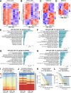
Fig 5. RNA-seq analysis of Yap-cKO CD4+ and CD8+ B16 TILs uncovers distinct gene expression changes that correlate with T-cell tumor infiltration.
CD4+ and CD8+ TILs and TDLNs were isolated from WT and Yap-cKO mice challenged with B16F10 tumors, and gene expression was analyzed by RNA-seq in the respective cells. RNA-seq data can be found at the NCBI GEO (Series Accession Number GSE139883) and in S1 and S2 Tables. The hyper-enrichment results are outlined in S3 Table. (A) Heatmap showing DEGs identified from Yap-cKO versus WT CD4+ TILs. (B) Yap-cKO versus WT CD8+ TIL DEG heatmap. (C) Yap-cKO versus WT CD4+ TDLN DEG heatmap. (D) Yap-cKO versus WT CD8+ TDLN DEG heatmap. (E) Hyper-enrichment analysis shows enrichment of induced gene sets observed in CD4+ Yap-cKO TILs. (F) Hyper-enrichment analysis shows enrichment of induced gene sets observed in CD8+ Yap-cKO TILs. (G) Hyper-enrichment analysis shows enrichment of repressed gene sets observed in CD4+ Yap-cKO TILs. (H) Hyper-enrichment analysis shows enrichment of repressed gene sets observed in CD8+ Yap-cKO TILs. (I) Gene expression changes identified in Yap-cKO CD4+ and CD8+ TILs correlate with genes reflecting tumor infiltration across many cancers in TCGA data. The heatmap is colored by the coefficient, and the text of each cell represents the adjusted _p_-value of the correlation. (J) Gene expression changes identified in Yap-cKO CD4+ and CD8+ T cells correlate with patient survival data available in TCGA across several cancers. Red: the average survival probability is higher for patients with high activity of the signature. Blue: the average survival probability is higher for patients with low activity of the signature. Each cell includes the _p_-value for the survival estimation. The distribution of _p_-values arising from the multiple survival analyses for each signature across TCGA datasets was compared to a uniform distribution using a Kolmogorov-Smirnov test. (K) Kaplan-Meier survival analysis showing the average survival probability of patients with LUAD that show low versus high Yap activity derived from the Yap-cKO CD4+ gene expression signature. (L) Kaplan-Meier survival analysis showing the average survival probability of patients with LUAD that show low versus high Yap activity derived from the Yap-cKO CD8+ gene expression signature. ACC, adrenocortical carcinoma; BLCA, bladder urothelial carcinoma; BRCA, breast invasive carcinoma; CD, cluster of differentiation; CESC, cervical squamous cell carcinoma and endocervical adenocarcinoma; CHOL, cholangiocarcinoma; cKO, conditional knockout; COAD, colon adenocarcinoma; DEG, differentially expressed gene; DLBC, lymphoid neoplasm diffuse large B-cell lymphoma; DLN, draining lymph node; ESCA, esophageal carcinoma; GBM, glioblastoma multiforme; GEO, Gene Expression Omnibus; HNSC, head and neck squamous cell carcinoma; KICH, kidney chromophobe; KIRC, kidney renal clear cell carcinoma; KIRP, kidney renal papillary cell carcinoma; k.s., Kolmogorov-Smirnov; LGG, brain lower grade glioma; LIHC, liver hepatocellular carcinoma; LUAD, lung adenocarcinoma; LUSC, lung squamous cell carcinoma; MESO, mesothelioma; NCBI, National Center for Biotechnology Information; OV, ovarian serous cystadenocarcinoma; PAAD, pancreatic adenocarcinoma; PCPG, pheochromocytoma and paraganglioma; PRAD, prostate adenocarcinoma; READ, rectum adenocarcinoma; RNA-seq, RNA sequencing; SARC, sarcoma; SKCM, skin cutaneous melanoma; STAD, stomach adenocarcinoma; TCGA, The Cancer Genome Atlas; TDLN, tumor-draining lymph node; TGCT, testicular germ cell tumors; THCA, thyroid carcinoma; THYM, thymoma; TIL, tumor-infiltrating lymphocyte; UCEC, uterine corpus endometrial carcinoma; UCS, uterine carcinosarcoma; UVM, uveal melanoma; WT, wild type; Yap, Yes-associated protein.
Similar articles
- YAP Attenuates CD8 T Cell-Mediated Anti-tumor Response.
Lebid A, Chung L, Pardoll DM, Pan F. Lebid A, et al. Front Immunol. 2020 Apr 8;11:580. doi: 10.3389/fimmu.2020.00580. eCollection 2020. Front Immunol. 2020. PMID: 32322254 Free PMC article. - Timely expression and activation of YAP1 in granulosa cells is essential for ovarian follicle development.
Lv X, He C, Huang C, Wang H, Hua G, Wang Z, Zhou J, Chen X, Ma B, Timm BK, Maclin V, Dong J, Rueda BR, Davis JS, Wang C. Lv X, et al. FASEB J. 2019 Sep;33(9):10049-10064. doi: 10.1096/fj.201900179RR. Epub 2019 Jun 14. FASEB J. 2019. PMID: 31199671 Free PMC article. - Adoptive transfer of siRNA Cblb-silenced CD8+ T lymphocytes augments tumor vaccine efficacy in a B16 melanoma model.
Hinterleitner R, Gruber T, Pfeifhofer-Obermair C, Lutz-Nicoladoni C, Tzankov A, Schuster M, Penninger JM, Loibner H, Lametschwandtner G, Wolf D, Baier G. Hinterleitner R, et al. PLoS One. 2012;7(9):e44295. doi: 10.1371/journal.pone.0044295. Epub 2012 Sep 4. PLoS One. 2012. PMID: 22962608 Free PMC article. - STC1 competitively binding βPIX enhances melanoma progression via YAP nuclear translocation and M2 macrophage recruitment through the YAP/CCL2/VEGFA/AKT feedback loop.
Ren Z, Xu Z, Chang X, Liu J, Xiao W. Ren Z, et al. Pharmacol Res. 2024 Jun;204:107218. doi: 10.1016/j.phrs.2024.107218. Epub 2024 May 18. Pharmacol Res. 2024. PMID: 38768671 - YAP mediates the interaction between the Hippo and PI3K/Akt pathways in mesangial cell proliferation in diabetic nephropathy.
Qian X, He L, Hao M, Li Y, Li X, Liu Y, Jiang H, Xu L, Li C, Wu W, Du L, Yin X, Lu Q. Qian X, et al. Acta Diabetol. 2021 Jan;58(1):47-62. doi: 10.1007/s00592-020-01582-w. Epub 2020 Aug 20. Acta Diabetol. 2021. PMID: 32816106
Cited by
- Erdr1 Drives Macrophage Programming via Dynamic Interplay with YAP1 and Mid1.
Wang Y. Wang Y. Immunohorizons. 2024 Feb 1;8(2):198-213. doi: 10.4049/immunohorizons.2400004. Immunohorizons. 2024. PMID: 38392560 Free PMC article. - Cell softness renders cytotoxic T lymphocytes and T leukemic cells resistant to perforin-mediated killing.
Zhou Y, Wang D, Zhou L, Zhou N, Wang Z, Chen J, Pang R, Fu H, Huang Q, Dong F, Cheng H, Zhang H, Tang K, Ma J, Lv J, Cheng T, Fiskesund R, Zhang X, Huang B. Zhou Y, et al. Nat Commun. 2024 Feb 15;15(1):1405. doi: 10.1038/s41467-024-45750-w. Nat Commun. 2024. PMID: 38360940 Free PMC article. - New insights into the ambivalent role of YAP/TAZ in human cancers.
Luo J, Deng L, Zou H, Guo Y, Tong T, Huang M, Ling G, Li P. Luo J, et al. J Exp Clin Cancer Res. 2023 May 22;42(1):130. doi: 10.1186/s13046-023-02704-2. J Exp Clin Cancer Res. 2023. PMID: 37211598 Free PMC article. Review. - Extracellular vesicles in fatty liver promote a metastatic tumor microenvironment.
Wang Z, Kim SY, Tu W, Kim J, Xu A, Yang YM, Matsuda M, Reolizo L, Tsuchiya T, Billet S, Gangi A, Noureddin M, Falk BA, Kim S, Fan W, Tighiouart M, You S, Lewis MS, Pandol SJ, Di Vizio D, Merchant A, Posadas EM, Bhowmick NA, Lu SC, Seki E. Wang Z, et al. Cell Metab. 2023 Jul 11;35(7):1209-1226.e13. doi: 10.1016/j.cmet.2023.04.013. Epub 2023 May 11. Cell Metab. 2023. PMID: 37172577 Free PMC article. - Expected and unexpected effects after systemic inhibition of Hippo transcriptional output in cancer.
Baroja I, Kyriakidis NC, Halder G, Moya IM. Baroja I, et al. Nat Commun. 2024 Mar 27;15(1):2700. doi: 10.1038/s41467-024-46531-1. Nat Commun. 2024. PMID: 38538573 Free PMC article. Review.
References
Publication types
MeSH terms
Substances
LinkOut - more resources
Full Text Sources
Other Literature Sources
Molecular Biology Databases
Research Materials


