The glomerular crescent: triggers, evolution, resolution, and implications for therapy - PubMed (original) (raw)
Review
The glomerular crescent: triggers, evolution, resolution, and implications for therapy
Lidia Anguiano et al. Curr Opin Nephrol Hypertens. 2020 May.
Abstract
Purpose of review: Crescents are classical histopathological lesions found in severe forms of rapidly progressive glomerulonephritis, also referred to as crescentic glomerulonephritis (CGN). Crescent formation is a consequence of diverse upstream pathomechanisms and unraveling these mechanisms is of great interest for improving the management of patients affected by CGN. Thus, in this review, we provide an update on the latest insight into the understanding on how crescents develop and how they resolve.
Recent findings: Cellular crescents develop from activated parietal epithelial cells (PECs) residing along Bowman's capsule and their formation has as a consequence the decline in glomerular filtration rate (GFR). Cellular crescents can be reversible, but when multilevel growth of PECs associate with an epithelial--mesenchymal transition-like change in cell phenotype, fibrous crescents form, and crescents become irreversible also in terms of GFR recovery. Different molecular pathways trigger the activation of PECs and are a prime therapeutics target in CGN. First, crescent formation requires also vascular injury causing ruptures in the glomerular basement membrane that trigger plasmatic coagulation within Bowman's space. This vascular necrosis can be triggered by different upstream mechanisms, such as small vessel vasculitides, immune complex glomerulonephritis, anti-GBM disease, and C3 glomerulonephritis, that all share complement activation but involve diverse upstream immune mechanisms outside the kidney accessible for therapeutic intervention.
Summary: Knowing the upstream mechanisms that triggered crescent formation provides a tool for the development of therapeutic interventions for CGN.
Figures
Box 1
no caption available
FIGURE 1
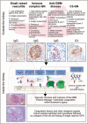
Evolution of crescent formation. The crescent is an unspecific histopathological lesion that can be triggered by a variety of different underlying disorders. This figure illustrates four of those and highlights first the extrarenal pathomechanisms leading to glomerular injury. The different patterns of immunoglobulin and complement deposits help to dissect these entities. Indeed, all of the aforementioned extrarenal drivers of injury to the glomerular microvasculature involve local complement activation as a shared molecular target for therapeutic interventions. Whenever, microvascular injury leads to rupture of the glomerular basement membrane (GBM), the leakage of plasma proteins drives parietal epithelial cell hyperplasia as the key cellular component of the crescent. Single nephron GFR decreases because of tuft collapse, rupture of the Bowman capsule, and influx of immune cells and fibroblasts are all secondary events that may or may not occur in individual glomeruli. Periglomerular immune cell infiltrates or fibrotic encasting of the activated parietal cells (fibrocellular crescents) are subsequent events that may affect the dynamics and prognosis of the disease. ANCA, antineutrophil cytoplasmic antibody; GFR = glomerular filtration rate; Ig, immunoglobulin; MPO, myeloperoxidase; PR3, proteinase 3.
Similar articles
- Glomerular expression of C-C chemokines in different types of human crescentic glomerulonephritis.
Liu ZH, Chen SF, Zhou H, Chen HP, Li LS. Liu ZH, et al. Nephrol Dial Transplant. 2003 Aug;18(8):1526-34. doi: 10.1093/ndt/gfg172. Nephrol Dial Transplant. 2003. PMID: 12897090 - Local macrophage proliferation in the pathogenesis of glomerular crescent formation in rat anti-glomerular basement membrane (GBM) glomerulonephritis.
Lan HY, Nikolic-Paterson DJ, Mu W, Atkins RC. Lan HY, et al. Clin Exp Immunol. 1997 Nov;110(2):233-40. doi: 10.1111/j.1365-2249.1997.tb08322.x. Clin Exp Immunol. 1997. PMID: 9367407 Free PMC article. - Novel therapeutic perspectives for crescentic glomerulonephritis through targeting parietal epithelial cell activation and proliferation.
Huang Y, Zhao X, Zhang Q, Yang X, Hou G, Peng C, Jia M, Zhou L, Yamamoto T, Zheng J. Huang Y, et al. Expert Opin Ther Targets. 2023 Jan;27(1):55-69. doi: 10.1080/14728222.2023.2177534. Epub 2023 Feb 16. Expert Opin Ther Targets. 2023. PMID: 36738160 Review. - [Crescent: structure, evolution, pathogenesis].
Modesto-Segonds A, Droz D, Rostaing L, Suc JM. Modesto-Segonds A, et al. Nephrologie. 1992;13(6):247-50. Nephrologie. 1992. PMID: 1299781 Review. French.
Cited by
- Targeting of oxidized Macrophage Migration Inhibitory Factor (oxMIF) with antibody ON104 attenuates the severity of glomerulonephritis.
Ferhat M, Mayer J, Costa LH, Prendecki M, Tarazona AAP, Schinagl A, Kerschbaumer RJ, Tam FWK, Landlinger C, Thiele M. Ferhat M, et al. PLoS One. 2024 Oct 7;19(10):e0311837. doi: 10.1371/journal.pone.0311837. eCollection 2024. PLoS One. 2024. PMID: 39374239 Free PMC article. - Single-cell RNA sequencing data locate ALDH1A2-mediated retinoic acid synthetic pathway to glomerular parietal epithelial cells.
Liu WB, Fermin D, Xu AL, Kopp JB, Xu Q. Liu WB, et al. Exp Biol Med (Maywood). 2024 Sep 18;249:10167. doi: 10.3389/ebm.2024.10167. eCollection 2024. Exp Biol Med (Maywood). 2024. PMID: 39360029 Free PMC article. Review. - [Renal involvement in systemic diseases].
Kain R. Kain R. Pathologie (Heidelb). 2024 Jul;45(4):261-268. doi: 10.1007/s00292-024-01338-1. Epub 2024 May 28. Pathologie (Heidelb). 2024. PMID: 38805092 Free PMC article. Review. German. - Glomerular crescents are associated with the risk of type 2 diabetic kidney disease progression: a retrospective cohort study.
Bae S, Yun D, Lee SW, Jhee JH, Lee JP, Chang TI, Oh J, Kwon YJ, Kim SG, Lee H, Kim DK, Joo KW, Moon KC, Chin HJ, Han SS. Bae S, et al. BMC Nephrol. 2024 May 20;25(1):172. doi: 10.1186/s12882-024-03578-y. BMC Nephrol. 2024. PMID: 38769500 Free PMC article. - Kidney Failure in Pauci-immune Crescentic Glomerulonephritis: Rationale for Immunosuppression to Improve Kidney Outcome.
Aqeel F, Geetha D. Aqeel F, et al. Curr Rheumatol Rep. 2024 Aug;26(8):290-301. doi: 10.1007/s11926-024-01150-z. Epub 2024 May 6. Curr Rheumatol Rep. 2024. PMID: 38709420 Review.
References
- Bajema IM, Wilhelmus S, Alpers CE, et al. Revision of the International Society of Nephrology/Renal Pathology Society classification for lupus nephritis: clarification of definitions, and modified National Institutes of Health activity and chronicity indices. Kidney Int 2018; 93:789–796. - PubMed
- Sethi S, Fervenza FC. Standardized classification and reporting of glomerulonephritis. Nephrol Dial Transplant 2019; 34:193–199. - PubMed
- This consensus statement presents a novel approach how to standardize a kidney biopsy report to determine the cause of the glomerulonephritis starting from the pattern of immune deposits.
- Romagnani P, Rinkevich Y, Dekel B. The use of lineage tracing to study kidney injury and regeneration. Nat Rev Nephrol 2015; 11:420–431. - PubMed
Publication types
MeSH terms
LinkOut - more resources
Full Text Sources
Research Materials
Miscellaneous

