Mechanisms of Cellular Senescence: Cell Cycle Arrest and Senescence Associated Secretory Phenotype - PubMed (original) (raw)
Review
Mechanisms of Cellular Senescence: Cell Cycle Arrest and Senescence Associated Secretory Phenotype
Ruchi Kumari et al. Front Cell Dev Biol. 2021.
Abstract
Cellular senescence is a stable cell cycle arrest that can be triggered in normal cells in response to various intrinsic and extrinsic stimuli, as well as developmental signals. Senescence is considered to be a highly dynamic, multi-step process, during which the properties of senescent cells continuously evolve and diversify in a context dependent manner. It is associated with multiple cellular and molecular changes and distinct phenotypic alterations, including a stable proliferation arrest unresponsive to mitogenic stimuli. Senescent cells remain viable, have alterations in metabolic activity and undergo dramatic changes in gene expression and develop a complex senescence-associated secretory phenotype. Cellular senescence can compromise tissue repair and regeneration, thereby contributing toward aging. Removal of senescent cells can attenuate age-related tissue dysfunction and extend health span. Senescence can also act as a potent anti-tumor mechanism, by preventing proliferation of potentially cancerous cells. It is a cellular program which acts as a double-edged sword, with both beneficial and detrimental effects on the health of the organism, and considered to be an example of evolutionary antagonistic pleiotropy. Activation of the p53/p21WAF1/CIP1 and p16INK4A/pRB tumor suppressor pathways play a central role in regulating senescence. Several other pathways have recently been implicated in mediating senescence and the senescent phenotype. Herein we review the molecular mechanisms that underlie cellular senescence and the senescence associated growth arrest with a particular focus on why cells stop dividing, the stability of the growth arrest, the hypersecretory phenotype and how the different pathways are all integrated.
Keywords: DNA damage response (DDR); DREAM complex; cell cycle arrest; cellular senescence; senescence associated secretory phenotype (SASP).
Copyright © 2021 Kumari and Jat.
Conflict of interest statement
The authors declare that the research was conducted in the absence of any commercial or financial relationships that could be construed as a potential conflict of interest.
Figures
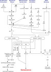
FIGURE 1
Signals and pathways involved in mediating senescence mediated cell cycle arrest. The figure shows the different intrinsic and extrinsic stimuli capable of inducing cellular senescence. Key pathways involved in manifesting cell cycle arrest in senescence such as p53/p21WAF1/CIP1 and p16INK4A/RB tumor suppressor pathways, DDR, AMPK, p38/MAPK, PI3K/AKT/mTOR are illustrated. It indicates how different pathways are interconnected and how the assembly of repressive DREAM complex triggers senescence and its disruption leads to cell cycle progression. ROS, reactive oxygen species; DDR, DNA damage response; DREAM, dimerization partner (DP), RB-like, E2F, and MuvB core complex.
FIGURE 2
Schematic of the different mechanisms involved in Senescence Associated Secretory Phenotype (SASP) regulation. This figure shows the different pathways involved in regulating SASP. Most of the pathways converge to activate the transcription factors NF-κB and c/EBPβ in senescent cells. The autocrine feed forward signaling of different pro-inflammatory cytokines such as IL-1A, IL-6, and IL-8 is illustrated. ZCAN4 promotes the expression of inflammatory cytokines via NF-κB. TAK1 activates p38/MAPK, a kinase that subsequently engages PI3K/Akt/mTOR pathway. mTOR is capable of activating the NF-κB signaling directly as well as indirectly via IL-1A. GATA-4 links autophagy and DNA damage response to SASP via IL-1A and TRAF3IP2. NAD+, ROS, and DNA from damaged mitochondria are also involved in regulating SASP. Increase in transcription of LINE-1, a retrotransposable element in senescent cells facilitates accumulation of cDNA in the cytoplasm which leads to the activation of cGAS/STING pathway. In addition to LINE-1, CCFs, and DNA from damaged mitochondria are recognized by cGAS to generate cGAMP which subsequently activates STING to induce expression of SASP factors. The triggers for SASP activation can originate within the cell such as DNA damage, CCFs, cytosolic DNA or act on membrane receptors such as HMGB1, IL-1A, IL-6, and IL-8. Degradation of the inhibitor IκBα which sequesters NF-κB in cytosol, leads to nuclear translocation of NF-κB leading to expression of SASP genes. Recruitment of the chromatin reader BRD4 to newly activated super-enhancers adjacent to key SASP genes is needed for the SASP and downstream paracrine signaling. CCFs, cytoplasmic chromatin fragments; ROS, reactive oxygen species.
Similar articles
- Cellular senescence and aging: the role of B-MYB.
Mowla SN, Lam EW, Jat PS. Mowla SN, et al. Aging Cell. 2014 Oct;13(5):773-9. doi: 10.1111/acel.12242. Epub 2014 Jul 1. Aging Cell. 2014. PMID: 24981831 Free PMC article. Review. - Distinct mechanisms mediating therapy-induced cellular senescence in prostate cancer.
Kallenbach J, Atri Roozbahani G, Heidari Horestani M, Baniahmad A. Kallenbach J, et al. Cell Biosci. 2022 Dec 15;12(1):200. doi: 10.1186/s13578-022-00941-0. Cell Biosci. 2022. PMID: 36522745 Free PMC article. Review. - The senescence-associated secretory phenotype and its physiological and pathological implications.
Wang B, Han J, Elisseeff JH, Demaria M. Wang B, et al. Nat Rev Mol Cell Biol. 2024 Dec;25(12):958-978. doi: 10.1038/s41580-024-00727-x. Epub 2024 Apr 23. Nat Rev Mol Cell Biol. 2024. PMID: 38654098 Review. - Quantifying Senescence-Associated Phenotypes in Primary Multipotent Mesenchymal Stromal Cell Cultures.
Nadeau S, Cheng A, Colmegna I, Rodier F. Nadeau S, et al. Methods Mol Biol. 2019;2045:93-105. doi: 10.1007/7651_2019_217. Methods Mol Biol. 2019. PMID: 31020633 - Key elements of cellular senescence involve transcriptional repression of mitotic and DNA repair genes through the p53-p16/RB-E2F-DREAM complex.
Kandhaya-Pillai R, Miro-Mur F, Alijotas-Reig J, Tchkonia T, Schwartz S, Kirkland JL, Oshima J. Kandhaya-Pillai R, et al. Aging (Albany NY). 2023 May 22;15(10):4012-4034. doi: 10.18632/aging.204743. Epub 2023 May 22. Aging (Albany NY). 2023. PMID: 37219418 Free PMC article.
Cited by
- Unraveling the Cave: A Seventy-Year Journey into the Caveolar Network, Cellular Signaling, and Human Disease.
D'Alessio A. D'Alessio A. Cells. 2023 Nov 22;12(23):2680. doi: 10.3390/cells12232680. Cells. 2023. PMID: 38067108 Free PMC article. Review. - Aging Hallmarks and Progression and Age-Related Diseases: A Landscape View of Research Advancement.
Tenchov R, Sasso JM, Wang X, Zhou QA. Tenchov R, et al. ACS Chem Neurosci. 2024 Jan 3;15(1):1-30. doi: 10.1021/acschemneuro.3c00531. Epub 2023 Dec 14. ACS Chem Neurosci. 2024. PMID: 38095562 Free PMC article. Review. - Umbilical cord blood-derived exosomes attenuate dopaminergic neuron damage of Parkinson's disease mouse model.
Ye J, Sun X, Jiang Q, Gui J, Feng S, Qin B, Xie L, Guo A, Dong J, Sang M. Ye J, et al. J Nanobiotechnology. 2024 Sep 14;22(1):567. doi: 10.1186/s12951-024-02773-1. J Nanobiotechnology. 2024. PMID: 39277761 Free PMC article. - Design, synthesis, molecular docking, and in vitro studies of 2-mercaptoquinazolin-4(3_H_)-ones as potential anti-breast cancer agents.
Alossaimi MA, Riadi Y, Alnuwaybit GN, Md S, Alkreathy HM, Elekhnawy E, Geesi MH, Alqahtani SM, Afzal O. Alossaimi MA, et al. Saudi Pharm J. 2024 Mar;32(3):101971. doi: 10.1016/j.jsps.2024.101971. Epub 2024 Feb 3. Saudi Pharm J. 2024. PMID: 38357701 Free PMC article. - Exercise Improves Redox Homeostasis and Mitochondrial Function in White Adipose Tissue.
Matta L, de Faria CC, De Oliveira DF, Andrade IS, Lima-Junior NC, Gregório BM, Takiya CM, Ferreira ACF, Nascimento JHM, de Carvalho DP, Bartelt A, Maciel L, Fortunato RS. Matta L, et al. Antioxidants (Basel). 2022 Aug 29;11(9):1689. doi: 10.3390/antiox11091689. Antioxidants (Basel). 2022. PMID: 36139762 Free PMC article.
References
Publication types
LinkOut - more resources
Full Text Sources
Other Literature Sources
Research Materials
Miscellaneous

