Quantitative analysis reveals expansion of human hematopoietic repopulating cells after short-term ex vivo culture - PubMed (original) (raw)
Quantitative analysis reveals expansion of human hematopoietic repopulating cells after short-term ex vivo culture
M Bhatia et al. J Exp Med. 1997.
Abstract
Ex vivo culture of human hematopoietic cells is a crucial component of many therapeutic applications. Although current culture conditions have been optimized using quantitative in vitro progenitor assays, knowledge of the conditions that permit maintenance of primitive human repopulating cells is lacking. We report that primitive human cells capable of repopulating nonobese diabetic (NOD)/severe combined immunodeficiency (SCID) mice (SCID-repopulating cells; SRC) can be maintained and/or modestly increased after culture of CD34+CD38- cord blood cells in serum-free conditions. Quantitative analysis demonstrated a 4- and 10-fold increase in the number of CD34+CD38- cells and colony-forming cells, respectively, as well as a 2- to 4-fold increase in SRC after 4 d of culture. However, after 9 d of culture, all SRC were lost, despite further increases in total cells, CFC content, and CD34+ cells. These studies indicate that caution must be exercised in extending the duration of ex vivo cultures used for transplantation, and demonstrate the importance of the SRC assay in the development of culture conditions that support primitive cells.
Figures
Figure 1
Increase in cell number after in vitro culture of CD34+ CD38− and CD34+CD38+ cells. Purified cells were counted and seeded (25–2,000) in wells containing serum free media (day 0). Cells were harvested from individual wells after 4 and 9 d, counted, and the mean fold increase in absolute cell number of CD34+CD38− (solid bar) and CD34+CD38+ (shaded bar) cells was calculated.
Figure 2
Analysis of CD34 and CD38 expression of highly purified populations before and after in vitro culture. A representative experiment (n = 3) of CD34 and CD38 cell surface expression performed on initially purified CD34+CD38− and CD34+CD38+ cells, and purified cells after 4 and 9 d of culture in serum-free conditions. The entire contents of individual wells was collected at 4 and 9 d (5,000–9,000 cells), stained with monoclonal antibodies, and analyzed using flow cytometric analysis.
Figure 3
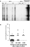
Quantitative analysis of SRC after ex vivo culture. (A) Representative Southern blot analysis of individual NOD/SCID mice transplanted with expanded cells from replicate wells at days 4 and 9. Lane 1 represents one mouse transplanted with the contents of one well containing 2,200 expanded cells, lanes 2–4 represent three individual mice transplanted with 730 expanded cells recovered from one well, lanes 5–12 represent eight mice transplanted with 275 expanded cells recovered from one well, all at day 4. At day 9, two mice were transplanted with 4,500 and 1,250 expanded cells (lanes 13–14 and 15–16, respectively). DNA was extracted from the murine BM 8 wk after transplant and hybridized with a human chromosome 17–specific α-satellite probe. (B) Summary of the level of human cell engraftment in the BM of 81 mice transplanted with expanded CD34+CD38− cells at 4 d from 10 CB samples.
Figure 4
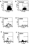
Multilineage differentiation of human CD34+CD38− cells in NOD/SCID mice after 4 d of ex vivo culture. A representative mouse was transplanted with 1,200 expanded CD34+CD38− CB cells after 4 d of ex vivo culture. Mouse BM was extracted 8 wk after transplant into NOD/SCID mice and analyzed by multiparameter flow cytometry. (A) Histogram of CD45 (human-specific panleukocyte marker) expression indicating that 0.4% of the cells present in the murine BM are human. Subsequent analysis of lineage markers was done on CD45+ cells within gate R1. (B) Isotype control for nonspecific IgG staining of mouse BM. (C–F ) Analysis for the presence of human B cell lineage cells using pan–B cell marker CD19 (C ) and mature B cell marker CD20 (D), and for the presence of human myeloid cells expressing myeloid marker CD33 (E ) and monocytic marker CD14 (F ). Isotype control for lineage markers is indicated by shaded regions of histogram.
Similar articles
- Dissociation between stem cell phenotype and NOD/SCID repopulating activity in human peripheral blood CD34(+) cells after ex vivo expansion.
Danet GH, Lee HW, Luongo JL, Simon MC, Bonnet DA. Danet GH, et al. Exp Hematol. 2001 Dec;29(12):1465-73. doi: 10.1016/s0301-472x(01)00750-0. Exp Hematol. 2001. PMID: 11750106 - Absence of CD34 on some human SCID-repopulating cells.
Dick JE. Dick JE. Ann N Y Acad Sci. 1999 Apr 30;872:211-7; discussion 217-9. doi: 10.1111/j.1749-6632.1999.tb08466.x. Ann N Y Acad Sci. 1999. PMID: 10372124 Review. - Integrative molecular and developmental biology of adult stem cells.
Bunting KD, Hawley RG. Bunting KD, et al. Biol Cell. 2003 Dec;95(9):563-78. doi: 10.1016/j.biolcel.2003.10.001. Biol Cell. 2003. PMID: 14720459 Review.
Cited by
- Preclinical Development of Autologous Hematopoietic Stem Cell-Based Gene Therapy for Immune Deficiencies: A Journey from Mouse Cage to Bed Side.
Garcia-Perez L, Ordas A, Canté-Barrett K, Meij P, Pike-Overzet K, Lankester A, Staal FJT. Garcia-Perez L, et al. Pharmaceutics. 2020 Jun 13;12(6):549. doi: 10.3390/pharmaceutics12060549. Pharmaceutics. 2020. PMID: 32545727 Free PMC article. Review. - Expansion of engrafting human hematopoietic stem/progenitor cells in three-dimensional scaffolds with surface-immobilized fibronectin.
Feng Q, Chai C, Jiang XS, Leong KW, Mao HQ. Feng Q, et al. J Biomed Mater Res A. 2006 Sep 15;78(4):781-91. doi: 10.1002/jbm.a.30829. J Biomed Mater Res A. 2006. PMID: 16739181 Free PMC article. - Generation of hematopoietic repopulating cells from human embryonic stem cells independent of ectopic HOXB4 expression.
Wang L, Menendez P, Shojaei F, Li L, Mazurier F, Dick JE, Cerdan C, Levac K, Bhatia M. Wang L, et al. J Exp Med. 2005 May 16;201(10):1603-14. doi: 10.1084/jem.20041888. Epub 2005 May 9. J Exp Med. 2005. PMID: 15883170 Free PMC article. - cDNA cloning of FRIL, a lectin from Dolichos lablab, that preserves hematopoietic progenitors in suspension culture.
Colucci G, Moore JG, Feldman M, Chrispeels MJ. Colucci G, et al. Proc Natl Acad Sci U S A. 1999 Jan 19;96(2):646-50. doi: 10.1073/pnas.96.2.646. Proc Natl Acad Sci U S A. 1999. PMID: 9892687 Free PMC article. - Expansion of human SCID-repopulating cells under hypoxic conditions.
Danet GH, Pan Y, Luongo JL, Bonnet DA, Simon MC. Danet GH, et al. J Clin Invest. 2003 Jul;112(1):126-35. doi: 10.1172/JCI17669. J Clin Invest. 2003. PMID: 12840067 Free PMC article.
References
- Moore MA. Expansion of myeloid stem cells in culture. Semin Hematol. 1995;32:183–200. - PubMed
- Williams DA. Ex vivo expansion of hematopoietic stem and progenitor cells–robbing Peter to pay Paul? . Blood. 1993;81:3169–3172. - PubMed
- Brugger W, Heimfeld S, Berenson RJ, Mertelsmann R, Kanz L. Reconstitution of hematopoiesis after high-dose chemotherapy by autologous progenitor cells generated ex vivo. N Engl J Med. 1995;333:283–287. - PubMed
- Chang J, Coutinho L, Morgenstern G, Scarffe JH, Deakin D, Harrison C, Testa NG, Dexter TM. Reconstitution of haemopoietic system with autologous marrow taken during relapse of acute myeloblastic leukaemia and grown in long-term culture. Lancet. 1986;1:294–295. - PubMed
- Barnett MJ, Eaves CJ, Phillips GL, Gascoyne RD, Hogge DE, Horsman DE, Humphries RK, Klingemann HG, Lansdorp PM, Nantel SH. Autografting with cultured marrow in chronic myeloid leukemia: results of a pilot study. Blood. 1994;84:724–732. - PubMed
Publication types
MeSH terms
Substances
LinkOut - more resources
Full Text Sources
Other Literature Sources
Medical
Research Materials
Miscellaneous


