Caenorhabditis elegans Akt/PKB transduces insulin receptor-like signals from AGE-1 PI3 kinase to the DAF-16 transcription factor - PubMed (original) (raw)
Caenorhabditis elegans Akt/PKB transduces insulin receptor-like signals from AGE-1 PI3 kinase to the DAF-16 transcription factor
S Paradis et al. Genes Dev. 1998.
Abstract
A neurosecretory pathway regulates a reversible developmental arrest and metabolic shift at the Caenorhabditis elegans dauer larval stage. Defects in an insulin-like signaling pathway cause arrest at the dauer stage. We show here that two C. elegans Akt/PKB homologs, akt-1 and akt-2, transduce insulin receptor-like signals that inhibit dauer arrest and that AKT-1 and AKT-2 signaling are indispensable for insulin receptor-like signaling in C. elegans. A loss-of-function mutation in the Fork head transcription factor DAF-16 relieves the requirement for Akt/PKB signaling, which indicates that AKT-1 and AKT-2 function primarily to antagonize DAF-16. This is the first evidence that the major target of Akt/PKB signaling is a transcription factor. An activating mutation in akt-1, revealed by a genetic screen, as well as increased dosage of wild-type akt-1 relieves the requirement for signaling from AGE-1 PI3K, which acts downstream of the DAF-2 insulin/IGF-1 receptor homolog. This demonstrates that Akt/PKB activity is not necessarily dependent on AGE-1 PI3K activity. akt-1 and akt-2 are expressed in overlapping patterns in the nervous system and in tissues that are remodeled during dauer formation.
Figures
Figure 1
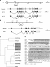
akt-1 and akt-2 encode Akt/PKB serine/threonine kinases. (A, top) Genetic and physical map of the akt-1 region; akt-1 is contained on cosmid C12D8. (Bottom) Exon/intron structure of akt-1. Coding regions are solid boxes, noncoding regions are open boxes, and introns are lines. The pleckstrin homology domain is indicated by hatched boxes (Musacchio et al. 1993), the kinase domain (Hanks and Hunter 1995) is indicated in gray. (B, top) Genetic and physical map of the akt-2 region; akt-2 is contained on cosmid R03E1. (Bottom) Exon/intron stucture of akt-2. All symbols are as in A. (C) Dendogram of Akt/PKB and PKC protein kinase families. PILEUP (GCG) was used to align the entire coding sequences of the indicated proteins. (Ce) C. elegans proteins; (r) rat; (h) human; (m) mouse; (b) bovine; (D) D. melanogaster. The GenBank accession numbers for the proteins used in the PILEUP are contained in parentheses: CePKC2a(U82935), rPKCβ1(M19007), hAkt/PKBα(M63167), mAkt/PKB(M94335), bAkt/PKB(X61036), hAkt/PKBβ2(M95936), rAkt/PKBγ(D49836), and Dakt1 (Z26242). The accession numbers for the proteins reported in the paper are contained in parentheses: AKT-1a (AF072379), AKT-1b (AF072380), and AKT-2 (AF072381). As an outgroup, rPKCβ1 (the closest non-Akt/PKB homolog to both akt-1a and hAkt/PKBα) and CePKC2a (the closest C. elegans homolog to rPKCβ1) were included in the PILEUP. The Akt/PKB homologs described in this report are indicated by the gray box. (D) AKT-1a, AKT-1b, AKT-2, and human Akt/PKBα (M63167) were aligned using PILEUP. Identical residues are indicated by dots; gaps introduced to align the sequence are indicated by dashes. The pleckstrin homology domain (Musacchio et al. 1993) is indicated by the amino-terminal shaded areas; the kinase domain (Hanks and Hunter 1995) is indicated by the carboxy-terminal shaded areas. The mg144 Ala-183–Thr substitution is indicated as a T above the AKT-1a sequence (SEAAKRD). The AKT-1 and AKT-2 phosphorylation sites that correspond to the hAkt/PKBα Thr-308 and Ser-473 phosphorylation sites (Alessi et al. 1996a) are indicated as dots above the amino acid residue that is phosphorylated.
Figure 2
AKT-1/GFP and AKT-2/GFP expression. (A–D) AKT-1/GFP expression; (E) AKT-2/GFP expression. (A) AKT-1/GFP expression in the head of an L2 animal. Expression in the anterior and posterior bulb of the pharynx (ant.ph. and post.ph.) is shown; expression in the isthmus of the pharynx is also observed. Many neurons in the head express AKT-1/GFP; expression in the neuronal nuclei is not visible. Also, expression can be seen in sensory axons (sens.axon) that proceed to the nose of the animal. (B) AKT-1/GFP expression in the ventral nerve cord (VNC) of an L1 animal (anterior is to the left). Both cell bodies (nuclei do not appear to express AKT-1/GFP) and axons of the VNC are clearly visible. Animal appears twisted because of coinjection with the rol-6 marker. (C) AKT-1/GFP expression in the tail of an L1 animal. The rectal gland cell (RGC), neurons of the tail with axons, and hypodermal cells (hyp) all clearly express AKT-1/GFP. AKT-1/GFP expression is not visible in the nuclei. (D) AKT-1/GFP expression in the spermatheca (sp) of an adult animal. (E) AKT-2/GFP expression in the head of an L4 animal. Expression in the anterior and posterior bulb of the pharynx (ant.ph. and post.ph.) is indicated; expression in the isthmus of the pharynx is also visible. Also shown is AKT-2/GFP expression in many neurons in the head; the nuclei appear to be excluded from AKT-2/GFP expression. AKT-2/GFP expression in the VNC, tail, and spermatheca was similar to that observed for AKT-1/GFP.
Figure 3
Model for regulation of dauer formation by AGE-1 PI3K activation of AKT-1/AKT-2. (A) Normal growth occurs in conditions of low pheromone as a result of DAF-2 insulin receptor-like signaling and converging TGF-β signaling; DAF-16 is possibly inactivated by phosphorylation in these growth conditions. (See text for details.) (B) Dauer arrest occurs in conditions of high pheromone that cause lack of DAF-2 insulin receptor-like signaling and converging TGF-β signaling; DAF-16 may repress transcription in these conditions. (See text for details.) (C) Location and sequence context of consensus Akt/PKB phosphorylation sites in DAF-16 and the human homologs FKHRL1, FKHR, and AFX. daf-16 is differentially spliced to produce two gene products (DAF-16a and DAF-16b) that differ in the amino-terminal one-third of the protein but are identical (DAF-16ab) for the remainder of the protein. The consensus Akt/PKB phosphorylation site, RXRXXS/THyd (Alessi et al. 1996b), is boxed in gray and the amino acid residues identical in that site in all proteins are in boldface type.Other amino acid residues flanking these sites are also identical or show conservative substitutions in all proteins but are not in boldface type. The sites are located in the same relative regions of each protein, near the amino terminus, at the carboxy-terminal region of the Fork head DNA-binding domain (but downstream of the DNA recognition helix) and downstream of the Fork head domain. Note that two adjacent Akt/PKB consensus sites occur within the Fork head domain of DAF-16 and are shown aligned with a single Akt/PKB consensus site in FKHRL1, etc.
Similar articles
- A PDK1 homolog is necessary and sufficient to transduce AGE-1 PI3 kinase signals that regulate diapause in Caenorhabditis elegans.
Paradis S, Ailion M, Toker A, Thomas JH, Ruvkun G. Paradis S, et al. Genes Dev. 1999 Jun 1;13(11):1438-52. doi: 10.1101/gad.13.11.1438. Genes Dev. 1999. PMID: 10364160 Free PMC article. - Activated AKT/PKB signaling in C. elegans uncouples temporally distinct outputs of DAF-2/insulin-like signaling.
Gami MS, Iser WB, Hanselman KB, Wolkow CA. Gami MS, et al. BMC Dev Biol. 2006 Oct 4;6:45. doi: 10.1186/1471-213X-6-45. BMC Dev Biol. 2006. PMID: 17020605 Free PMC article. - The C. elegans PTEN homolog, DAF-18, acts in the insulin receptor-like metabolic signaling pathway.
Ogg S, Ruvkun G. Ogg S, et al. Mol Cell. 1998 Dec;2(6):887-93. doi: 10.1016/s1097-2765(00)80303-2. Mol Cell. 1998. PMID: 9885576 - The Fork head transcription factor DAF-16 transduces insulin-like metabolic and longevity signals in C. elegans.
Ogg S, Paradis S, Gottlieb S, Patterson GI, Lee L, Tissenbaum HA, Ruvkun G. Ogg S, et al. Nature. 1997 Oct 30;389(6654):994-9. doi: 10.1038/40194. Nature. 1997. PMID: 9353126 - Worming pathways to and from DAF-16/FOXO.
Mukhopadhyay A, Oh SW, Tissenbaum HA. Mukhopadhyay A, et al. Exp Gerontol. 2006 Oct;41(10):928-34. doi: 10.1016/j.exger.2006.05.020. Epub 2006 Jul 12. Exp Gerontol. 2006. PMID: 16839734 Review.
Cited by
- Natural genetic variation drives microbiome selection in the Caenorhabditis elegans gut.
Zhang F, Weckhorst JL, Assié A, Hosea C, Ayoub CA, Khodakova AS, Cabrera ML, Vidal Vilchis D, Félix MA, Samuel BS. Zhang F, et al. Curr Biol. 2021 Jun 21;31(12):2603-2618.e9. doi: 10.1016/j.cub.2021.04.046. Epub 2021 May 27. Curr Biol. 2021. PMID: 34048707 Free PMC article. - Nuclear Localization Marker of FOXO3a: Can it be Used to Predict Doxorubicin Response?
Gong C, Khoo US. Gong C, et al. Front Oncol. 2013 Jun 5;3:149. doi: 10.3389/fonc.2013.00149. eCollection 2013. Front Oncol. 2013. PMID: 23761862 Free PMC article. No abstract available. - PTEN negatively regulates MAPK signaling during Caenorhabditis elegans vulval development.
Nakdimon I, Walser M, Fröhli E, Hajnal A. Nakdimon I, et al. PLoS Genet. 2012;8(8):e1002881. doi: 10.1371/journal.pgen.1002881. Epub 2012 Aug 16. PLoS Genet. 2012. PMID: 22916028 Free PMC article. - Noncanonical control of C. elegans germline apoptosis by the insulin/IGF-1 and Ras/MAPK signaling pathways.
Perrin AJ, Gunda M, Yu B, Yen K, Ito S, Forster S, Tissenbaum HA, Derry WB. Perrin AJ, et al. Cell Death Differ. 2013 Jan;20(1):97-107. doi: 10.1038/cdd.2012.101. Epub 2012 Aug 31. Cell Death Differ. 2013. PMID: 22935616 Free PMC article. - daf-28 encodes a C. elegans insulin superfamily member that is regulated by environmental cues and acts in the DAF-2 signaling pathway.
Li W, Kennedy SG, Ruvkun G. Li W, et al. Genes Dev. 2003 Apr 1;17(7):844-58. doi: 10.1101/gad.1066503. Epub 2003 Mar 21. Genes Dev. 2003. PMID: 12654727 Free PMC article.
References
- Alessi DR, Caudwell FB, Andjeclkovic M, Hemmings BA, Cohen P. Molecular basis for the substrate specificity of protein kinase B; comparison with MAPKAP kinase-1 and p70 S6 kinase. FEBS Lett. 1996b;399:333–338. - PubMed
- Alessi DR, James SR, Downes CP, Holmes AB, Gaffney PRJ, Reese CB, Cohen P. Characterization of a 3-phosphoinositide-dependent protein kinase which phosphorylates and activates protein kinase Bα. Curr Biol. 1997;7:261–269. - PubMed
- Avruch J. Insulin signal transduction through protein kinase cascades. Mol Cell Biochem. 1998;182:31–48. - PubMed
- Bargmann CI, Horvitz HR. Control of larval development by Chemosensory neurons in Caenorhabditis elegans. Science. 1991;251:1243–1246. - PubMed
Publication types
MeSH terms
Substances
LinkOut - more resources
Full Text Sources
Other Literature Sources
Molecular Biology Databases
Miscellaneous


