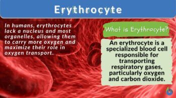
Erythrocyte Definition and Examples - Biology Online Dictionary (original) (raw)
Erythrocyte
n., plural: erythrocytes
[əˈɹɪθɹəˌsaɪt]
Definition: A red blood cell
Table of Contents
Toggle
- Erythrocyte Definition
- Erythrocyte Structure
- Life Cycle Of Erythrocytes
- Functions
- Tests
- Science underlying Hemoglobin
- Quiz
- Further Reading
- References
Erythrocytes (red blood cells or RBCs) are the myeloid series of specialized cells that play an integral role in the circulatory system. They are highly specialized, biconcave-shaped, and rich in a red pigment called hemoglobin. With an optimized surface-to-volume ratio and devoid of most organelles, these selfless envoys through their remarkable flexibility and red blood cell deformability efficiently facilitate the exchange of respiratory gases like inhaled oxygen and carbon dioxide.
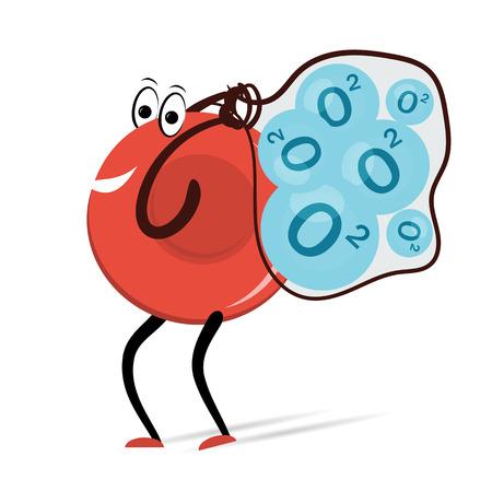
Figure 1: A cartoon showing red blood cells carrying oxygen molecules. Image Credit: 123RF
The erythrocytes are a unique type of blood cell that is characterized by the absence of the ability to undergo cell division and the presence of hemoglobin (a unique iron-containing molecule). Some important points to note about erythrocytes are:
- They are called by different names like:
- Erythroid Cells
- Red Blood Cells (RBCs)
- Haematids
- Red Cells
- Red Blood Corpuscles
- They are highly differentiated.
- They don’t have cell organelles (sometimes called “anucleate” as they don’t possess a nucleus at maturity). This modality in the structure supports the maximization of space in the erythrocytes for the accommodation of hemoglobin molecules.
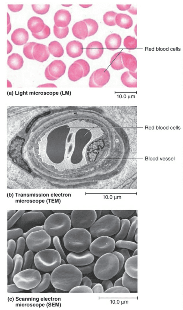
Figure 2: Structure of RBCs as observed under 3 different types of microscope. Image Credit: Human Anatomy - They are devoid of the ability to divide and form new erythrocytes.
- Their physiology supports oxygen transport throughout the human body tissues.
- They are produced specifically in the red bone marrow (BM) from hematopoietic stem cells (HSCs). The process of production of erythrocytes is called “erythropoiesis” and takes place as HSCs give rise to myeloid progenitors or erythrocyte precursors. So, erythrocytes are “myeloid in origin” or belong to the myeloid series of blood cells. There are several changes (especially morphological) that cumulatively give rise to mature erythrocytes.
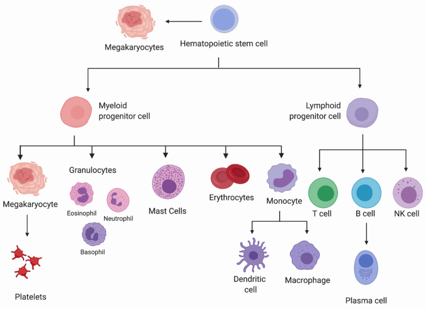
Figure 3: We can notice the myeloid origin or erythrocytes in the flowchart. Image Credit: Yasharah Raza - Their average lifespan is 100-120 days (nearly 3.5-4 months).
- The production of human red blood cells in adults is estimated to be approximately 2,400,000 new cells per second.
- Their communications and interactions are wide-scoped; with endothelial cells (as in sickle cell patients), platelets (clotting cells), macrophages (a type of white blood cells), and even bacteria via membrane proteins.
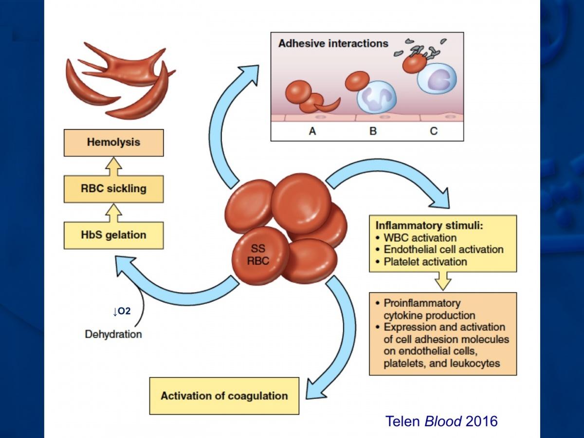
Figure 4: Interaction of RBCs in sickle cell anemia. Image Credit: Duke University - There are reports that RBCs can synthesize nitric oxide.
- They are biconcave in shape (for the provision of a larger “surface to volume ratio”) that eventually increases the oxygen-carrying capacity.
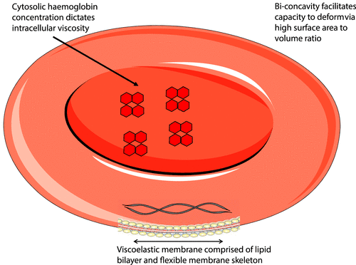
Figure 5: Role of biconcave shape in increasing the surface area. Image Credit: American Journal of Physiology - They are filled with hemoglobin (Hb) which renders them with the ability to oxygen supply.
- The richness of Hb in the RBCs’ cytoplasm imparts a red color to blood. There is an estimate of about 270,000,000 Hb molecules in each RBC.
- The flexible nature of erythrocytes which allows their easy circulation in the circulatory system and bloodstream is due to the presence of unique lipids and proteins in the RBC membrane.
- The primary function of erythrocytes is the transportation of oxygen from the lungs to all tissues and parts of the body.
- The hematocrit value (i.e. the volume of packed erythrocytes) usually varies from men to women. For men, it ranges between 42-54% of total volume while in women, it varies from 37-47%. These values are usually lower in children. These values are higher for people living at high altitudes with low oxygen tension. Since the erythrocytes are more or less uniform in volume, we usually determine the hematocrit value by evaluating the number of erythrocytes (RBCs) per unit of blood, which are usually 4,000,000 to 6,000,000 per cubic millimeter.
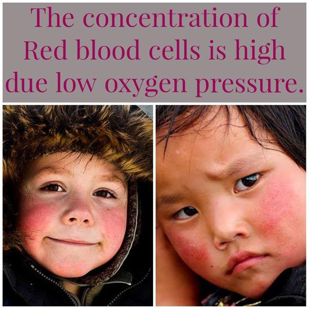
Figure 6: We all have noticed the red cheeks of people living in high altitudes. This increased production of erythrocytes (evident as red cheeks) is a physiological mechanism to compensate for the low oxygen pressure/tension at higher altitudes. Image Credit: Quora - When understanding their representation in the total blood volume, erythrocytes represent nearly half of the total blood cells ranging between 40 to 45 percent.
- In contrast to the uptake of oxygen from the lungs by erythrocytes in mammals, oxygen is extracted from gills in fishes.
- There is a common medical terminology called “packed red blood cells/pRBC” which stands for the erythrocytes that are donated, processed, and thereby eventually stored in hospital blood banks for treatments like blood transfusion.
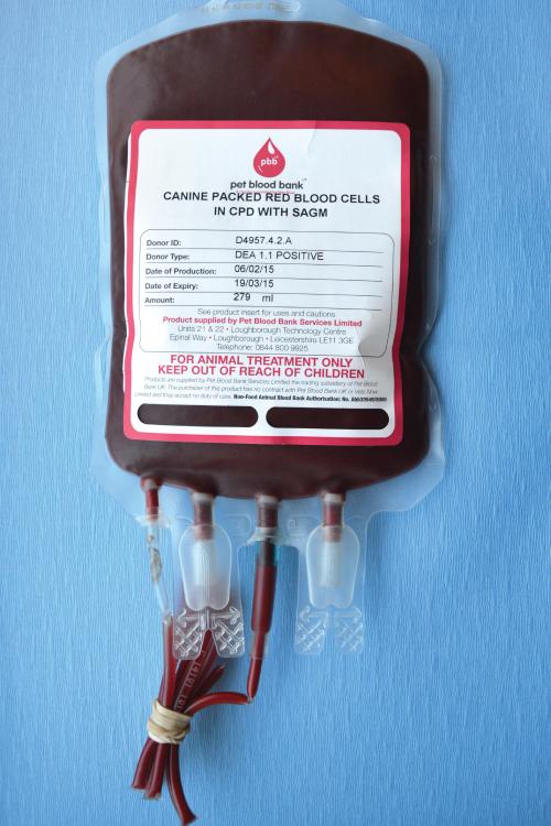
Figure 7: Packed RBC (pRBC) for research and blood transfusions. Image Credit: Veterian Key - Within the membrane of red blood cells (RBCs), a bilayer composed of lipid molecules with both hydrophilic and hydrophobic properties is connected to a framework of skeletal proteins using transmembrane proteins.
Erythrocytes Etymology
The word erythrocyte is derived from two Greek words;
- Erythros meaning “red”
- Kytos means “_hollow vessel_”
The word cyte is usually translated to “cell” in modern language.
Watch this vid about erythrocytes:
Biology definition:
An erythrocyte is a blood cell with hemoglobin-rich cytoplasm whose main function is to transport respiratory gases, particularly oxygen and carbon dioxide. Oxygen molecules move into the erythrocytes from the air inspired via the respiratory organs (e.g. the lungs of terrestrial vertebrates or the gills of fish). The erythrocytes deliver the oxygen (via the circulatory system) to hypoxic tissues needing oxygenation. With the release of oxygen molecules, most carbon dioxide molecules (from tissues diffusing into the blood plasma) move into the erythrocytes to be transported and eventually released outside via the respiratory system.
Most vertebrates have red blood cells that remain nucleated. However, in mammals including humans, the mature red blood cells are biconcave and anucleated. In humans, carbon dioxide molecules from the erythrocytes are released as bicarbonate ions into the plasma and then converted back to carbon dioxide upon reaching the alveolar space in the lungs to be expired. The crocodile icefish of the family Channichthyidae is the only vertebrate group to naturally lack erythrocytes and obtain oxygen from their oxygen-rich aquatic habitat via passive transport.
Synonym: red blood cell; red blood corpuscle; haematid; erythroid cell Compare: leukocyte See also blood cells.
Erythrocyte Structure
The erythrocyte structure is a physiological marvel. Some basic pointers to their structure are:
- Diameter: 7-8 µm
- Biconcave shape (resembling a donut)
- Thicker periphery than their central parts
- Maximum total surface areas of the cell membrane (facilitating easy oxygen transport and other gas exchange)
- Absence of a nucleus (hence the absence of DNA) and other cellular organelles
- Presence of only 2 structures namely cytoplasm and red cell membrane.
Let’s discuss the cytoplasm and cell membrane in detail.
Cytoplasm
The cytoplasm of mature erythrocytes has the presence of no organelles. When we observe erythrocytes under a microscope after staining blood samples with hematoxylin and eosin (H&E), we see them as intensely stained red-orange cells. One point that we should note here is that hematoxylin alone doesn’t stain erythrocytes as hematoxylin specifically stains the nucleus of cells which is absent in erythrocytes.

Figure 8: Staining of different stages as erythrocyte progresses to mature. Image Credit: Yale
Cell membrane
Here are some important notes about the cell membrane of erythrocytes.
- There is a lipid bilayer membrane that covers the erythrocyte’s cytoplasm.
- Proteins: There are two types of cell membrane proteins called integral as well as peripheral proteins.
- Peripheral proteins: Peripheral proteins found on the membrane’s inner surface help in providing an extremely elastic property to the erythrocytes.
- Integral Proteins: these are proteins that span the entire membrane.
- Carbohydrates: the ABO blood grouping used for type for blood group characterization is based on the presence or absence of certain types of carbohydrate molecules that act as antigens on the red blood cell membrane; blood types thus are called the A, B, O, and AB blood types. Expression of blood group antigens by these integral proteins leads to exploiting ABO blood groups for medical purposes.
- Besides this ABO blood typing, the erythrocytes cell membrane can have a Rhesus (Rh) antigen (RH factor protein). The presence and absence of Rh on a person’s erythrocytes lead to the blood type being Rh positive (A+/B+/O+/AB+) or Rh negative (A-/B-/O-/AB-). Both ABO and Rh surface antigens on the erythrocyte cell membranes are integral for blood transfusions.

Figure 9: Different antigens for different blood group types. Image Credit: NCBI
- Function: The erythrocyte’s cell membrane not only supports the internal structure till the time that the cell reaches maturity by also helps in the binding of hemoglobin molecules.
- Permeability: The erythrocyte’s cell membrane is impermeable to Hb but freely permeable to:
- Water
- Oxygen
- Carbon dioxide
- Glucose
- Urea
- Major Ions:
- Within cell: Potassium
- In plasma and extracellular fluids: Sodium
- Osmosis: Erythrocytes are subject to osmotic effects.
- On suspension in hypotonic solutions: Increased volume & transition to a more spheroid shape
- On suspension in hypertonic solutions: Loss of water leading to even more shrinkage

Figure 10: Osmotic effects on RBCs. Image Credit: BC Open Textbooks
- Process of Hemolysis: Upon damage to the erythrocyte’s cell membrane or alterations in the membrane skeleton, there is a leak of cell constituents like hemoglobin (Hb) and other dissolved contents. This paves the way for the ghosting of membranous structures. This is usually a result of physical damage or osmotic effects. In extreme conditions, it can also lead to hemolytic anemia (low oxygen transport capacity)
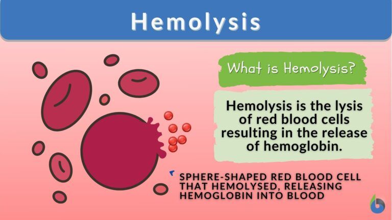
Figure 11: The process of Hemolysis is discussed in detail in the article here. Image Prepared by Maria Victoria Gonzaga for Biology online
Life Cycle Of Erythrocytes
There are three main phases in the life cycle of erythrocytes; synthesis, lifetime, and senescence.
Synthesis Phase/Erythropoiesis
- Erythrocytes are synthesized by the process called “erythropoiesis”.
- It takes about 7 days for erythrocytes to be produced from red bone marrow cells (committed stem cells for RBC production) of the larger bones.
- NOTE: A point to note here is that in the embryo, these erythrocytes are rather produced in the liver and not in the bone marrow cells of bones.
- A specific hormone called erythropoietin (EPO) stimulates this synthesis process from its location in the kidney.
- The idea of reticulocytes: While erythrocytes are being produced in the bone marrow, they are called reticulocytes just before and after they leave the bone marrow. These reticulocytes comprise only 1% of circulating erythrocytes.

Figure 12: Role of EPO in the synthesis of RBCs. Image Credit: Colostate
Lifetime Phase
- The active lifetime of an erythrocyte is nearly 100–120 days.
- Erythrocytes are continually on the move during their lifetimes. They move by the force of:
* Blood flow push: in arteries (arterial blood)
* Blood flow pull: in veins (venous blood)
* Combination of both blood flow push and pull- in microvessels (capillaries) - Erythrocytes Recycling: The site of erythrocytes recycling is bone marrow.
Senescence Phase/Erythrocyte Destruction
- This is the aging phase of erythrocytes where they undergo a lot of physical changes like in their plasma membranes.
- These changes make them highly susceptible to being selectively recognized by a type of leucocytes called macrophages.
- What follows next is the phagocytosis process by the mononuclear phagocytes and their system that’s constituted by the liver, lymph nodes, and spleen.
- By the process of phagocytosis, old and damaged erythrocytes are continually removed from the bloodstream. This is also called “eryptosis” meaning erythrocyte’s programmed death. For example, Exposure to PS (phosphatidylserine) causes adhesion of erythrocytes to vascular endothelial cells thus interrupting their normal transport and movement.

Figure 13: Different factors responsible for eryptosis. Image Credit: Sumiah A. Alghareeb - The rate of eryptosis is usually the same as the rate of erythropoiesis. This balances the total circulating erythrocyte count (RBC count).
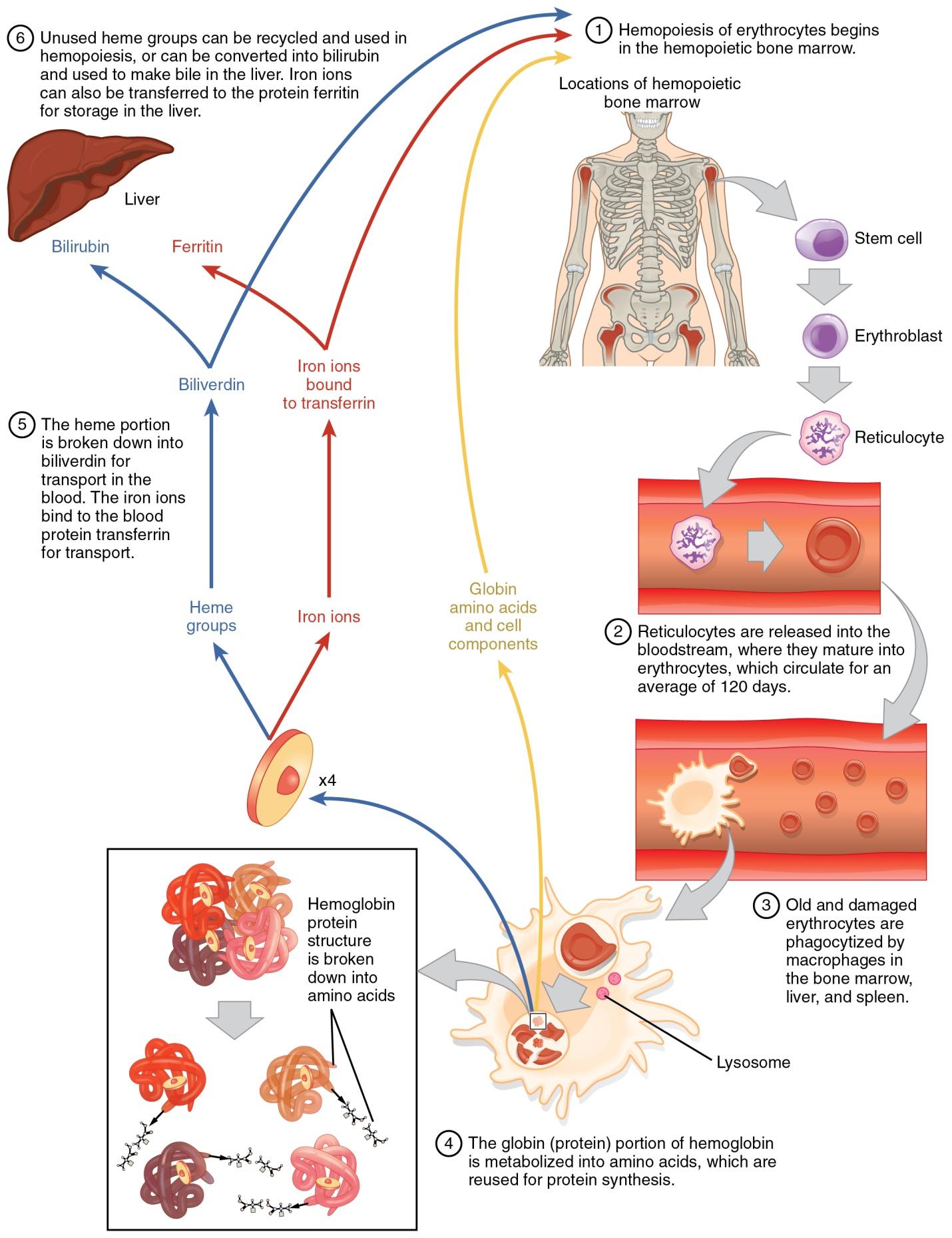
Figure 14: Life cycle of erythrocytes. Image Credit: Open Stax College
Erythropoiesis
Erythropoiesis is the process of production of erythrocytes that takes place within the red bone marrow as a “part of hematopoiesis”. Initially, hematopoiesis generates an erythroid stem cell known as CFU-E (Colony Forming Unit – Erythroid), which initiates the process of erythropoiesis primarily under the influence of the erythropoietin hormone. These CFU-E cells are localized within erythroid islands within the bone marrow and undergo replication and differentiation to ultimately form mature erythrocytes. During the differentiation process, several generations of cells are produced, including proerythroblasts, erythroblasts, reticulocytes, and finally, erythrocytes. With each generation, the cells histologically resemble erythrocytes more closely.
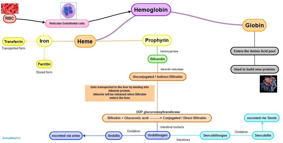
Erythrocyte destruction
The breakdown products from eryptosis are recirculated in the body.
- Heme of the Hb: Broken down into biliverdin and Fe3+ ion (iron ion).
- Biliverdin: Reduced to bilirubin (a yellow pigment)
- Bilirubin: Release into plasma; via albumin, it is recirculated to liver
- Iron: Released into plasma; recirculated by transferrin (a carrier protein)
- Polypeptide globin chains: Degraded into amino acids
There are some diseases and disorders which are marked by an abnormally high rate of eryptosis like:
- Sepsis
- Wilson’s disease
- Hemolytic uremic syndrome
- Iron deficiency
- Malaria
- Phosphate depletion
- Beta-thalassemia (characterized by abnormal hemoglobin/abnormal ratio of hemoglobin subunits)
- Sickle cell disease/ sickle cell anemia (iron deficiency anemia) – Anemia can be caused due to different reasons like:
* Due to blood loss
* Due to defective/reduced erythrocyte production (aplastic anemia)
* Due to excessive destruction of erythrocytes (hemolytic anemia)
* Due to mutation in hemoglobin genes - Glucose-6-phosphate dehydrogenase deficiency
Note: Polycythemia (characterized by an elevated RBC count)
Functions
Erythrocytes perform many invaluable functions for the proper functioning of the body.
Oxygen Transport
A unique protein called hemoglobin aids the process of oxygen (O2) transport in the body. Oxygen binds to hemoglobin in the lungs. This leads to the formation of a very strong bond allowing erythrocytes to efficiently transport O2 molecules. As erythrocytes circulate through blood vessels, they deliver O2 to organs and tissues in need. By providing cells with the necessary O2, erythrocytes ensure energy production and survival of the tissues and organs.
CO2 Transport
Erythrocytes have a crucial role in the transport of carbon dioxide produced (CO2) in our bodies. During cellular metabolism, CO2 is produced as a waste product. Erythrocytes aid in removing CO2 from tissues and carrying it back to the lungs for elimination. CO2 diffuses from cells into nearby capillaries. Within erythrocytes, CO2 combines with water to form bicarbonate ions (HCO3–). This reaction is facilitated by an enzyme called “carbonic anhydrase” present in erythrocytes.
Bicarbonate ions are more soluble and can be transported easily in the blood plasma. Some of the bicarbonate ions remain within erythrocytes, while others are transported in the plasma. As erythrocytes reach the lungs, carbonic anhydrase converts bicarbonate ions into CO2 and water. The released CO2 is exhaled from the lungs, thus completing the process of CO2 transport by erythrocytes.
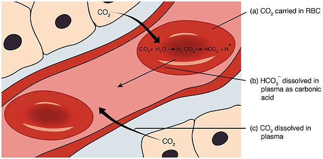
Figure 16: The role of the enzyme carbonic anhydrase is extremely important for the proper functioning of RBCs as it facilitates the transport of carbon dioxide from all bodily tissues to the lungs for exhalation or expulsion. Image Credit: OpenStax College
CO Transport
Erythrocytes can inadvertently transport carbon monoxide (CO) when it binds to hemoglobin, leading to a compromised ability to deliver oxygen to tissues.
Carbon monoxide poisoning occurs when an excessive amount of CO binds to hemoglobin. This impairs the normal oxygen-carrying function of erythrocytes.
When CO is inhaled, it readily binds to hemoglobin within erythrocytes.
Hemoglobin has a higher affinity for carbon monoxide (CO) compared to oxygen. This leads to the formation of a stable compound known as carboxyhemoglobin.
This binding of CO leads to oxygen deprivation in tissues.
The presence of carboxyhemoglobin in erythrocytes can have severe health consequences, as it interferes with normal oxygen delivery and can result in CO poisoning.
Tests
Anyone trying to understand the overall health of the body in the past 3-4 months, they can get their RBCs tested. Since the average lifespan of mature RBCs after being released in the bloodstream is 100 to 120 days, these cells can help in the appropriate description of the health state.
Glycated hemoglobin test: This HbA1c test is performed for people suffering from diabetes. It is usually recommended to get tested every 3 months to properly track the blood glucose levels. Since after every 3.5-4 months, the old erythrocytes are recycled by special cells called the macrophages in the lymph nodes, spleen, and liver, it’s important that the cycle of clinical tests in kept in sync with the natural biological renewal cycle of erythrocytes.
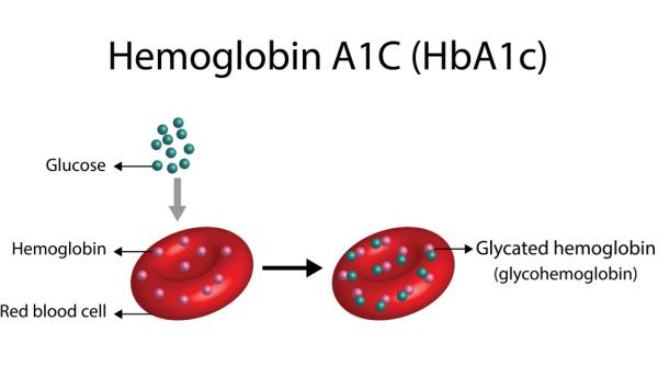
Figure 17: HbA1C test is the recommended test for regularly tracking diabetes. Image Credit: BeatO
More of the erythrocyte-related tests and their medical significance are listed in the table below.
| Table 1: A compiled list of different erythrocyte assessments for clinical purposes. Many diseases and disorders are diagnosed with a peripheral blood smear. | ||
|---|---|---|
| Erythrocyte Test | Interpretation | Use in Medical Conditions |
| Hemoglobin (Hb) | Measures oxygen-carrying capacity of the blood | Assess anemia, monitor response to treatment, screen for blood disorders |
| Hematocrit (Hct) | Measures the proportion of red blood cells in the blood | Evaluate hydration status, assess anemia, monitor blood disorders |
| Red Blood Cell Count (RBC) | Measures the number of red blood cells in a given volume of blood | Diagnose anemia, monitor response to treatment, assess blood disorders |
| Mean Corpuscular Volume (MCV) | Measures the average size of red blood cells | Classify anemias as microcytic, normocytic, or macrocytic |
| Mean Corpuscular Hemoglobin (MCH) | Measures the average amount of hemoglobin in red blood cells | Evaluate anemias and assess hemoglobin content in red blood cells |
| Mean Corpuscular Hemoglobin Concentration (MCHC) | Measures the concentration of hemoglobin in red blood cells | Diagnose and classify anemias based on hemoglobin content |
| Red Cell Distribution Width (RDW) | Measures the variation in the size of red blood cells | Assess anemias and determine the underlying cause |
| Erythrocyte Sedimentation Rate (ESR) | Measures the rate at which red blood cells settle in a tube | Detect and monitor inflammation, infection, and certain autoimmune conditions |
| Reticulocyte Count | Measures the percentage of immature red blood cells (reticulocytes) | Evaluate bone marrow function, assess response to anemia treatment |
| Coombs Test | Detects antibodies or proteins on the surface of red blood cells | Diagnose and monitor autoimmune hemolytic anemia and blood transfusion reactions |
Data Source: Dr. Harpreet of Biology Online
Warning/Caution Note: None of these tests should be performed or interpreted at home. Recommendations and advice from physicians are mandatory.
NOTE IT!
Science underlying Hemoglobin
Hemoglobin makes a notable presence in the erythrocyte’s cytoplasm. Hemoglobin is a tetrameric protein constituted by:
- 4 polypeptide chains: Also known as globin proteins or globin chains. Since there are 4 types of chains α, β, γ, and δ, we get 3 main classes of Hb (HbA, HbA2, and HbF). HbA is the most common in human adults.
- 4 carbon-based (C-based) heme molecules: The iron atom in each globin bound to heme plays an integral role in the binding of gases like O2 and CO2.
There are 2 main types of Hb:
- HbA (adult hemoglobin)
- HbF (fetal hemoglobin)
Hemoglobin binds reversibly with oxygen molecules. And this reversible binding allows to and fro transport of oxygen (O2) and carbon dioxide (CO2). 1 Hb holds the potential to transport 4 molecules of O2 and CO2.
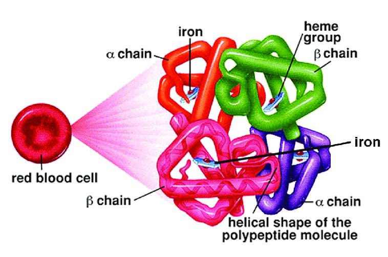
Figure 18: Structure of hemoglobin. Image Credit: Towards AI
The intricate arrangement of porphyrin and protein within the structure of hemoglobin creates an optimal setting for the iron atom, allowing it to effectively bind and release oxygen by physiological requirements. Hemoglobin exhibits such a strong affinity for oxygen that approximately 95% of its binding sites become saturated with oxygen within the lungs, where the oxygen pressure is relatively high. However, as oxygen tension decreases in the tissues, oxygen dissociates from hemoglobin, enabling its diffusion across the red blood cell membrane and plasma, ultimately reaching the sites where it is utilized. This process ensures a proportional distribution of oxygen to meet the body’s needs.
Take the Erythrocyte – Biology Quiz!
Further Reading
References
- Zivot, A., Lipton, J. M., Narla, A., & Blanc, L. (2018). Erythropoiesis: insights into pathophysiology and treatments in 2017. Molecular Medicine, 24, 1-15.
- Diez-Silva, M., Dao, M., Han, J., Lim, C. T., & Suresh, S. (2010). Shape and Biomechanical Characteristics of Human Red Blood Cells in Health and Disease. MRS Bulletin, 35(5), 382–388. https://doi.org/10.1557/mrs2010.571
- Liu, S., Grigoryan, M. M., Vasilevko, V., Sumbria, R. K., Paganini-Hill, A., Cribbs, D. H., & Fisher, M. J. (2014). Comparative analysis of H&E and Prussian blue staining in a mouse model of cerebral microbleeds. The Journal of histochemistry and Cytochemistry: official journal of the Histochemistry Society, 62(11), 767–773.
- Dunn, J. O., Mythen, M. G., & Grocott, M. P. (2016). Physiology of oxygen transport. Bja Education, 16(10), 341-348.
- Kinoshita, H., Türkan, H., Vucinic, S., Naqvi, S., Bedair, R., Rezaee, R., & Tsatsakis, A. (2020). Carbon monoxide poisoning. Toxicology reports, 7, 169-173.
- Ahmed, M. H., Ghatge, M. S., & Safo, M. K. (2020). Hemoglobin: structure, function, and allostery. Vertebrate and invertebrate respiratory proteins, lipoproteins, and other body fluid proteins, 345-382.
©BiologyOnline.com. Content provided and moderated by Biology Online Editors.