Cilia And Flagella (original) (raw)
Last Updated : 29 May, 2026
Cilia and flagella are hair-like cellular organelles that extend from the surface of cells, playing a crucial role in movement and sensory functions. These structures are microscopic and cannot be seen with the naked eye. They are mainly composed of microtubules made of the protein tubulin.

Although both structures share a similar basic composition, they differ in terms of length, number, structure, and function. Cilia are generally short and numerous, while flagella are long and few in number.
Cilia
Cilia are short, thin, hair-like projections present on the surface of certain eukaryotic cells. They help in locomotion or in moving substances over the cell surface. For example, in Paramecium, cilia beat rhythmically to help the organism move in water. In humans, cilia are present in the respiratory tract, where they help in clearing mucus and dust particles.
Characteristics of Cilia
- Cilia are short, microscopic, hair-like structures present on the surface of many eukaryotic cells. They are specialised cellular organelles that help in movement, transportation of substances, and sensory functions.
- Cilia are usually present in large numbers on a single cell and beat in a coordinated rhythmic manner. Their movement helps move fluids, mucus, food particles, or even the entire cell from one place to another.
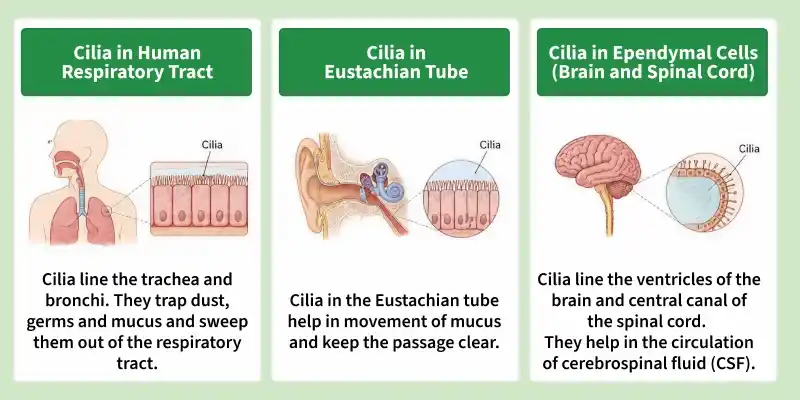
- Cilia are commonly found in organisms such as protozoans and in certain tissues of the human body, including the respiratory tract and fallopian tubes.
- The singular form of cilia is **cilium.
- These structures are found only in eukaryotic cells and are absent in prokaryotic organisms such as bacteria.
Structure of Cilia
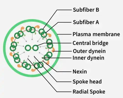
Each cilium is covered externally by the plasma membrane, which surrounds its internal structural components. The core structure of a cilium is called the axoneme, which is composed mainly of microtubules made up of the protein tubulin. The axoneme shows a characteristic 9+2 arrangement of microtubules, which is a distinctive feature of motile cilia and flagella in eukaryotic cells.
**1. Axoneme
The central core of a cilium is called the axoneme.
- It contains microtubules arranged in a characteristic 9+2 pattern.
- Nine peripheral doublet microtubules surround two central single microtubules.
- This arrangement is typical of motile cilia.
**2. Plasma Membrane
- The entire cilium is covered by the plasma membrane.
- It protects the internal structure.
**3. Basal Body
- Present at the base of the cilium.
- Anchors the cilium to the cell.
- Structurally similar to a centriole.
- Has a 9+0 arrangement of microtubules.
Functions of Cilia
- Cilia perform several important functions in different organisms and tissues of the body.
- One of the major functions of cilia is locomotion in unicellular organisms such as Paramecium.
- In humans, cilia play an important protective role in the respiratory tract through a mechanism known as mucociliary clearance.
- Cilia are also involved in the movement of cerebrospinal fluid within the ventricles of the brain.
- In the female reproductive system, cilia present in the fallopian tubes help in the transport of the ovum from the ovary toward the uterus.
- Apart from movement and transport, some cilia also perform sensory functions.
- Sensory cilia are important in several organs and contribute to functions related to sensation and cellular communication.
Flagella
Flagella are long, slender, whip-like appendages that project from the surface of certain cells and are primarily responsible for locomotion and movement. They are specialised cellular structures that enable organisms or cells to move from one place to another in liquid environments. They are fewer in number compared to cilia. For example, the sperm cell uses a flagellum for movement, and many bacteria also use flagella for locomotion.

Characteristics of Flagella
- The singular form of flagella is flagellum.
- Flagella are present in both prokaryotic and eukaryotic cells.
- They are long, thin, hair-like structures that extend outward from the cell surface.
- They are usually one or few in number, unlike cilia, which are numerous.
- Their primary function is locomotion and movement of the cell.
- Flagella may also help in sensory functions and environmental response.
- The structure and mechanism of movement of flagella differ between prokaryotes and eukaryotes.
- Prokaryotic flagella rotate like propellers, while eukaryotic flagella move in a whip-like or undulating manner.
Structure of Flagella
Eukaryotic flagella are structurally more complex and are found in organisms such as protozoans, algae, and animal sperm cells. These flagella are covered by the plasma membrane and internally contain microtubules arranged in a characteristic 9+2 pattern.
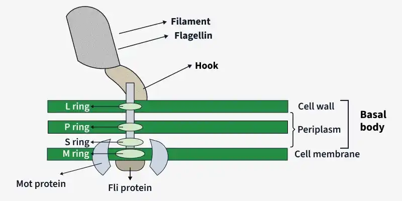
A typical prokaryotic flagellum consists of three main parts:
- **Filament: The filament is the long, external, whip-like portion of the flagellum that extends outside the bacterial cell. It is composed of many molecules of the protein flagellin arranged in a helical pattern. The filament acts as the main locomotory structure.
- **Hook: The hook is a short, curved structure that connects the filament to the basal body. It functions like a flexible joint and helps transmit rotational movement from the basal body to the filament.
- **Basal Body: The basal body is the innermost portion of the flagellum embedded within the cell wall and plasma membrane. It acts as the motor region of the flagellum. The basal body contains ring-like structures that help the flagellum rotate.
Functions of Flagella
- They help in locomotion and movement of cells and microorganisms.
- They assist bacteria in moving toward nutrients and away from harmful substances.
- They help sperm cells swim toward the ovum during fertilisation.
- In some organisms, they assist in feeding and circulation of surrounding fluids.
- They may also function as sensory organelles that detect environmental changes.