Myeloperoxidase-Specific Plasma Cell Depletion by Bortezomib Protects from Anti-Neutrophil Cytoplasmic Autoantibodies–Induced Glomerulonephritis (original) (raw)
Abstract
Anti-neutrophil cytoplasmic autoantibodies (ANCA) cause vasculitis and necrotizing crescentic glomerulonephritis (NCGN). Steroids and cytotoxic drugs reduce mortality but can cause significant adverse events. The proteasome inhibitor bortezomib (BTZ) prevents glomerulonephritis in mouse models of lupus but its efficacy in ANCA-associated glomerulonephritis is unknown. We induced anti-MPO IgG-mediated NCGN by transplanting wild-type bone marrow (BM) into irradiated MPO-deficient mice immunized with MPO. Four weeks after BM transplantation, we treated mice with steroid/cyclophosphamide (S/CYC) or BTZ. Compared with untreated control mice, both S/CYC and BTZ significantly reduced urine abnormalities, NCGN, and infiltration of neutrophils and macrophages. Response to BTZ depended on timing of administration: BTZ abrogated NCGN if begun 3 weeks, but not 5 weeks, after BM transplantation. BTZ treatment significantly reduced total and MPO-specific plasma cells in both the spleen and bone marrow, resulting in significantly reduced anti-MPO titers. Furthermore, BTZ affected neither B cells nor total CD4 and CD8 T cells, including their naive and effector subsets. In contrast, S/CYC reduced the total number of cells in the spleen, including total and MPO-specific plasma cells and B cells. In contrast to BTZ, S/CYC did not affect total and MPO-specific plasma cells in the bone marrow. Three of 23 BTZ-treated mice died within 36 hours after BTZ administration. In summary, BTZ depletes MPO-specific plasma cells, reduces anti-MPO titers, and prevents NCGN in mice.
Anti-neutrophil cytoplasmic antibodies (ANCA) to either proteinase 3 or myeloperoxidase (MPO), and subsequently also to lysosomal-associated membrane protein-2, are found in patients with small-vessel vasculitis and necrotizing crescentic glomerulonephritis (NCGN).1–3 ANCA activate polymorphonuclear neutrophils (PMN) and monocytes in vitro.4–6 ANCA pathogenicity was documented by MPO-ANCA transfer from mother to newborn, and in several animal models.7–12 Randomized controlled trials established standard treatment protocols for patients with generalized ANCA vasculitis that are based on steroids in combination with cytotoxic agents.13–15 Severe renal and pulmonary manifestations mandate the additional removal of ANCA by plasma exchange.16 These protocols resulted in significantly reduced mortality from vasculitis, but added new treatment-associated side effects that harbor their own morbidity and mortality hazards.17,18 Thus, clinicians developed more specific approaches. B cells can present antigens, provide co-stimulatory signals to T cells, and produce antibodies when differentiated into plasma cells. B cell–targeted therapies include mycophenolate mofetil and rituximab.19–21 Rituximab data from reports and small patient series were particularly encouraging and randomized trials are ongoing. However, novel treatments should first be established in experimental ANCA vasculitis models. With use of these model systems, TNFα, complement, chemokines, and integrins were identified as potential targets.22–26 We optimized an MPO-ANCA mouse model and showed that the complement receptor C5a and the PI3Kγ isoform are novel targets.27,28 We used this model to assess NCGN with the 26S proteasome inhibitor bortezomib (BTZ), compared with standard steroid/cyclophosphamide (S/CYC) treatment. BTZ is approved for use in multiple myeloma and was recently effective in preventing glomerulonephritis in the NZB/W and MRL/lpr lupus mouse model.29
RESULTS
BTZ and S/CYC Combined Treatment Prevents ANCA-Induced Necrotizing Crescentic Glomerulonephritis
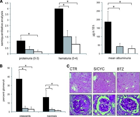
Anti-MPO–mediated NCGN was induced by immunizing MPO-deficient mice with murine MPO, followed by irradiation and transplantation of wild-type (WT) bone marrow (BM). Four weeks after BM transplantation, mice were left untreated or received combined S/CYC treatment, and BTZ, respectively (n = 8 in each group). Mice were sacrificed 8 weeks after transplantation, or whenever a mouse appeared too sick to survive until the next day. All mice (100%) in the control group developed proteinuria and hematuria, whereas dipstick analysis and albuminuria by ELISA were significantly less in both treatment groups (Figure 1A). Moreover, all mice (8 of 8) in the control group developed NCGN, whereas only 5 of 8 in the S/CYC and 1 of 6 in the BTZ group showed these lesions. Two of 8 BTZ-treated mice died within 36 hours after the first BTZ dose and were omitted from urine and histology analysis. The adverse events are discussed in more detail below.
Figure 1.
BTZ and S/CYC treatment prevents ANCA-induced necrotizing crescentic glomerulonephritis. Urine and renal histology in controls (black columns, CTR) or in mice treated with S/CYC (gray) and BTZ (white), respectively. Mice were sacrificed after 4 weeks of treatment. Panel (A) shows dipstick analysis and albuminuria ELISA. Panel (B) depicts renal tissue analysis; glomerular crescents and necrosis were expressed as the mean percentage of glomeruli with crescents and necrosis. Typical examples for each treatment group are depicted with ×20 magnification in the upper row and ×40 magnification in the lower row in panel (C). *P < 0.05.
When we analyzed the percentage of glomeruli with crescent or necrosis formation in each animal, we observed a significant reduction with both treatment protocols compared with control animals (Figure 1B). Typical examples of the light microscopy findings are depicted. Immunohistology for IgG, IgA, IgM, and C3 deposition was very weak and did not differ between the three groups (data not shown).
BTZ and S/CYC Treatment Diminished Glomerular PMN and Macrophage Influx in Mice
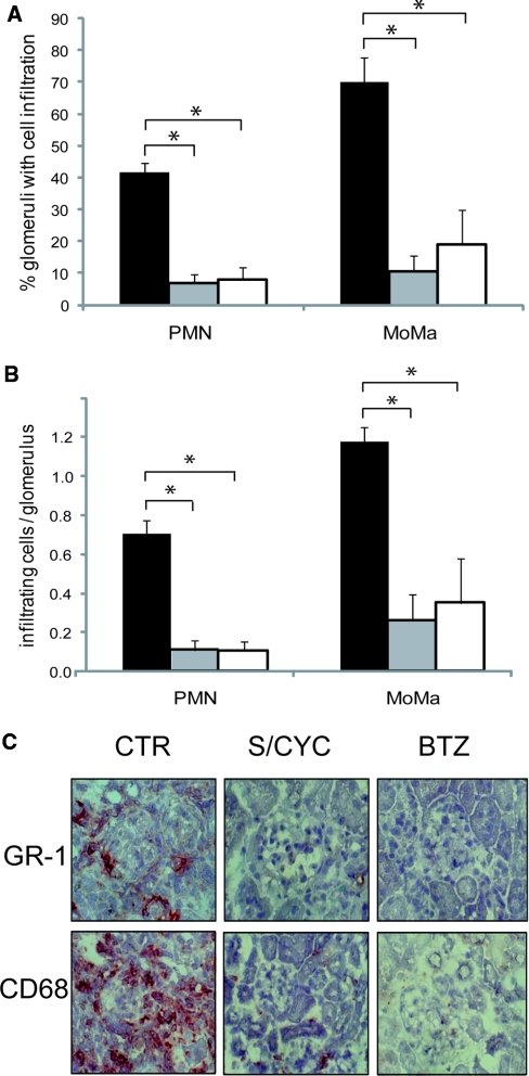
Strong infiltration of neutrophils and macrophages occurred in the control group (Figure 2, A and B). When we analyzed the results with respect to the percentage of glomeruli that showed leukocyte infiltration (Figure 2A), or to the number of infiltrating cells (Figure 2B), we observed a significant reduction in PMN and macrophage influx in both active treatment arms.
Figure 2.
BTZ and S/CYC treatments diminish glomerular PMN and macrophage influx. Panel (A) shows the percentage of glomeruli with PMN or macrophage infiltration and panel (B) the absolute number of these cells per glomerulus. Representative cross sections with GR-1 staining for PMN and CD68 staining for macrophages are depicted in panel (C). The groups consist of controls (CTR, black columns), S/CYC-treated mice (gray), and BTZ-treated mice (white). *P < 0.05.
BTZ Strongly Reduces Anti-MPO Antibody Titer
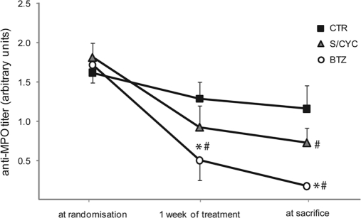
We next assessed the treatment effects on anti-MPO titers by ELISA. Serum samples were obtained at randomization, after 1 week of treatment, and at the time of death or sacrifice. The anti-MPO antibody titers were significantly reduced by BTZ, compared with the untreated control mice (Figure 3). S/CYC reduced the anti-MPO titer at the end of treatment when compared with the titer at randomization. However, the differences from the control animals were NS.
Figure 3.
BTZ reduces anti-MPO antibody titer. Results are shown in arbitrary units (405) nm. The anti-MPO titer was measured at randomization, after 1 week of treatment, and at sacrifice after 3 to 4 weeks of treatment. * indicates a significant difference compared with the control group at the indicated time point. # indicates a significant difference compared with the titer at randomization. P < 0.05.
The Effect of BTZ and S/CYC Treatment on Plasma Cells in Spleen and Bone Marrow
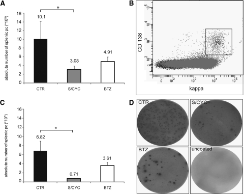
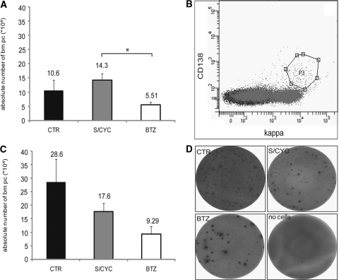
We then studied the effect of treatment on plasma cells in spleen and BM. We first assessed the absolute number of splenic plasma cells by flow cytometry and by ELISPOT analysis. Flow cytometry is based on the characteristic CD138 surface expression together with cytoplasmic κ light chain staining. ELISPOT detects IgG and IgM secreting plasma cells. Compared with the controls, significant plasma cell reduction occurred with S/CYC treatment. Splenic plasma cells were reduced to approximately 50% by BTZ; however, this effect did not reach statistical significance. Typical examples for flow cytometry and ELISPOT analysis are included (Figure 4). We next assessed BM plasma cells by the same two methods (Figure 5). BTZ reduced BM plasma cells in the flow cytometry assay by approximately 50% and in the ELISPOT assay by 70%. Depletion by BTZ was significantly stronger than that by S/CYC (P < 0.05). Typical examples for flow cytometry and ELISPOT analysis are depicted. We observed that S/CYC treatment led to a strong reduction in spleen weight, as well as an absolute reduction in splenic cell number. Spleens of vehicle-control treated mice weighed 169 ± 22 mg, 80 ± 11 mg in S/CYC treated mice, and 176 ± 13 mg in BTZ-treated animals (P < 0.05 for S/CYC). The absolute splenic cell number was 8.6 ± 1.3 × 10⁷ cells for controls, 3.2 ± 0.5 × 10⁷ cells for S/CYC, and 12.2 ± 1.2 × 10⁷ cells for BTZ (P < 0.05 for S/CYC).
Figure 4.
BTZ does not reduce the number of total splenic plasma cells. Panel (A) shows the effect of S/CYC (gray columns) and BTZ (white) in comparison to untreated control mice (CTR, black) on the absolute number of splenic plasma cells analyzed by flow cytometry. Panel (B) depicts a typical experiment in a control mouse. The gated plasma cells express CD138 and cytoplasmic Ig κ light chains (cIg-κ). Panel (C) gives the absolute number of IgG and IgM secreting splenic plasma cells by ELISPOT with a typical example in panel (D). *P < 0.05.
Figure 5.
BTZ and S/CYC do not reduce the number of BM plasma cells. Panel (A) shows the effect of S/CYC (gray columns) and BTZ (white) in comparison to control mice (CTR, black) on the absolute number of BM plasma cells analyzed by flow cytometry. Panel (B) depicts a typical experiment in a control mouse. The gated plasma cells express CD138 and cytoplasmic Ig κ light chains (cIg-κ). Panel (C) gives the absolute number of IgG and IgM secreting BM plasma cells by ELISPOT with a typical example in panel (D). *P < 0.05.
BTZ Reduces MPO-Specific Plasma Cells in Spleen and Bone Marrow
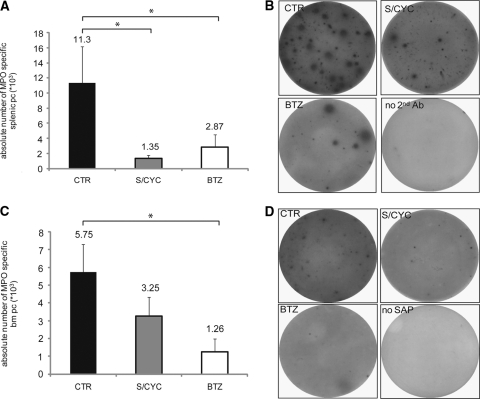
We then established the ELISPOT analysis on murine MPO-coated screen plates to detect MPO-specific plasma cells. Compared with the controls, significant MPO-specific plasma cell reduction in spleen and BM occurred with BTZ (Figure 6). S/CYC treatment significantly reduced splenic MPO-specific plasma cells, but not BM MPO-specific plasma cells. Typical examples for ELISPOT analysis are included. Again, the reduction in MPO-specific splenic plasma cells by S/CYC should be interpreted in the context of a strong reduction in spleen weight.
Figure 6.
BTZ reduces MPO-specific plasma cells in spleen and BM. Panel (A) shows the effect of S/CYC (gray columns) and BTZ (white) in comparison to control mice (CTR, black) on the absolute number of MPO-specific splenic plasma cells. Panel (C) shows MPO-specific BM plasma cells. Panel (B) (splenic) and panel (D) (BM) depict typical experiments. No SAP means that the substrate streptavidin-AP was omitted. *P < 0.05.
BTZ Does Not Affect the Number of B Lymphocytes in Spleen and Bone Marrow
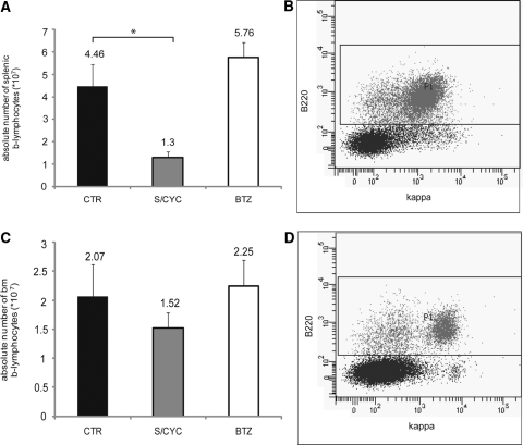
We next performed flow cytometry to assay B220 (CD45R) positive B lymphocytes (Figure 7). We found that BTZ did not affect B lymphocytes in spleen or BM. In contrast, the S/CYC combination significantly reduced splenic B lymphocytes. Typical flow cytometry experiments are given.
Figure 7.
BTZ does not affect the number of B cells in spleen and bone marrow. Panel (A) shows the effect of S/CYC (gray columns) and BTZ (white) in comparison to control mice (CTR, black) on the absolute number of splenic B220-positive B lymphocytes analyzed by flow cytometry. Panel (B) depicts a typical experiment in a control mouse. Panel (C) shows the effects on the absolute number of BM B lymphocytes with a typical example in panel (D). *P < 0.05.
Early But Not Late BTZ Treatment Protects from ANCA-Induced NCGN
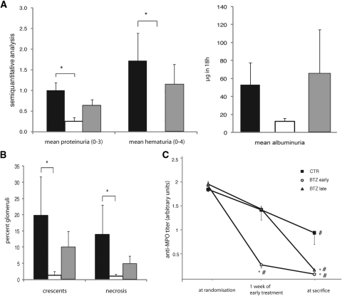
We performed a second set of experiments to evaluate the effects of an early (3 weeks after BM transplantation) versus late BTZ treatment strategy (5 weeks after BM transplantation). On the basis of the urine analysis, glomerulonephritis was evident in approximately 50% of the animals when early, and in approximately 90% when late, treatment was begun. Seven of 7 mice in the control group developed proteinuria and 5 of 7 mice developed hematuria. Proteinuria and hematuria were detected in 4 of 7 and 1 of 7 of the early BTZ treatment group and in 6 of 7 and 5 of 7 of the late BTZ treatment group. Urine analysis is shown in Figure 8A. Similar to our first set of experiments, one mouse in the late BTZ group died within 36 hours after the first BTZ dose. This animal was omitted from urine and histology analysis. The adverse events are discussed below. At the time of sacrifice, 7 of 7 mice in the control group developed NCGN, whereas 2 of 7 in the early BTZ treatment group and 5 of 7 in the late BTZ treatment group showed lesions. Compared with control animals, analysis of the percentage of glomeruli with crescents or necrosis indicates a significant reduction with the early, but not with the late, BTZ treatment protocol (Figure 8B). By ELISA we observed that early and late BTZ treatment resulted in significantly reduced circulating anti-MPO antibody titers at the time of sacrifice (Figure 8C).
Figure 8.
Early but not late BTZ treatment protects from ANCA-induced NCGN. Urine dipstick analysis and albuminuria were measured at sacrifice (A) and renal tissue was examined (B). Black columns depict controls (CTR), white columns early BTZ, and dark gray columns late BTZ treatment. Glomerular crescents and necrosis were expressed as the mean percentage of glomeruli with crescents and necrosis. The treatment effect on circulating anti-MPO titers was assessed by ELISA. Results are shown in arbitrary units at outside diameter 405 nm. The anti-MPO titer was measured at random, 1 week after early BTZ treatment had started, and at sacrifice. * indicates a significant difference compared with the control group at the indicated time point. # indicates a significant difference compared with the titer at randomization. P < 0.05.
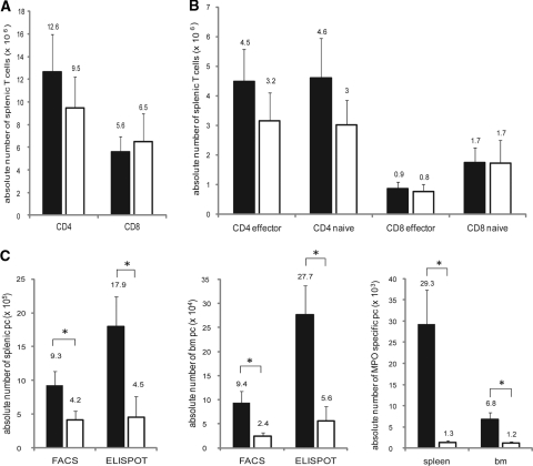
We used the control and the early BTZ treatment group to evaluate treatment effects on T cells. Estimating total, naive, and effector CD4 and CD8 cell numbers by flow cytometry in the spleen of control and BTZ-treated mice indicates that BTZ did affect neither total splenic T cell numbers (Figure 9A) nor the corresponding naive and effector subsets (Figure 9B).
Figure 9.
Early BTZ treatment has no effect on splenic T cells and reduces total and MPO-specific plasma cells. Panel (A) shows the effect of early BTZ treatment on the absolute number of splenic CD4 and CD8 T cells, panel (B) the effect on the CD4 and CD8 effector and naive subtypes, and panel (C) the effect of early BTZ treatment on the absolute number of splenic and BM total and MPO-specific plasma cells analyzed by flow cytometry and ELISPOT. White columns depicts early BTZ treatment and black columns the control group. *P < 0.05.
We analyzed plasma cells in the early BTZ treatment group and observed statistically significant total and MPO-specific splenic and BM plasma cell reduction by BTZ (Figure 9C). Combining the ELISPOT data obtained in mice treated 3 and 4 weeks after BM transplantation (i.e., from the first and second experimental set) showed that BTZ reduced splenic total plasma cells by 67% and splenic MPO-specific plasma cells by 90% (P < 0.05 for both). In addition, BTZ reduced BM total plasma cells by 74% and BM MPO-specific plasma cells by 81% (P < 0.05). These data, expressed as a ratio between MPO-specific plasma cell reduction and total plasma cell reduction, were 1.3 for spleen and 1.1 for BM, suggesting a slightly greater BTZ effect on MPO-specific plasma cells compared with total plasma cells.
We included 4 of 8 mice of the late BTZ treatment group that were sacrificed at the end of the 8-week study period in the analysis of MPO-specific plasma cells by ELISPOT. These four mice had received 3 weeks of BTZ. We observed significant MPO-specific plasma cell reduction that was similar to the early BTZ treatment group (spleen: 1.3 ± 0.6 for early and 0.9 ± 0.8 for late; BM: 1.2 ± 0.4 for early and 1.4 ± 0.6 for late BTZ). We failed to detect plasma cells in the kidney either by flow cytometry or by immunohistochemistry. In contrast, plasma cells were easily detected in spleens and BM by both methods (data not shown).
Adverse Events
In the first set of experiments, we monitored blood cell counts before treatment, after 1 week of treatment, and at the end of treatment. The data are summarized in Table 1. S/CYC, but not BTZ, treatment resulted in lymphopenia. One week after treatment, BTZ-treated mice developed lower platelet counts compared with controls. The platelet number normalized thereafter. It was also noted that BTZ-treated mice showed anemia at sacrifice. We emphasize this fact because one had expected that improvement in disease should be accompanied by improvement in anemia. Most remarkably, 3 of 23 mice that were treated with BTZ died within 36 hours after the first drug dose. We immediately examined these mice. Histology revealed toxic lung injury with edema and hemorrhage. For lack of any alternative explanation, we attributed the deaths to toxic BTZ-related effects. However, when we injected an additional small series of 12 nonimmunized mice (6 C57BL/6J [B6] and 6 Balb/c mice) with BTZ, we observed no acute adverse events.
Table 1.
Analysis of peripheral blood cell counts before, 1 week after start of treatment and at sacrifice
| CTR | S/CYC | BTZ | |||||||
|---|---|---|---|---|---|---|---|---|---|
| Before Treatment | 1 Week of Treatment | At Sacrifice | Before Treatment | 1 Week of Treatment | At Sacrifice | Before Treatment | 1 Week of Treatment | At Sacrifice | |
| Erythrocytes × 1012 | 9.5 ± 0.5 | 7.1 ± 0.4b | 6.7 ± 1.1b | 10.4 ± 0.7 | 8.7 ± 0.8 | 8.4 ± 0.8 | 11.1 ± 0.3 | 10.3 ± 0.8a | 6.6 ± 1.0b |
| Thrombocytes × 109 | 1120 ± 187 | 1109 ± 202 | 1460 ± 182 | 987 ± 137 | 1302 ± 87a | 893 ± 141 | 1102 ± 160 | 639 ± 162a | 1118 ± 314 |
| Leukocytes × 109 | 8.3 ± 2.0 | 8.2 ± 0.7 | 8.1 ± 2.0 | 8.4 ± 1.7 | 7.8 ± 0.9 | 4.9 ± 0.8 | 8.7 ± 1.1 | 7.2 ± 1.2 | 7.7 ± 2.3 |
| Lymphocytes × 109 | 5.6 ± 0.9 | 5.3 ± 0.5 | 4.7 ± 1.3 | 5.1 ± 0.7 | 5.1 ± 0.6 | 2.8 ± 0.4b | 5.9 ± 0.8 | 5.0 ± 0.7 | 4.9 ± 1.2 |
| Granulocytes × 109 | 2.7 ± 0.8 | 2.1 ± 0.3 | 2.6 ± 0.8 | 2.4 ± 0.7 | 1.9 ± 0.3 | 2.1 ± 0.2 | 2.0 ± 0.3 | 1.7 ± 0.4 | 2.2 ± 1.0 |
| Monocytes × 109 | 1.0 ± 0.3 | 0.8 ± 0.1 | 0.7 ± 0.2 | 0.9 ± 0.3 | 0.8 ± 0.1 | 0.6 ± 0.1 | 0.8 ± 0.1 | 0.5 ± 0.2 | 0.6 ± 0.2 |
DISCUSSION
This study is the first to examine an experimental treatment, compared with an established clinical treatment, in an ANCA-induced nephritis animal model. Our main findings are that both BTZ and a conventional combined treatment with steroids and cyclophosphamide strongly abrogated NCGN. However, both treatment regimens had different capacities for therapeutic depletion of plasma cells and anti-MPO antibodies, as well as induction of adverse reactions. BTZ treatment significantly reduced total and MPO-specific splenic and BM plasma cells with a slightly more pronounced effect on the MPO-specific plasma cells. This effect resulted in a significant reduction in anti-MPO antibody titers. Furthermore, BTZ left B cells, total CD4 and CD8 T cells, and their naive and effector subsets unperturbed. In contrast, S/CYC caused spleen shrinkage, resulting in reduced total spleen cells, including total and MPO-specific plasma cells and B cells. Furthermore, S/CYC had no effect on total and MPO-specific bone marrow plasma cells. A caveat should be added at this point because 3 of 23 BTZ-treated mice died within 36 hours after the first dosage. Our data demonstrate the usefulness of an ANCA vasculitis animal model for testing efficiency and side effects of novel treatment strategies.
Severe ANCA vasculitis in patients and in our animal model is life-threatening when not treated. Combined treatment with steroids and cytotoxic drugs became the standard treatment that has strongly reduced disease-associated mortality, however, at the cost of substantial treatment-associated morbidity and mortality.13–15,17,18,30 Novel therapeutic approaches need to be developed that can be used in addition, or alternatively, when side effects of standard protocols required their discontinuation.
On the basis of the underlying mechanisms that are at work in ANCA vasculitis, we hypothesized that targeting ANCA-producing plasma cells by BTZ provides a novel treatment strategy that can be tested in a mouse model. B cell–mediated mechanisms, including ANCA generation, are important in ANCA vasculitis. For example, passive anti-MPO antibody transfer was sufficient to induce vasculitis in Rag2−/− mice that lack functional T and B cells, but also in a newborn after MPO-ANCA transfer from his mother.7,12 Xiao and co-workers extended their initial findings by showing that the transfer of splenocytes from MPO-immunized mice induced NCGN, whereas transfer of purified T cells did not.31 Recently, the importance of central and peripheral deletion of anti-MPO–specific B cells was shown in a transgenic mouse model 32. Drugs that target B cells were recently introduced and evaluated in ANCA vasculitis patients. The most promising data were obtained with rituximab, which binds to CD20. CD20 is expressed on immature and mature B cells, but not on plasma blasts and plasma cells, mediating proliferation and differentiation.33 However, the molecule is not found on mature bone marrow plasma cells and is only heterogeneously expressed by plasmablasts/immature plasma cells.34 Rituximab results in depletion of CD20 positive cells. Two very recent randomized controlled trials report that rituximab induced disease remission similar to cyclophosphamide and resulted in strong reduction of B cells and circulating ANCA.35,36 However, both the standard steroid/cyclophosphamide combination and rituximab are associated with treatment failures, relapses, and adverse events. Depleting plasma cells may provide an additional, and more specific, treatment approach.
The proteasome inhibitor BTZ targets plasmablasts and mature plasma cells effectively and specifically; however, it leaves total B cell, T cell, and dendritic cell populations unperturbed in mice.29 The United States Food and Drug Administration (FDA) approved the drug for clinical use in patients with multiple myeloma in 2008.37 BTZ prevents degradation of proteins marked by ubiquitination by inhibiting the 26S proteasome. Substrates enter the proteasome through a gated pore, a process that involves ATP-stimulated events.38 BTZ's main effects are NF-κB inhibition, stabilization of critical cell cycle proteins such as p27 and p53, induction of apoptosis, and induction of cell death by the activation of the terminal unfolded protein response.39 Because of its well-documented effect on plasma cells in multiple myeloma, BTZ has implications for antibody-mediated immune diseases as well.
Persistent antibody titers were observed despite long-term immunosuppressive treatment or autologous stem cell transplantation in several autoimmune diseases.40–42 Studies showed that long-lived plasma cells survive for decades, even in the absence of memory B cells and antigen exposure, and that these cells were resistant to radiation, cyclophosphamide, and mitomycin C.43 In NZB/W mice, both nondividing long-lived plasma cells and short-lived plasma cells contributed to anti-DNA antibody generation.43 Given the BTZ effects on plasma cells in multiple myeloma, the drug was recently explored in autoimmune lupus mouse models. Neubert et al. found that both short- and long-lived plasma cells were decreased by >60% in spleens and >95% in BM after 48 hours of BTZ treatment, whereas dexamethasone and cyclophoshamide had only minor effects.29 Importantly, BTZ decreased anti-dsDNA antibodies and prevented lupus nephritis. Our experiments in another antibody-dependent model, namely, in a MPO-ANCA mouse model, show that BTZ reduces the total IgG- and IgM-secreting plasma cell pool in spleen and BM by 67% and 74%, respectively. Probably more importantly, we found significant depletion of MPO-specific plasma cells (90% for spleen and 81% for BM). Subsequently, this effect led to a fall in anti-MPO antibody titers, compared with controls. Compared with S/CYC, the BTZ effect on MPO-specific plasma cells and the subsequent fall of anti-MPO titers was stronger and occurred in the splenic and BM compartment. T-cell participation in ANCA-vasculitis mouse models was recently implicated.44,45 We found that BTZ spared total CD4 and CD8 splenic T cells as well as the naive and effector T cell subsets, and B lymphocytes. Furthermore, and in contrast to BTZ, S/CYC strongly decreased spleen weight, diminished the number of total splenic cells, including total and MPO-specific plasma cells, and splenic B lymphocytes. The data indicate that BTZ was more efficient and specific compared with S/CYC. Interestingly, plasma cell depletion by BTZ was slightly more pronounced for MPO-specific plasma cells, compared with total IgG- and IgM-secreting plasma cells. Conceivably, BTZ activates the terminal unfolded protein response more strongly in the former and/or the latter cells feature a faster replenishment. These questions were not addressed by experiments. However, Neubert et al. showed partial replenishment of the plasma cell pool within 2 to 5 days after BTZ.29
Untreated control animals developed severe NCGN with strong neutrophil and macrophage influx. BTZ and S/CYC standard treatment, started 4 weeks after BM transplantation, showed equal efficiency in preventing disease. Interestingly, when we compared BTZ starting early (3 weeks) or late (5 weeks) after BM transplantation, we observed that both MPO-specific plasma cells and circulating anti-MPO titers were reduced in the early and the late treatment group to a similar extent. However, late treatment did not prevent disease progression. Thus, our data obtained after 3, 4, and 5 weeks of BTZ treatment suggest that, in already-established disease, mechanisms beside direct ANCA effects are initiated and cannot be effectively targeted solely by reduction of autoantibody levels. Our data suggest that BTZ provides an alternative to S/CYC that can be used in either refractory patients or in patients that have already received high cyclophosphamide doses. Whether the BTZ depleting effect on MPO-specific plasma cells would reduce relapses during the disease course is an interesting future hypothesis to be tested.
In our group of 23 BTZ-treated mice, we observed three acute deaths within 36 hours after the first BTZ dose. Histology suggested that toxic pulmonary effects were at work. No such events were described in MRL/lpr and NZB/W F1 lupus mice that were treated in an identical manner, and observed during longer periods up to 10 weeks.29 We followed up our observation with limited additional studies in mice and were not able to repeat the events. The incidence of serious side effects in patients is <5%. Thrombocytopenia and peripheral neuropathy are the most clinically relevant adverse events.37,46 However, isolated reports exist where patients developed severe pulmonary complications. Miyakoshi et al. reported that 4 of 13 BTZ-treated Japanese myeloma patients developed acute pulmonary complications with wheezing, infiltrates, and edema; two of them died.47 The authors point out that the larger clinical BTZ studies were mostly performed in Europe and in the United States and that different patient ethnic backgrounds may be responsible. An additional African-American patient was described soon after.48 Similar genetically related issues could pertain to different mouse strains. Alternatively, BTZ dosage issues or pulmonary disease manifestations, such as in ANCA vasculitis, may predispose to toxic BTZ effects. On the basis of the reports from the literature, we used 0.75 mg/kg body wt BTZ because this dosage was shown to significantly reduce short- and long-lived plasma cells, activate the terminal unfolded protein response, abrogate NF-κB, and impair dendritic cell maturation and functions.29,49,50 Future studies should explore reduced dosage protocols. This issue of adverse effects certainly needs close follow-up and clinicians need to be alert.
To our knowledge, this study is the first to examine the efficacy of the standard human treatment in mice in a comparative fashion. Nonetheless, our primary hypothesis was that BTZ depletes anti-MPO–specific plasma cells, reduces anti-MPO titers, and prevents NCGN in mice in a fashion that is better than standard therapy. BTZ could expand our arsenal to treat patients with ANCA-induced vasculitis and glomerulonephritis. However, BTZ timing and possible adverse events need further attention. These animal studies suggest that clinical studies in patients failing currently accepted treatments are warranted.
CONCISE METHODS
Materials
BTZ was obtained from Janssen-Cilag (Neuss, Germany), dexamethasone from Merck (Darmstadt, Germany), and cyclophosphamide from Baxter (Unterschleissheim, Germany). Borgal was from Intervet (Unterschleissheim, Germany) and the urine dipsticks from Roche Diagnostics Corporation (Indianapolis, IN). The antibodies to Gr-1 (clone RB6-8C5), CD68 (clone FA11), and CD138 (clone 281–2) were from BD Biosciences (Heidelberg, Germany). Fluorescein isothiocyanate–conjugated (FITC-conjugated) goat anti-mouse IgG was from Molecular Probes Invitrogen (Carlsbad, CA), and FITC-conjugated goat anti-mouse C3, IgG, IgM, IgA, and MPO were from ICN/Cappel (Aurora, OH). Goat anti-mouse IgG and IgM and biotinylated goat anti-mouse IgG and IgM for ELISPOT were from Southern Biotech, Biozol (Eching, Germany), and streptavidin-AP and NBT/BCIP were from Roche Diagnostics (Mannheim, Germany). Antibodies for FACS analysis (CD16/CD32, CD45R [B220], CD138, κ light chain, CD8, CD11c, and CD62L) were from BD Biosciences. Anti-CD4 was from eBiosciences (Frankfurt, Germany), and anti-CD44 was kindly provided from the German Rheumatological Research Center (Berlin, Germany). Multiscreen plates for ELISPOT analysis were purchased from Millipore (Schwalbach, Germany), and BD Falcon cell strainers were from BD Biosciences. PBS, RPMI, and P/S were purchased from PAA (Cölbe, Germany), FCS from Biochrom (Berlin, Germany), and saponin and BSA from Sigma (München, Germany).
Mice and Immunization Protocol
C57BL/6J (B6) mice breeding pairs were purchased from Jackson Laboratories (Bar Harbor, ME). Mice lacking MPO (MPO−/− mice) were the sixth-generation progeny of a backcross into B6 mice. MPO−/− mice (8 to 10 weeks old) were used for immunization. Nine- to 10-week-old B6 WT mice were used as bone-marrow donors. Local authorities approved all animal experiments, which followed American Physiologic Society guidelines for animal care.
The purification of mouse MPO and the immunization of MPO−/− mice were performed as described previously.9 Briefly, murine MPO was purified from WEHI-3 cells by dounce homogenization, concanavalin A affinity chromatography, ion exchange, and gel filtration chromatography. MPO−/− mice were immunized intraperitoneally with 10 μg of purified murine MPO in complete Freund adjuvant and boosted at day 28 with 10 μg of purified murine MPO in incomplete Freund adjuvant. Development of antibodies was monitored by anti-MPO ELISA.
Bone-Marrow Transplantation in Mice
After immunization, the MPO−/− mice were kept at sterile housing conditions with food and water ad libitum (sterile water with trimethoprim and sulfadoxine, Borgal). After immunization, mice were γ-irradiated with 900 rad of whole-body dose and reconstituted with BM cells from WT of the same genetic background. BM cells were harvested from femura and tibiae, erythrocytes were lysed, and 1.5 × 10⁷ BM cells were intravenously injected.
Treatment Protocols
In our first set of experiments treatment started 4 weeks after BM transplantation. Mice were treated up to 4 weeks and sacrificed thereafter. The control group (CTR) received food and water ad libitum (n = 8). One treatment arm received the steroid dexamethasone daily (1 mg/kg body wt) orally and cyclophosphamide in 0.9% saline once weekly (30 mg/kg body wt) intraperitoneally (n = 8). The other treatment arm received the proteasome inhibitor BTZ in 0.9% saline two times weekly (0.75 mg/kg body wt) intravenously (n = 8). We performed a second set of experiments to test early versus late BTZ treatment. BTZ administration started 3 weeks after BM transplantation in the early treatment group (n = 7) and 5 weeks after BM transplantation (n = 8) in the late treatment group. Randomization was performed at 3 weeks after BM transplantation (i.e., when early treatment was started) based on anti-MPO titer and hematuria. At that time point, 5 of 7 mice in the control group, 4 of 7 in the early BTZ treatment group, and 6 of 8 in the late BTZ treatment group showed signs of glomerulonephritis. The average hematuria was 1.2 (range 0 to 4) in each group. In both experimental sets, mice were sacrificed at 8 weeks after BM transplantation or earlier if they showed signs of apathy, defense reactions, reduced food or water intake, dyspnea, motion abnormality, or abnormal posture. Three CTR, one S/CYC, and three BTZ mice during the first set of experiments and one CTR, one early BTZ-treated, and four late BTZ-treated mice showed at least one of the above-mentioned signs.
Functional Evaluation of Renal Injury
Mice were placed in metabolic cages 1 day before sacrifice and urine was collected for 18 hours overnight. Urine was tested by dipstick for hematuria and proteinuria and the extent is expressed as the mean on a scale of 0 to 4 for hematuria and 0 to 3 for proteinuria. Albuminuria was determined by an albumin-specific ELISA (CellTrend, Luckenwalde, Germany).
Histologic Evaluation of Renal Injury
Samples of kidney tissue were collected at the time of sacrifice, fixed in 10% formalin, and embedded in paraffin using routine protocols. Four-micrometer coronal sections of specimens were stained with hematoxylin and eosin (H&E), periodic acid–Schiff (PAS), and evaluated by light microscopy. The extent of glomerular crescents and necrosis was expressed as the mean percent of glomeruli with crescents and necrosis in each animal. For immunofluorescence microscopy to detect glomerular localization of immune determinants, 4-μm frozen sections were stained with fluoresceinated antibodies. The glomerular IgG deposition was performed by using FITC-conjugated goat anti-mouse IgG. Deposition of mouse complement C3, IgM, IgA, and MPO was visualized with a FITC-conjugated goat anti-mouse C3, IgM, IgA, and MPO. For detection of leukocytes, sections of snap-frozen tissue were stained with rat antibodies to neutrophils (anti–Gr-1), monocytes/macrophages (anti-CD68), and plasma cells (anti-CD138), respectively. Rat antibody binding was detected using peroxidase-labeled secondary rabbit anti-rat IgG and tertiary goat anti-rabbit IgG antibodies followed by 3-amino-9-ethylcarbazole and hydrogen peroxide. Sections were counterstained with hematoxylin.
Isolation of Cells from Spleen and Bone Marrow
Single-cell solutions from spleen and BM (femora and tibiae) were obtained by disrupting whole organs through a 70-μm cell filter using a syringe plunger and PBS. The single-cell suspension was washed once with cold RPMI/10% FCS/1% PS. Number of viable cells were counted using a Neubauer hemocytometer.
Detection of Antibody-Secreting Cells by Enzyme-Linked Immunosorbent Spot (ELISPOT) Assay
For quantification of IgG and IgM secreting cells we coated 96-well Multi Screen plates with 5 μg/ml goat anti-mouse IgG and IgM in sodium carbonate buffer pH 9 overnight at 4°C and then blocked with RPMI/10% FCS/1% PS and PBS/3% BSA for 2 hours at 37°C. Single-cell suspensions from spleens and BMs in RPMI/10% FCS/ 1% PS were incubated in duplicate in serial dilutions of 1:5 on coated plates for 3 hours at 37°C in a 5% CO2-containing incubator. Plates were washed and incubated in 1 μg/ml biotinylated goat anti-mouse IgG and IgM for 2 hours at 37°C. Bound IgG and IgM was detected by addition of streptavidin-AP and of NBT/BCIP in Tris/MgCl/NaCl buffer as staining solution. For assessment of anti-MPO (IgG and IgM) secreting cells, we coated plates with 5 μg/ml MPO in sodium carbonate buffer pH 9.
The number of spots was counted employing a video-based automatic ELISPOT reader (CTL ImmunoSpot S4 Analyzer). The analysis was performed with ImmunoSpot Software 5.0 (CTL-Europe, Bonn, Germany).
Flow Cytometric Analysis
Flow cytometric analysis of splenocytes and BM cells was performed using fluorochrome-conjugated monoclonal antibodies to mouse CD45R (B220), CD138, κ light chain, CD4, CD8, CD44, CD62L, and CD11c. To detect plasma cells, we performed Fc-block with rat anti-mouse CD16/CD32 at 4°C and surface staining with PE-labeled anti-CD138. Cells were then fixed with 1.5% PFA for 20 minutes at room temperature. After washing the cells with PBS/1% BSA /0.1% saponin, we performed intracellular κ light chain staining with FITC-labeled anti–κ light chain in PBS/1% BSA/0.5% saponin. We used the same protocol for the detection of renal plasma cells. To detect B lymphocytes, we performed surface staining with Pacific blue–labeled anti-CD45R. To detect splenic T cells, we performed surface staining with PE-labeled anti-CD8 and e450-labeled anti-CD4. CD4 and CD8 naïve subsets were characterized by CD44low/CD62Lhigh staining. Effector subsets were characterized by CD44high/CD62Llow staining. Splenic single-cell suspensions were additionally stained for CD11c to exclude dendritic cells from the analysis. Samples were analyzed using a FACS CANTO II flow cytometer and FACSDiva software (BD Biosciences).
Statistical Analyses
Results are given as means ± SEM. Comparisons were made using ANOVA with Fisher post hoc analysis. Differences were considered significant at P < 0.05.
DISCLOSURES
None.
Acknowledgments
We thank Sylvia Krüger, Gisela Philipp, and Baerbel Kuhlmann for excellent technical assistance and Martin Szyska und Katrin Moser from the German Rheumatological Research Center (DRFZ) for sharing the flow cytometry and ELISPOT methodology. DFG grants (KE 576/7-1 and SCHR 771/5-1) and an ECRC grant supported this study.
Footnotes
Published online ahead of print. Publication date available at www.jasn.org.
REFERENCES
- 1.van der Woude FJ, Rasmussen N, Lobatto S, Wiik A, Permin H, van Es LA, van der Giessen M, van der Hem GK, The TH: Autoantibodies against neutrophils and monocytes: Tool for diagnosis and marker for disease activity in Wegener's granulomatosis. Lancet 1(8426): 425–429, 1985 [DOI] [PubMed] [Google Scholar]
- 2.Falk RJ, Jennette JC: Anti-neutrophil cytoplasmic autoantibodies with specificity for myeloperoxidase in patients with systemic vasculitis and idiopathic necrotizing and crescentic glomerulonephritis. N Engl J Med 318: 1651–1657, 1988 [DOI] [PubMed] [Google Scholar]
- 3.Kain R, Matsui K, Exner M, Binder S, Schaffner G, Sommer EM, Kerjaschki D: A novel class of autoantigens of anti-neutrophil cytoplasmic antibodies in necrotizing and crescentic glomerulonephritis: The lysosomal membrane glycoprotein h-lamp-2 in neutrophil granulocytes and a related membrane protein in glomerular endothelial cells. J Exp Med 181: 585–597, 1995 [DOI] [PMC free article] [PubMed] [Google Scholar]
- 4.Falk RJ, Terrell RS, Charles LA, Jennette JC: Anti-neutrophil cytoplasmic autoantibodies induce neutrophils to degranulate and produce oxygen radicals in vitro. Proc Natl Acad Sci U S A 87: 4115–4119, 1990 [DOI] [PMC free article] [PubMed] [Google Scholar]
- 5.Savage CO, Pottinger BE, Gaskin G, Pusey CD, Pearson JD: Autoantibodies developing to myeloperoxidase and proteinase 3 in systemic vasculitis stimulate neutrophil cytotoxicity toward cultured endothelial cells. Am J Pathol 141: 335–342, 1992 [PMC free article] [PubMed] [Google Scholar]
- 6.Kettritz R, Jennette JC, Falk RJ: Crosslinking of ANCA-antigens stimulates superoxide release by human neutrophils. J Am Soc Nephrol 8: 386–394, 1997 [DOI] [PubMed] [Google Scholar]
- 7.Xiao H, Heeringa P, Hu P, Liu Z, Zhao M, Aratani Y, Maeda N, Falk RJ, Jennette JC: Antineutrophil cytoplasmic autoantibodies specific for myeloperoxidase cause glomerulonephritis and vasculitis in mice. J Clin Invest 110: 955–963, 2002 [DOI] [PMC free article] [PubMed] [Google Scholar]
- 8.Pfister H, Ollert M, Froehlich LF, Quintanilla-Martinez L, Colby TV, Specks U, Jenne DE: Antineutrophil cytoplasmic autoantibodies (ANCA) against the murine homolog of proteinase 3 (Wegener autoantigen) are pathogenic in vivo. Blood 104: 1411–1418, 2004 [DOI] [PubMed] [Google Scholar]
- 9.Schreiber A, Xiao H, Falk RJ, Jennette JC: Bone marrow-derived cells are sufficient and necessary targets to mediate glomerulonephritis and vasculitis induced by anti-myeloperoxidase antibodies. J Am Soc Nephrol 17: 3355–3364, 2006 [DOI] [PubMed] [Google Scholar]
- 10.Kain R, Exner M, Brandes R, Ziebermayr R, Cunningham D, Alderson CA, Davidovits A, Raab I, Jahn R, Ashour O, Spitzauer S, Sunder-Plassmann G, Fukuda M, Klemm P, Rees AJ, Kerjaschki D: Molecular mimicry in pauci-immune focal necrotizing glomerulonephritis. Nat Med 14: 1088–1096, 2008 [DOI] [PMC free article] [PubMed] [Google Scholar]
- 11.Little MA, Smyth L, Salama AD, Mukherjee S, Smith J, Haskard D, Nourshargh S, Cook HT, Pusey CD: Experimental autoimmune vasculitis: An animal model of anti-neutrophil cytoplasmic autoantibody-associated systemic vasculitis. Am J Pathol 174: 1212–1220, 2009 [DOI] [PMC free article] [PubMed] [Google Scholar]
- 12.Schlieben DJ, Korbet SM, Kimura RE, Schwartz MM, Lewis EJ: Pulmonary-renal syndrome in a newborn with placental transmission of ANCAs. Am J Kidney Dis 45: 758–761, 2005 [DOI] [PubMed] [Google Scholar]
- 13.Jayne D, Rasmussen N, Andrassy K, Bacon P, Tervaert JW, Dadoniene J, Ekstrand A, Gaskin G, Gregorini G, de Groot K, Gross W, Hagen EC, Mirapeix E, Pettersson E, Siegert C, Sinico A, Tesar V, Westman K, Pusey C: A randomized trial of maintenance therapy for vasculitis associated with antineutrophil cytoplasmic autoantibodies. N Engl J Med 349: 36–44, 2003 [DOI] [PubMed] [Google Scholar]
- 14.Pagnoux C, Mahr A, Hamidou MA, Boffa JJ, Ruivard M, Ducroix JP, Kyndt X, Lifermann F, Papo T, Lambert M, Le Noach J, Khellaf M, Merrien D, Puechal X, Vinzio S, Cohen P, Mouthon L, Cordier JF, Guillevin L: Azathioprine or methotrexate maintenance for ANCA-associated vasculitis. N Engl J Med 359: 2790–2803, 2008 [DOI] [PubMed] [Google Scholar]
- 15.de Groot K, Harper L, Jayne DR, Flores Suarez LF, Gregorini G, Gross WL, Luqmani R, Pusey CD, Rasmussen N, Sinico RA, Tesar V, Vanhille P, Westman K, Savage CO: Pulse versus daily oral cyclophosphamide for induction of remission in antineutrophil cytoplasmic antibody-associated vasculitis: A randomized trial. Ann Intern Med 150: 670–680, 2009 [DOI] [PubMed] [Google Scholar]
- 16.Jayne DR, Gaskin G, Rasmussen N, Abramowicz D, Ferrario F, Guillevin L, Mirapeix E, Savage CO, Sinico RA, Stegeman CA, Westman KW, van der Woude FJ, de Lind van Wijngaarden RA, Pusey CD: Randomized trial of plasma exchange or high-dosage methylprednisolone as adjunctive therapy for severe renal vasculitis. J Am Soc Nephrol 18: 2180–2188, 2007 [DOI] [PubMed] [Google Scholar]
- 17.Hoffman GS, Kerr GS, Leavitt RY, Hallahan CW, Lebovics RS, Travis WD, Rottem M, Fauci AS: Wegener granulomatosis: An analysis of 158 patients. Ann Intern Med 116: 488–498, 1992 [DOI] [PubMed] [Google Scholar]
- 18.Little MA, Nightingale P, Verburgh CA, Hauser T, De Groot K, Savage C, Jayne DR, Harper L: Early mortality in systemic vasculitis: Relative contribution of adverse events and active vasculitis. Ann Rheum Dis 69: 1036–1043, 2010 [DOI] [PubMed] [Google Scholar]
- 19.Joy MS, Hogan SL, Jennette JC, Falk RJ, Nachman PH: A pilot study using mycophenolate mofetil in relapsing or resistant ANCA small vessel vasculitis. Nephrol Dial Transplant 20: 2725–2732, 2005 [DOI] [PubMed] [Google Scholar]
- 20.Walsh M, Jayne D: Rituximab in the treatment of anti-neutrophil cytoplasm antibody associated vasculitis and systemic lupus erythematosus: Past, present and future. Kidney Int 72: 676–682, 2007 [DOI] [PubMed] [Google Scholar]
- 21.Hu W, Liu C, Xie H, Chen H, Liu Z, Li L: Mycophenolate mofetil versus cyclophosphamide for inducing remission of ANCA vasculitis with moderate renal involvement. Nephrol Dial Transplant 23: 1307–1312, 2008 [DOI] [PubMed] [Google Scholar]
- 22.Little MA, Bhangal G, Smyth CL, Nakada MT, Cook HT, Nourshargh S, Pusey CD: Therapeutic effect of anti-TNF-alpha antibodies in an experimental model of anti-neutrophil cytoplasm antibody-associated systemic vasculitis. J Am Soc Nephrol 17: 160–169, 2006 [DOI] [PubMed] [Google Scholar]
- 23.Huugen D, Xiao H, van Esch A, Falk RJ, Peutz-Kootstra CJ, Buurman WA, Tervaert JW, Jennette JC, Heeringa P: Aggravation of anti-myeloperoxidase antibody-induced glomerulonephritis by bacterial lipopolysaccharide: Role of tumor necrosis factor-alpha. Am J Pathol 167: 47–58, 2005 [DOI] [PMC free article] [PubMed] [Google Scholar]
- 24.Huugen D, van Esch A, Xiao H, Peutz-Kootstra CJ, Buurman WA, Tervaert JW, Jennette JC, Heeringa P: Inhibition of complement factor C5 protects against anti-myeloperoxidase antibody-mediated glomerulonephritis in mice. Kidney Int 71: 646–654, 2007 [DOI] [PubMed] [Google Scholar]
- 25.van der Veen BS, Petersen AH, Belperio JA, Satchell SC, Mathieson PW, Molema G, Heeringa P: Spatiotemporal expression of chemokines and chemokine receptors in experimental anti-myeloperoxidase antibody-mediated glomerulonephritis. Clin Exp Immunol 158: 143–153, 2009 [DOI] [PMC free article] [PubMed] [Google Scholar]
- 26.Kuligowski MP, Kwan RY, Lo C, Wong C, James WG, Bourges D, Ooi JD, Abeynaike LD, Hall P, Kitching AR, Hickey MJ: Antimyeloperoxidase antibodies rapidly induce alpha-4-integrin-dependent glomerular neutrophil adhesion. Blood 113: 6485–6494, 2009 [DOI] [PubMed] [Google Scholar]
- 27.Schreiber A, Xiao H, Jennette JC, Schneider W, Luft FC, Kettritz R: C5a receptor mediates neutrophil activation and ANCA-induced glomerulonephritis. J Am Soc Nephrol 20: 289–298, 2009 [DOI] [PMC free article] [PubMed] [Google Scholar]
- 28.Schreiber A, Rolle S, Peripelittchenko L, Rademann J, Schneider W, Luft FC, Kettritz R: Phosphoinositol 3-kinase-gamma mediates antineutrophil cytoplasmic autoantibody-induced glomerulonephritis. Kidney Int 77: 118–128, 2010 [DOI] [PubMed] [Google Scholar]
- 29.Neubert K, Meister S, Moser K, Weisel F, Maseda D, Amann K, Wiethe C, Winkler TH, Kalden JR, Manz RA, Voll RE: The proteasome inhibitor bortezomib depletes plasma cells and protects mice with lupus-like disease from nephritis. Nat Med 14: 748–755, 2008 [DOI] [PubMed] [Google Scholar]
- 30.Haubitz M, Ehlerding C, Kamino K, Koch KM, Brunkhorst R: Reduced gonadal toxicity after i.v. cyclophosphamide administration in patients with nonmalignant diseases. Clin Nephrol 49: 19–23, 1998 [PubMed] [Google Scholar]
- 31.Jennette JC, Xiao H, Heeringa P, Hu P, Liu Z, Falk RJ: Adoptive transfer of T lymphocytes alone from MPO knockout mice immunized with MPO can not induce necrotizing and crescentic glomerulonephritis. Kidney Blood Press Res 26: 254–255, 2003 [Google Scholar]
- 32.Bunch DO, Silver JS, Majure MC, Sullivan P, Alcorta DA, Chin H, Hogan SL, Lindstrom YI, Clarke SH, Falk RJ, Nachman PH: Maintenance of tolerance by regulation of anti-myeloperoxidase B cells. J Am Soc Nephrol 19: 1763–1773, 2008 [DOI] [PMC free article] [PubMed] [Google Scholar]
- 33.Salama AD, Pusey CD: Drug insight: Rituximab in renal disease and transplantation. Nat Clin Pract Nephrol 2: 221–230, 2006 [DOI] [PubMed] [Google Scholar]
- 34.Medina F, Segundo C, Campos-Caro A, Gonzalez-Garcia I, Brieva JA: The heterogeneity shown by human plasma cells from tonsil, blood, and bone marrow reveals graded stages of increasing maturity, but local profiles of adhesion molecule expression. Blood 99: 2154–2161, 2002 [DOI] [PubMed] [Google Scholar]
- 35.Jones RB, Cohen Tervaert JW, Hauser T, Luqmani R, Morgan MD, Peh CA, Savage CO, Segelmark M, Tesar V, van Paassen P, Walsh D, Walsh M, Westman K, Jayne DRW, the European Vasculitis Study G: Rituximab versus cyclophosphamide in ANCA-associated renal vasculitis. N Engl J Med 363: 211–220, 2010 [DOI] [PubMed] [Google Scholar]
- 36.Stone JH, Merkel PA, Spiera R, Seo P, Langford CA, Hoffman GS, Kallenberg CGM, St. Clair EW, Turkiewicz A, Tchao NK, Webber L, Ding L, Sejismundo LP, Mieras K, Weitzenkamp D, Ikle D, Seyfert-Margolis V, Mueller M, Brunetta P, Allen NB, Fervenza FC, Geetha D, Keogh KA, Kissin EY, Monach PA, Peikert T, Stegeman C, Ytterberg SR, Specks U, the, R-ITNRG: Rituximab versus cyclophosphamide for ANCA-associated vasculitis. N Engl J Med 363: 221–232, 2010 [DOI] [PMC free article] [PubMed] [Google Scholar]
- 37.San Miguel JF, Schlag R, Khuageva NK, Dimopoulos MA, Shpilberg O, Kropff M, Spicka I, Petrucci MT, Palumbo A, Samoilova OS, Dmoszynska A, Abdulkadyrov KM, Schots R, Jiang B, Mateos MV, Anderson KC, Esseltine DL, Liu K, Cakana A, van de Velde H, Richardson PG: Bortezomib plus melphalan and prednisone for initial treatment of multiple myeloma. N Engl J Med 359: 906–917, 2008 [DOI] [PubMed] [Google Scholar]
- 38.Besche HC, Peth A, Goldberg AL: Getting to first base in proteasome assembly. Cell 138: 25–28, 2009 [DOI] [PMC free article] [PubMed] [Google Scholar]
- 39.Goldberg AL: Functions of the proteasome: From protein degradation and immune surveillance to cancer therapy. Biochem Soc Trans 35: 12–17, 2007 [DOI] [PubMed] [Google Scholar]
- 40.McSweeney PA, Nash RA, Sullivan KM, Storek J, Crofford LJ, Dansey R, Mayes MD, McDonagh KT, Nelson JL, Gooley TA, Holmberg LA, Chen CS, Wener MH, Ryan K, Sunderhaus J, Russell K, Rambharose J, Storb R, Furst DE: High-dose immunosuppressive therapy for severe systemic sclerosis: Initial outcomes. Blood 100: 1602–1610, 2002 [PMC free article] [PubMed] [Google Scholar]
- 41.Izumi N, Fuse I, Furukawa T, Uesugi Y, Tsuchiyama J, Toba K, Togashi K, Yamada K, Ohtake S, Saitoh Y, Yanagisawa N, Aizawa Y: Long-term production of pre-existing alloantibodies to E and c after allogenic BMT in a patient with aplastic anemia resulting in delayed hemolytic anemia. Transfusion 43: 241–245, 2003 [DOI] [PubMed] [Google Scholar]
- 42.Traynor AE, Schroeder J, Rosa RM, Cheng D, Stefka J, Mujais S, Baker S, Burt RK: Treatment of severe systemic lupus erythematosus with high-dose chemotherapy and haemopoietic stem-cell transplantation: A phase I study. Lancet 356: 701–707, 2000 [DOI] [PubMed] [Google Scholar]
- 43.Hoyer BF, Moser K, Hauser AE, Peddinghaus A, Voigt C, Eilat D, Radbruch A, Hiepe F, Manz RA: Short-lived plasmablasts and long-lived plasma cells contribute to chronic humoral autoimmunity in NZB/W mice. J Exp Med 199: 1577–1584, 2004 [DOI] [PMC free article] [PubMed] [Google Scholar]
- 44.Gan PY, Steinmetz OM, Tan DS, O'Sullivan KM, Ooi JD, Iwakura Y, Kitching AR, Holdsworth SR: Th17 cells promote autoimmune anti-myeloperoxidase glomerulonephritis. J Am Soc Nephrol 21: 925–931, 2010 [DOI] [PMC free article] [PubMed] [Google Scholar]
- 45.Ruth AJ, Kitching AR, Kwan RY, Odobasic D, Ooi JD, Timoshanko JR, Hickey MJ, Holdsworth SR: Anti-neutrophil cytoplasmic antibodies and effector CD4+ cells play nonredundant roles in anti-myeloperoxidase crescentic glomerulonephritis. J Am Soc Nephrol 17: 1940–1949, 2006 [DOI] [PubMed] [Google Scholar]
- 46.Richardson PG, Sonneveld P, Schuster MW, Irwin D, Stadtmauer EA, Facon T, Harousseau JL, Ben-Yehuda D, Lonial S, San Miguel JF, Cavenagh JD, Anderson KC: Safety and efficacy of bortezomib in high-risk and elderly patients with relapsed multiple myeloma. Br J Haematol 137: 429–435, 2007 [DOI] [PubMed] [Google Scholar]
- 47.Miyakoshi S, Kami M, Yuji K, Matsumura T, Takatoku M, Sasaki M, Narimatsu H, Fujii T, Kawabata M, Taniguchi S, Ozawa K, Oshimi K: Severe pulmonary complications in Japanese patients after bortezomib treatment for refractory multiple myeloma. Blood 107: 3492–3494, 2006 [DOI] [PubMed] [Google Scholar]
- 48.Boyer JE, Batra RB, Ascensao JL, Schechter GP: Severe pulmonary complication after bortezomib treatment for multiple myeloma. Blood 108: 1113, 2006 [DOI] [PubMed] [Google Scholar]
- 49.Maseda D, Meister S, Neubert K, Herrmann M, Voll RE: Proteasome inhibition drastically but reversibly impairs murine lymphocyte development. Cell Death Differ 15: 600–612, 2008 [DOI] [PubMed] [Google Scholar]
- 50.Zinser E, Rossner S, Littmann L, Luftenegger D, Schubert U, Steinkasserer A: Inhibition of the proteasome influences murine and human dendritic cell development in vitro and in vivo. Immunobiology 214: 843–851, 2009 [DOI] [PubMed] [Google Scholar]
