Phenotype and genotype of pancreatic cancer cell lines - PubMed (original) (raw)
Review
Phenotype and genotype of pancreatic cancer cell lines
Emily L Deer et al. Pancreas. 2010 May.
Erratum in
- Phenotype and Genotype of Pancreatic Cancer Cell Lines: Erratum.
[No authors listed] [No authors listed] Pancreas. 2018 Jul;47(6):e37. doi: 10.1097/MPA.0000000000001087. Pancreas. 2018. PMID: 29894427 No abstract available.
Abstract
The dismal prognosis of pancreatic adenocarcinoma is due in part to a lack of molecular information regarding disease development. Established cell lines remain a useful tool for investigating these molecular events. Here we present a review of available information on commonly used pancreatic adenocarcinoma cell lines as a resource to help investigators select the cell lines most appropriate for their particular research needs. Information on clinical history; in vitro and in vivo growth characteristics; phenotypic characteristics, such as adhesion, invasion, migration, and tumorigenesis; and genotypic status of commonly altered genes (KRAS, p53, p16, and SMAD4) was evaluated. Identification of both consensus and discrepant information in the literature suggests careful evaluation before selection of cell lines and attention be given to cell line authentication.
Figures
Figure 1
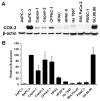
Relative expression of COX-2 in PA cell lines. Basal expression of COX-2 was determined by Western blot analysis in PA cell line lysates and quantified by densitometry. A: Representative Western blot. All cell lines were acquired from the American Type Culture Collection (ATCC, Manassas, VA) and propagated in ATCC recommended media. Cells were grown to 80% confluence before preparation of cell lysates. Proteins were separated by SDS-polyacrylamide gel electrophoresis and transferred to PVDF membranes using standard protocols. The membranes were probed with antibodies to COX-2 (mouse monoclonal, Cayman Chemical, Ann Arbor, MI) and β-actin (rabbit monoclonal, Cell Signaling Technology, Danvers, MA) or GAPDH (mouse monoclonal, Novus Biologicals, Littleton, CO) as total protein loading controls. B: Densitometric analyses. Autoradiographs of Western blots were quantified using ImageJ software (National Institutes of Health,
). COX-2 intensity was first normalized to the corresponding loading control (β-actin or GAPDH) and then normalized to the BxPC-3 ratio for each blot. Data represents the combined results of four independent experiments (mean ± SEM).
Similar articles
- Genomic sequencing of key genes in mouse pancreatic cancer cells.
Wang Y, Zhang Y, Yang J, Ni X, Liu S, Li Z, Hodges SE, Fisher WE, Brunicardi FC, Gibbs RA, Gingras MC, Li M. Wang Y, et al. Curr Mol Med. 2012 Mar;12(3):331-41. doi: 10.2174/156652412799218868. Curr Mol Med. 2012. PMID: 22208613 Free PMC article. - Clinical Effect of Driver Mutations of KRAS, CDKN2A/P16, TP53, and SMAD4 in Pancreatic Cancer: A Meta-Analysis.
Gu Y, Ji Y, Jiang H, Qiu G. Gu Y, et al. Genet Test Mol Biomarkers. 2020 Dec;24(12):777-788. doi: 10.1089/gtmb.2020.0078. Genet Test Mol Biomarkers. 2020. PMID: 33347393 - [Genetic aspects of pancreatic cancer].
Grigor'eva IN, Efimova OV, Suvorova TS, Tov NL. Grigor'eva IN, et al. Eksp Klin Gastroenterol. 2014;(10):70-6. Eksp Klin Gastroenterol. 2014. PMID: 25911935 Review. Russian. - Clinical significance of the genetic landscape of pancreatic cancer and implications for identification of potential long-term survivors.
Yachida S, White CM, Naito Y, Zhong Y, Brosnan JA, Macgregor-Das AM, Morgan RA, Saunders T, Laheru DA, Herman JM, Hruban RH, Klein AP, Jones S, Velculescu V, Wolfgang CL, Iacobuzio-Donahue CA. Yachida S, et al. Clin Cancer Res. 2012 Nov 15;18(22):6339-47. doi: 10.1158/1078-0432.CCR-12-1215. Epub 2012 Sep 18. Clin Cancer Res. 2012. PMID: 22991414 Free PMC article. - Genetics and Biology of Pancreatic Ductal Adenocarcinoma.
Dunne RF, Hezel AF. Dunne RF, et al. Hematol Oncol Clin North Am. 2015 Aug;29(4):595-608. doi: 10.1016/j.hoc.2015.04.003. Epub 2015 Jun 10. Hematol Oncol Clin North Am. 2015. PMID: 26226899 Free PMC article. Review.
Cited by
- Asymmetrically Substituted Quadruplex-Binding Naphthalene Diimide Showing Potent Activity in Pancreatic Cancer Models.
Ahmed AA, Angell R, Oxenford S, Worthington J, Williams N, Barton N, Fowler TG, O'Flynn DE, Sunose M, McConville M, Vo T, Wilson WD, Karim SA, Morton JP, Neidle S. Ahmed AA, et al. ACS Med Chem Lett. 2020 Jul 16;11(8):1634-1644. doi: 10.1021/acsmedchemlett.0c00317. eCollection 2020 Aug 13. ACS Med Chem Lett. 2020. PMID: 32832034 Free PMC article. - Expression of CASC8 RNA in Human Pancreatic Cancer Cell Lines.
Burenina OY, Lazarevich NL, Kustova IF, Zatsepin TS, Rubtsova MP, Dontsova OA. Burenina OY, et al. Dokl Biochem Biophys. 2022 Aug;505(1):137-140. doi: 10.1134/S1607672922040020. Epub 2022 Aug 29. Dokl Biochem Biophys. 2022. PMID: 36038677 Free PMC article. - Multidrug transporter MRP4/ABCC4 as a key determinant of pancreatic cancer aggressiveness.
Sahores A, Carozzo A, May M, Gómez N, Di Siervi N, De Sousa Serro M, Yaneff A, Rodríguez-González A, Abba M, Shayo C, Davio C. Sahores A, et al. Sci Rep. 2020 Aug 26;10(1):14217. doi: 10.1038/s41598-020-71181-w. Sci Rep. 2020. PMID: 32848164 Free PMC article. - Antiproliferative and antimetabolic effects behind the anticancer property of fermented wheat germ extract.
Otto C, Hahlbrock T, Eich K, Karaaslan F, Jürgens C, Germer CT, Wiegering A, Kämmerer U. Otto C, et al. BMC Complement Altern Med. 2016 Jun 1;16:160. doi: 10.1186/s12906-016-1138-5. BMC Complement Altern Med. 2016. PMID: 27245162 Free PMC article. - Molecular mechanism for USP7-mediated DNMT1 stabilization by acetylation.
Cheng J, Yang H, Fang J, Ma L, Gong R, Wang P, Li Z, Xu Y. Cheng J, et al. Nat Commun. 2015 May 11;6:7023. doi: 10.1038/ncomms8023. Nat Commun. 2015. PMID: 25960197 Free PMC article.
References
- Douglas EJ, Fiegler H, Rowan A, et al. Array comparative genomic hybridization analysis of colorectal cancer cell lines and primary carcinomas. Cancer Res. 2004;64:4817–4825. - PubMed
- Larramendy ML, Lushnikova T, Bjorkqvist AM, et al. Comparative genomic hybridization reveals complex genetic changes in primary breast cancer tumors and their cell lines. Cancer Genet Cytogenet. 2000;119:132–138. - PubMed
- Arumugam T, Simeone DM, Van Golen K, et al. S100P promotes pancreatic cancer growth, survival, and invasion. Clin Cancer Res. 2005;11:5356–5364. - PubMed
- Marchesi F, Monti P, Leone BE, et al. Increased survival, proliferation, and migration in metastatic human pancreatic tumor cells expressing functional CXCR4. Cancer Res. 2004;64:8420–8427. - PubMed
Publication types
MeSH terms
Substances
LinkOut - more resources
Full Text Sources
Other Literature Sources
Medical
Research Materials
Miscellaneous
