The therapeutic effects of recombinant adenovirus RA538 on human gastric carcinoma cells in vitro and in vivo (original) (raw)
Original Articles Open Access
Copyright ©The Author(s) 2000. Published by Baishideng Publishing Group Inc. All rights reserved.
World J Gastroenterol. Dec 15, 2000; 6(6): 855-860
Published online Dec 15, 2000. doi: 10.3748/wjg.v6.i6.855
The therapeutic effects of recombinant adenovirus RA538 on human gastric carcinoma cells in vitro and in vivo
Jie Ping Chen, Cai Pu Xu, Department of Gastroenterology, Southwest Hospital, The Third Military Medical University, Chongqing 400038, China
Chen Lin, Xue Yan Zhang, Ming Wu, National Laboratory of Molecular Oncology, Department of Cell Biology, Cancer Institute, Peking Union Medical College (PUMC) & Chinese Academy of Medical Sciences (CAMS), Beijing 100021, China
Jie Ping Chen, graduated from Third Military Medical University as a Ph.D. postgraduate in 1998, postdoctoral fellow in Second Affiliated Hospital of Chongqing University of Medical Sciences, associate professor of gastroenterology, major in
Helicobacter pylori
infection and gastric cancer, having 30 papers published.
ORCID number: $[AuthorORCIDs]
Author contributions: All authors contributed equally to the work.
Supported by the National 863 Science and Technology Fund of China, No.Z20-01-02
Correspondence to: Dr. Jie Ping Chen, Department of Gastroenterology, Southwest Hospital, Third Military Medical University, Chongqing 400038, China. Email: jpchen@public.cta.cq.cn
Telephone: 0086-23-65318301 Ext.73094
Received: April 3, 2000
Revised: April 19, 2000
Accepted: April 26, 2000
Published online: December 15, 2000
Abstract
AIM: To evaluate the potential of RA-538 gene therapy for gastric carcinoma.
METHODS: Human gastric carcinoma cell line SGC7901 treated with Ad-RA538 or Ad-LacZ were analysed by X-gal stain, MTT, DNA ladder, Tunel, flow cytometric analysis, PCR, and Western Blot in vitro. The tumorigenicity and experimental therapy in nude mice model were assessed in vivo.
RESULTS: Ad-LacZ could efficiently transfer the LacZ gene into SGC7901 cells. X-gal-positive cells at MOI 25, 50, 100, and 200 were 90%, 100%, 100%, and 100% respectively. Ad RA538 could strongly inhibit cell growth and induced apoptosis in SGC7901 cells. The proliferation of the Ad-RA538-in fected SGC7901 cells was reduced by 76.3%. The mechanism of killing of gastric carcinoma cells by Ad-RA538 was found to be apoptosis by DNA ladder, Tunel and flow cytometric analysis. The tumorigenicity in nude mice using Ad-RA538 showed that all three mice failed to form tumor from 7 to 30 d compared with Ad-LacZ and parent SGC7901 cells. Experimental therapy on the nude mice model bearing subcutaneous tumor of SGC7901 cells showed that intratumor instillation of Ad-RA538 inhibited the growth of the tumors. Ad-RA538-treated tumors were inhibited by 60.66%, compared with that of the tumor injected with Ad-LacZ and mock.
CONCLUSION: The expression of Ad-RA538 can inhibit growth and induce apoptosis of gastric cancer cell in vitro and in vivo. Ad-RA538 can be used potentially in gene therapy for gastric carcinoma.
- Citation: Chen JP, Lin C, Xu CP, Zhang XY, Wu M. The therapeutic effects of recombinant adenovirus RA538 on human gastric carcinoma cells in vitro and in vivo. World J Gastroenterol 2000; 6(6): 855-860
- URL: https://www.wjgnet.com/1007-9327/full/v6/i6/855.htm
- DOI: https://dx.doi.org/10.3748/wjg.v6.i6.855
INTRODUCTION
Gastric carcinoma is one of the most common malignant tumors in the world. It is treatable by surgical resection in the early stages, but advanced gastric carcinoma does not usually respond to conventional therapy[1-6]. Therefore gene therapy represents an attractive alternative for the treatment of gastric carcinoma. Present studies, suggest that overexpression of oncogenies, with or without functional loss of tumor suppressor genes, is responsible for the progression of human malignancies through multistep processes[7-13]. On the basis of this multiple hit model of carcinogenesis, cancer gene therapy has rapidly developed as alternative to conventional cancer therapy. It is widely accepted that c-myc gene plays a pivotal role in regulating cell proliferation and differentiation. Some studies indicated that c-myc gene has a close relationship with carcinogenesis[14-16]. Using subtr active hybridization strategy, RA538 was isolated, a cDNA colon from a human eso phageal cancer cell line before and after RA-treatment[17]. It is proved that RA538 can induce differentiation and apoptosis of tumor cell and down-regulate c-myc gene expression[18-21]. In the present study, we treated a human gastric carcinoma cell line with adenovirus-recombinants carrying RA538 or LacZ gene.
MATERIALS AND METHODS
Cell lines and culture
Human gastric carcinoma cell line (SGC 7901) was obtained from the Academy of Military Medical Sciences, China. Human embryonic kidney cell line (293) was kindly provided by Professor Zhan Qi-Ming (Academy of Sciences, China). SGC 7901 cells were grown in RPMI 1640 medium supplemented with 10% fetal bovine serum. Two hundred and ninety-three cells were cultured in Dulbecco’s modified Eagle’s medium (DMEM) supplemented with 10% fetal bovine serum.
Recombinant adenovirus production
The recombinant, replication-deficient type 5 adenoviruses that have been deve loped for gene therapy contained deletions of the E1 and E3 regions. The vector was constructed by homologous recombination in 293 cells of the PJM17 plasmid containing the right end of Ad5 (Microbix Biosystems Inc., Canada) and a plasmid containing 0-16 m.u. of left end of pAd CMV (a gift from Zhang Wei-Wei). A recombination adenovirus vector, called Ad-RA538 containing the human RA-5 38 cDNA fragment, the total 3.8 kb of RA538 suppressor gene was constructed. Ad-RA538 and Ad-LacZ were constructed by National Laboratory of Molecular Oncology, Department of Cell Biology, Chinese Academy of Medical Sciences. The resulting vector was plaque-purified twice on 293 cells and propagated. Virus stocks were titered by plaque-forming assay on 293 cells. Ad-RA538 and Ad-LacZ were identified by polymerase chain reaction (PCR).
Recombinant adenovirus infections of the cell lines were carried out by dilution of viral stock to certain concentrations, addition of viral solution to cell monolayers (0.5 mL per 6 cm dish), and incubation at room temperature for 30 min with agitation every 10 min. This was followed by addition of culture medium and the return of the infected cells to the 37 °C incubator.
Adenovirus transduction efficiency
Gastric carcinoma cells were seeded in 6 cm culture plates at a density of 1 × 10⁶ cells/dish and cultured 12 h. Cells were infected with Ad-LacZ at a multiplicity of infection with 25, 50, 100 and 200 (MOI). After 48 h, the cells were washed with phosphate-buffered saline (PBS), treated with 5-bromo-4-chloro-3-indolyl-β-D-galactopyranoside (X-gal). X-gal-positive cells were revealed by a blue precipitate in the cell.
Assay of cell growth
Cells were seeded at 8 × 10³ cells/well in 96-well plates and were infected with either Ad-RA538, Ad-LacZ and PBS (Mock) at 25, 50, 100 and 200 MOI within 12-24 h. At different time after adenoviral infection, 3-2,5-diphenyl tetrazolium bromide (MTT) assay was performed. Cell viability is proportional to the absorbance at the test wave length (525 nm).
DNA extraction and gel electrophoresis[22]
Cells were lysed in gunaidine-isothiocyanate and DNA was extracted by benzyle chloride using Herrmann M’methord[5]. RNA was digested with 1 μg/mL RNaseA 50 μg of each DNA sample was loaded on to a 18 g/L agarose gel.
TUNEL assay
Air dried cell samples were fixed with parafor-maldehyde solution for 30 min. Slides were rinsed with PBS and incubated with blocking solution (0.3% H2O2 in methanol) for 30 min. Slides were rinsed with PBS and incubated in permeabilisation solution (0.1% Triton X-100 in 0.1% sodium citrate) for 2 min in ice. TUNEL reaction mixture were added with 50 μL in sample. Slides were incubated in a humidified chamber for 60 min at 37 °C, and analysed under a fluorescence microscope. Coverter-POD were added with 50 μL in sample. Slides were incubate in a humidified chamber for 30 min at 37 °C. Slides were rinsd with PBS for three times. DAB substate solution were added with 50 μL-100 μL for 15 min. Slides were rinsd with PBS for three times and were stained with hemotolin. Slides were mountd under glass coverslip and analysed under light microscope.
Flow cytometry
The cells were fixed in 70% ethanol, treated with 0.1 g/L Rnase A, and with 100 mg/L propidium iodide. Cell cycle phase distribution were analysed by flow cytometry (Coulter Epice, ELITE, ESP, USA).
Polymerase chain reaction
The polymerase chain reaction (PCR) was designed to amplify a 800 bp fragment of the adenovirus gene using primer (5’-primer TCGTTTCTCAGCAGCTGTTG; 3’-primer CATCTGAACTCAAAGCGTGG) and a 300 bp fragment of the RA538 gene using primer (5’-primer ATGGGTGAACAACAGAAGAG;3’-primer TTAGAGATTCAGATTTGGCT). Hot-start PCR amplification was performed. In a Pekrin-Elmer 2400 thermocydcer using the following program: 1 × 95 °C for 5 min; 30 × 95 °C for 1 min; 59 °C for 50 s; 72 °C for 50 s; and 1 × 72 °C for 7 min. The final concentration for all PCR components in a 25 μL volume was as follows: 100 μM of each of extracted genomic DNA and 1 unit of Taq polymerase in 1 ×Taq polymerase buffer. PCR products were run on 1% agarose gels.
Western blot analysis
Total cell lysated were prepared by lysing the cell with sodium dodecyle sulfate polyacrylamide gel electrophoresis (SDS-PAGE) sample buffer after rinsing the cells with PBS. For the SDS-PAGE analysis, each lane was loaded with cell lysates equivalent to 6 × 10⁴ cells (30 μL). The protein in the gel were transferred to nitrocellulose filter (NC filter, Bio-Rad Company, California, USA). The membranes were blocked with 0.5% dry milk in PBS. The primary antibodies used were mouse anti-human c-myc or β-actin monoclonal antibody (Santa Cruz, California, USA), and the secondary antibody was horseradish peroxidase-conjugated rabbit anti-mouse IgG (Amersham, Uppsala, Sweden). The membranes were hybridized according to the Amersham’s ECL protocol. The membrane was exposed to X-ray film.
Tumorigenicity assay of SGC7901 cell growth after treatment with Ad-RA538
The gastric cancer cells were infected with Ad-RA538 and Ad-LacZ at a dose of 100 MOI. An equal number of cells was treated with PBS and mock infection. Twenty-four hours after infection, the treated cells were harvested and rinsed with PBS. For each treatment, 10.6 cells in 0.1 mL were injected to BALB/c male nude mice. Tumor formation was evaluated after 4 wk.
Adenovirus treatment in vivo
Mice (BALB/c nude mice aged 5 wk) were inoculated with 106 SGC 7901 cells into the flank. Tumors were allowed to grow to 5 mm in diameter. The animals were divided into three groups: Ad-RA538 injection; Ad-LacZ injection and PBS injection. There were five nude mice in each group. Ad-RA538 or Ad-LacZ (1 × 10⁹ pfu/each/100 μL) or PBS/100 μL were directly injected into the tumor centre of each nude mouse at d1, d3 and d5. Tumor sizes were observed 4 wk after the injection, and estimated with calipers.
Statistics
Data are presented as means ± standard errors of the means. Comparisons among different groups of samples were made by two-tailed t test and χ² test.
RESULTS
Recombinant adenovirus prediction
Ad-RA538 and Ad-LacZ were propagated on 293 cells. Their titers were 3.0 × 10⁹ pfu/mL and 3.0 × 10¹¹ pfu/mL respectively. Ad-RA538 and Ad-LacZ were identified by PCR. Ad-RA538 showed expression of RA538 gene. Ad-LacZ showed expression of adenovirus gene.
Adenovirus transfection efficiency in SGC7901 cell line
The time course of β-galactosidase (β-Gal) expression was first determined by counting the percentage of X-Gal-positive cells at 48 h after infection with Ad-LacZ. Ad-LacZ could efficiently transfer the LacZ gene into SGC7901 cells. And X-gal-positive cells at MOI 25, 50, 100 and 200 were 90%, 100% 100% and 100% respectively.
Inhibition of SGC 7901 cells growth
The degree of growth inhibition was measured by MTT assay. The growth rates of Ad-RA538 infected SGC 7901 cells were inhibited by 76.3% (MOI 200, 8 d) as compared to Ad-LacZ and Mock (P < 0.01) (Table 1). Ad-RA538 induced apoptosis in SGC 7901 cells.
Table 1 Anti-proliferative effect of Ad-RA538 on SGC7901 cells (8 d, ¯x ± s).
| MOI | Ad-RA538 | Ad-LacZ | Mock | |||
|---|---|---|---|---|---|---|
| A | Survival (%) | A | Survival (%) | A | Survival (%) | |
| 25 | 1.062±0.05 | 95.0±4.5 | 1.120±0.077 | 100±6.9 | 1.31±0.065 | 100±6.3 |
| 50 | 0.941±0.067 | 84.0±6.0 | 0.984±0.024 | 87.5±2.1 | 1.29±0.034 | 100±5.2 |
| 100 | 0.534±0.091 | 47.4±8.1 a | 1.195±0.095 | 100±8.5 | 1.28±0.041 | 99.8±7.0 |
| 200 | 0.265±0.090 | 23.7±8.0 b | 1.241±0.091 | 100±8.0 | 1.23±0.057 | 98.0±4.5 |
DNA fragmentation
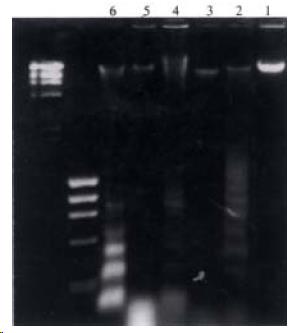
SGC7901 cells were treated with MOI 100 Ad-RA538 for 2, 4 and 6 d. Figure 1 shows that the electrophoresis pattern after treatment with Ad-RA538. DNA fragmentation became apparent at d2, d4 and d6. The peak of ladder pattern was detected at d6. Ladder pattern was not detected in Ad-LacZ treated cells.
Figure 1 DNA ladder of Ad-RA538 on SGC7901 cells. lane 1: Ad-lac Z (2d); lane 2: Ad-RA538 (2d);lane 3: Ad-lacZ (4d); lane 4: Ad RA538 (4d);lane 5: Ad-lacZ (6d); lane 6: Ad-RA538 (6d).
TUNEL assay
SGC 7901 cells treated with Ad-RA538 were assayed for apoptosis by TUNEL. SGC 7901 cells could induce apoptosis. The abnormal chromatin clumps, nuclear membrane wrinkling, nuclear collapse, cytoplasm bubble and cytomembrane wrinkling had appeared after treatment with Ad-RA538. Apoptotic cells were not shown by TUNEL assay in Ad-LacZ treated cells. In this kit terminal deoxynucleotidyl transferase, which catalyzes polymerization of nucleotides to free 3’-OH DNA ends in a template-independent manner, was used to label DNA strand breaks. After substrate reaction, stained cells can be analyzed under light microscope (Figure 2).
Figure 2 TUNEL assay of Ad-RA538 and Ad-LacZ on SGC 7901 cells.
Flow cytometric analysis
A flow cytometric analysis was performed on SGC 7901 cells infected with Ad-RA 538 and Ad-LacZ. Apoptotic cells of Ad-RA538 infected cells played a peak in the flow cytometry histogram. Apoptotic peak was 34.2% at d2. The cell cycle G-2M arrest were shown at d2, d4 and d6. The apoptosis peak and cell cycle arrest were not found in Ad-LacZ treated cells (Table 2).
Table 2 Flow cytometry analysis of cell cycle effects of Ad-RA538 on SGC7901 (%).
| Type of adenovirus | 12 h | 1 d | 2 d | 4 d | 6 d | ||||||||||
|---|---|---|---|---|---|---|---|---|---|---|---|---|---|---|---|
| G1 | G2M | S | G1 | G2M | S | G1 | G2M | S | G1 | G2M | S | G1 | G2M | S | |
| Ad-RA538 | 74.6 | 13.0 | 12.4 | 55.7 | 28.2 | 17.2 | 69.9 | 14.0a | 16.1 | 39.7 | 26.6b | 33.8 | 44.4 | 20.9c | 34.7 |
| Ad-LacZ | 66.5 | 15.3 | 18.2 | 59.2 | 18.1 | 22.7 | 59.5 | 22.8 | 17.3 | 71.0 | 12.4 | 16.6 | 73.3 | 8.4 | 18.3 |
c-myc expression in Ad-RA538 infected cells
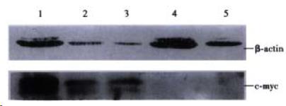
The expression of c-myc protein in SGC 7901 cells was detected by western blot analysis after Ad-RA538 infection for 1, 3, 5 or 7 d. Ad-RA538 may down-regulate expression of c-myc gene (Figure 3).
Figure 3 Western blot analysis c-myc and β-actin expression of SGC7901 cell infected with Ad-RA538. Lane 1: control, Lane 2-5: 1d, 3d, 5d, 7d.
Inhibition of tumor growth in vivo
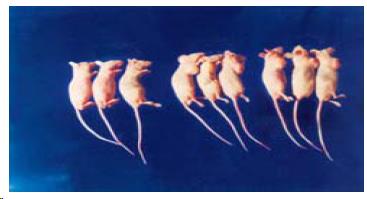
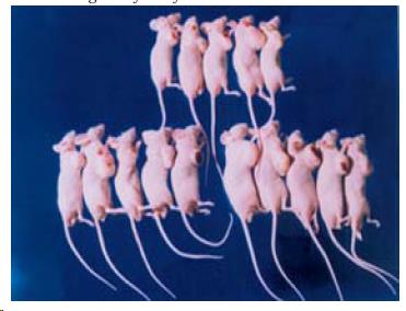
The tumori genicity in nude mice of using Ad-RA538 showed that three of these mice failed to form tumor from d7 to d30compared with Ad-LacZ and parent SGC 7901 cells (P < 0.01) (Figure 4). The tumorigenicity in nude mice using Ad-LacZ and parent SGC 7901 cells was 100%.
Figure 4 Tumorigenicity assay in nude mice follow AdRA538.
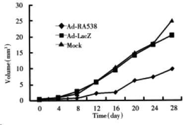
Experimental therapy on the nude mice model bearing subcutaneous tumor of SGC 7901 cell showed that intratumor instillation of Ad-RA538-treated tumor were inhibited by 60.66%, compared with that of the tumor injected with Ad-lacZ and mock (Figure 5, Figure 6).
Figure 5 In vivo therapy of Ad-RA538 on SGC7901 tumors in nude mice.
Figure 6 Experimental therapy on the nude mice model of AdRA538.
DISCUSSION
The ability to infect numerous different cell types and the absence of requirement for dividing cells make adenovirus an attractive candidate for in vivo gene therapy[23]. They can be grown to a very high titer, and can be easily concentrated to reach titers of 1013-1014 particles per milliliter. Adenoviral vectors are very effective agents for gene transfer with extremely high transduction efficiency for a wide variety of cell types. In this study, adenovirus vector has high transduction efficiency for SGC 7901 cells, and can mediate a high level of expression of interested gene in transducted cells. The percentage of X-gal staining positive SGC 7901 cells 48 h after infection with Ad-LacZ was 90% at MOI 25. Ad-RA538 could strongly inhibit growth and induce apoptosis of SGC 7901 cells. It may be related to high trasduction efficiency of adenovirus for SGC 7901 cells.
It is known that activation of proto-oncogene and inactivation of tumor suppressive gene are the most common genetic alternation in tumor[24-33]. Using subtractive hybridization strategy, RA538 was isolated, a cDNA colon from a human esophageal cancer cell line before and after retinoic acid (RA)treatment. RA can induce differentiation and apoptosis of tumor cell[34-37]. It is proved that RA538 can induce differentiation and apoptosis of tumor cell and down-regulate c-myc gene expression[17,18,38-41]. Reducing the expression of c-myc may effectively suppress the proliferation and malignant phenotype of cancer cells[42-48] and to counter c-myc gene amplification in gastric carcinoma cells[49]. The effects of Ad-RA538 on human gastric carcinoma cell line were examined both in vitro and in vivo. Ad-RA538, containing the human RA538 cDNA fragment, was constructed. Firstly, Ad-LacZ could transfer the LacZ gene into more than 90% of gastric carcinoma cells. Ad-RA538 could successfully inhibit expression of c-myc protein. These data showed the capability of adenovirus to transfer exogenous genes efficiently into gastric carcinoma cell line. Secondly, the growth inhibitory effect of Ad-RA538 was then examined. In c-myc gene amplification gastric carcinoma cell line SGC7901, the growth inhibition by infection with Ad-RA538 correlated with both transduction efficiency and adenovirus dose. The growth rates of Ad-RA538 infected SGC 7901 cells were inhibited by 76.3%. DNA fragmentation, TUNEL and flow cytometric analysis suggested that Ad-RA538 could strongly induce apoptosis of gastric carcinoma cells. Some reported that tumor suppressive gene BCL-XS could inhibit the proliferation and malignant phenotype of cancer cells[50], Bouillellet[51] reported that stra 6, a retinoic acid-responsive gene could induce apoptosis of tumor cells. These are the same with our studies about Ad-RA538. Our studies with the tumorigenicity in nude mice and experimental therapy on the nude mice model using Ad-RA538 also supported these findings and showed that Ad-RA538 may be useful to inhibit the growth of gastric tumors. Our studies suggested that adenoviral-mediated RA538 overexpression may result in the elimination of tumor cells by apoptosis, and inhibition of proliferation, thus reducing the tumor burden. In conclusion, these findings indicated that the Ad-RA538 might promote efficient tumor cell death and inhibit tumor cell proliferation. The use of these vectors may be a potential tool for reducing tumor growth in vivo and treatment of gastric carcinoma.
Footnotes
Edited by You DY and Ma JY
References
| 1. | Zhao XH, Xue Y, Qian GS. Chemo-endocrine therapy for advanced gastric carcinoma and it’s clinical evaluation. Xin Xiaohuabingxue Zazhi. 1994;2:30. [PubMed] [DOI] [Cited in This Article: ] |
|---|
| 4. | Wang GT, Zhu JS, Xu WY, Wang Y, Zhou AG. Clinical and experimental studies on Fuzheng anti cancer granula combined with chemotherapy in advanced gastric cancer. Huaren Xiaohua Zazhi. 1998;6:214-218. [PubMed] [DOI] [Cited in This Article: ] |
|---|
| 5. | Tong ZM. Relationship between lymph node metastasis and postoperative survival in gastric cancer. Huaren Xiaohua Zazhi. 1998;6:224-226. [PubMed] [DOI] [Cited in This Article: ] |
|---|
| 6. | Zhao ZS, Wang SW, Jiao XL, Chen JH. Influence of radical gastrectomy on single cancer cells to blood metastases. Huaren Xiaohua Zazhi. 1998;6:610-611. [PubMed] [DOI] [Cited in This Article: ] |
|---|
| 8. | Cheng LY, Gao Y, Yang JZ. Growth inihbition of human gastric carcinoma cells by DNA polymerase α antisenseoligodeoxynucleotide. Xin Xiaohuabingxue Zazhi. 1996;4:666-668. [PubMed] [DOI] [Cited in This Article: ] |
|---|
| 9. | Shi XY, Zhao FZ. Molecular biological study on gastric precancerous lesions. Huaren Xiaohua Zazhi. 1998;6:74-75. [PubMed] [DOI] [Cited in This Article: ] |
|---|
| 10. | Shi XQ, Li G, Li CS, Ni CR, Luan X, Qu Y. Oncogene ras, c-myc mRNA expression in gastric carcinoma and its clinicalsignificance. Huaren Xiaohua Zazhi. 1998;6:123-124. [PubMed] [DOI] [Cited in This Article: ] |
|---|
| 11. | Wang YK, Ji XL, Ma NX. Expressions of p53 bcl-2 and c-erbB-2 proteins in precarcinomatous gastric mucosa. Shijie Huaren Xiaohua Zazhi. 1999;7:114-116. [PubMed] [DOI] [Cited in This Article: ] |
|---|
| 12. | Zhao Y, Zhang XY, Shi XJ, Hu PZ, Zhang CS, Ma FC. Clinical significance of expressions of P16, p53 proteins and PCNA in gastric cancer. Shijie Huaren Xiaohua Zazhi. 1999;7:246-248. [PubMed] [DOI] [Cited in This Article: ] |
|---|
| 13. | Ryan KM, Birnie GD. Myc oncogenes: the enigmatic family. Biochem J. 1996;314:713-721. [PubMed] [DOI] [Cited in This Article: ] |
|---|
| 14. | Ryan KM, Birnie GD. Myc oncogenes: the enigmatic family. Biochem J. 1996;314:713-721. [PubMed] [DOI] [Cited in This Article: ] |
|---|
| 15. | Wang LD, Zhou Q, Wei JP, Yang WC, Zhao X, Wang LX, Zou JX, Gao SS, Li YX, Yang C. Apoptosis and its relationship with cell proliferation, p53, Waf1p21, bcl-2 and c-myc in esophageal carcinogenesis studied with a high-risk population in northern China. World J Gastroenterol. 1998;4:287-293. [PubMed] [DOI] [Cited in This Article: ] |
|---|
| 16. | Dai J, Yu SX, Qi XL, Bo AH, Xu YL, Guo ZY. Expression of bcl-2 and c-myc protein in gastric carcinoma and precancerouslesions. World J Gastroenterol. 1998;4:84-85. [PubMed] [DOI] [Cited in This Article: ] |
|---|
| 17. | Feng L, Wang XQ, Fu M, Wang ZH, Tian Y, Cai Y, Wu M. A strategy for isolating differentiation-inducing complementary DNAs from human esophageal cancer cell line treated with retinoic acid. Sci China B. 1992;35:445-454. [PubMed] [DOI] [Cited in This Article: ] |
|---|
| 18. | Yang X, Ding F, Zheng W. [Expression of cDNA RA538 induces terminal differentiation and apoptosis of its parental malignant cell line _in vitro_]. Zhongguo Yi Xue Ke Xue Yuan Xue Bao. 1994;16:251-254. [PubMed] [DOI] [Cited in This Article: ] |
|---|
| 19. | Chen JP. [In vitro and in vivo studies on the biologic effects and molecular mechanism of recombinant RA538 and antisense C-myc adenovirus on human gastric, esophageal and cancer cell lines with high-expression of Bcl-2 gene]. Sheng Li Ke Xue Jin Zhan. 1999;30:227-230. [PubMed] [DOI] [Cited in This Article: ] |
|---|
| 20. | Chen JP, Lin C, Xu CP, Zhang XY, Fu M, Deng YP, Cheng JK, Wu M. Studies on the molecular therapy with recombinant RA538 adenovirus in human gastric cancer cells. Disan Junyi Daxue Xuebao. 1999;21:309-313. [PubMed] [DOI] [Cited in This Article: ] |
|---|
| 21. | Chen J, Lin C, Xu C, Zhang X, Fu M, Wu M. The effects of recombinant RA538 and antisense c-myc adenovirus on tumor cells and the molecular mechanism concerned. Zhonghua Yi Xue Yi Chuan Xue Za Zhi. 2000;17:164-168. [PubMed] [DOI] [Cited in This Article: ] |
|---|
| 22. | Herrmann M, Lorenz HM, Voll R, Grünke M, Woith W, Kalden JR. A rapid and simple method for the isolation of apoptotic DNA fragments. Nucleic Acids Research. 1994;22:5506-5507. [PubMed] [DOI] [Cited in This Article: ] |
|---|
| 24. | Zhang SL, Chen HY, Zhang DQ, Liu WH. C_erbB_2 oncogene and MG_3c7 antigen experssion in gastric carcinoma. Xin Xiaohuabingxue Zazhi. 1995;3:195-196. [PubMed] [DOI] [Cited in This Article: ] |
|---|
| 25. | Meng FJ, Dai WS, Pan BR. _p_53 genes expression and its correlation with metastasis and prognosis in gastric carcinoma. Xin Xiaohuabingxue Zazhi. 1996;4:677-678. [PubMed] [DOI] [Cited in This Article: ] |
|---|
| 26. | Zhao LJ, Liu TQ, Xing GY. Expression of p53 protein and PCNA/cyclin in advanced gastric cancer. Xin Xiaohuabingxue Zazhi. 1996;4:38-40. [PubMed] [DOI] [Cited in This Article: ] |
|---|
| 27. | Song ZY, Xu RZ, Qian KD, Tang XQ, Zhao XY, Lin M. Abnormal expression of p16/CDKN2 gene at protein level in humangastric cancer. Xin Xiaohuabingxue Zazhi. 1997;5:139-140. [PubMed] [DOI] [Cited in This Article: ] |
|---|
| 28. | Mi JQ, Yang SQ, Shen MC. The expression of c-erbB-2 proto-oncogene product in gastric carcinoma and precancerouslesions. Xin Xiaohuabingxue Zazhi. 1997;5:152-153. [PubMed] [DOI] [Cited in This Article: ] |
|---|
| 29. | Shi XQ, Li G, Ni CR, Li CS, Luan X, Qu Y. Gastric carcinoma metastasis suppressor gene nm23_H1 mRNA and its significance. Xin Xiaohuabingxue Zazhi. 1997;5:377-378. [PubMed] [DOI] [Cited in This Article: ] |
|---|
| 30. | Wang JY, Jin ML, Lu YY, Li JY. Alteration of p53 suppression gene in gastric mucosa and carcinogenesis. Xin Xiaohuabingxue Zazhi. 1997;5:429-430. [PubMed] [DOI] [Cited in This Article: ] |
|---|
| 31. | Zou JX, Chen YL, Wang ZH, Wang LD, Zhou Q, Zhao X. Correlation between alteration of tumor suppressor gene p53, p16 and biological behavior of gastriccancer. Xin Xiaohuabingxue Zazhi. 1997;5:775-776. [PubMed] [DOI] [Cited in This Article: ] |
|---|
| 32. | Mao LZ, Wang SX, Ji WF, Ren JP, Du HZ, He RZ. Comparative studies on p53 and PCNA expressions in gastric carcinoma between young and aged patients. Huaren Xiaohua Zazhi. 1998;6:397-399. [PubMed] [DOI] [Cited in This Article: ] |
|---|
| 34. | Giandomenico V, Lancillotti F, Fiorucci G, Percario ZA, Rivabene R, Malorni W, Affabris E, Romeo G. Retinoic acid and IFN inhibition of cell proliferation is associated with apoptosis in squamous carcinoma cell lines: role of IRF-1 and TGase II-dependent pathways. Cell Growth Differ. 1997;8:91-100. [PubMed] [DOI] [Cited in This Article: ] |
|---|
| 36. | Chen Y, Xu CF. All trans retinoic acid induced differentiation in human gastric carcinoma cell line SGC-7901. Xin Xiaohuabingxue Zazhi. 1997;5:491-492. [PubMed] [DOI] [Cited in This Article: ] |
|---|
| 38. | Chen JP, Lin C, Xu CP, Wu M. Studies on the biologic effects and molecular mechanism of recombinant RA538, antisense c-myc adenovirus on human gastric, esophageal and high-expression of bcl-2 gene cancer cell lines in vitro and in vivo. Chongqing Yixue. 1998;23:430. [PubMed] [DOI] [Cited in This Article: ] |
|---|
| 39. | Chen JP, Lin C, Xu CP, Zhang XY, Fu M, Deng YP, Wei Y, Wu M. Transduction efficiency, biologic effects and mechanismof recombinant RA538, antisense C-myc adenovirus on different cell lines. Shijie Huaren Xiaohua Zazhi. 2000;8:266-270. [PubMed] [DOI] [Cited in This Article: ] |
|---|
| 40. | Chen JP, Lin C, Xu CP, Zhang XY, Fu M, Wei Y, Deng YP, Wu M. Transduction efficiency of recombinant RA538, antisense C-myc adenovirus on different cell lines and molecular mechanism. Zhongguo Zhongliu Shengwu Zhiliao Zazhi. 2000;7:63-64. [PubMed] [DOI] [Cited in This Article: ] |
|---|
| 41. | Chen JP, Lin C, Xu CP, Zhang XY, Wei Y, Wu M. Effects of recombinant RA538, antisense c-myc adenovirus on human normal cells. Disan Junyi Daxue Xuebao. 1999;21:712-715. [PubMed] [DOI] [Cited in This Article: ] |
|---|
| 43. | Leonetti C, D'Agnano I, Lozupone F, Valentini A, Geiser T, Zon G, Calabretta B, Citro G C, Zupi G. Antitumor effect of c-myc antisense phosphorothioate oligodeoxynucleotides on human melanoma cells in vitro and and in mice. J Natl Cancer Inst. 1996;88:419-429. [PubMed] [DOI] [Cited in This Article: ] |
|---|
| 44. | Chen JP, Lin C, Xu CP, Zhang XY, Fu M, Deng YP, Kui Y, Wu M. In vitro and in vivo molecular therapy with AS c-mycadenovirus for human gastric carcinoma cell line. Shijie Huaren Xiaohua Zazhi. 1999;7:482-486. [PubMed] [DOI] [Cited in This Article: ] |
|---|
| 45. | Chen JP, Lin C, Xu CP, Zhang XY, Fu M, Deng YP, Wei Y, Wu M. Gene the rapy with antisense c-myc adenovirus for human gastric carcinoma cell line in vitro and for implanted carcinoma in nude mice. J Med CPLA. 2000;15:111-114. [PubMed] [DOI] [Cited in This Article: ] |
|---|
| 46. | Liu Y, Lu MZ, Li QM, Wang YL. The expression of p53 C-myc and P-gp proteins in gastric cancer. Xin Xiaohuabingxue Zazhi. 1997;5:585-586. [PubMed] [DOI] [Cited in This Article: ] |
|---|
| 47. | Li XL, Hao YR, Zou JX, Yang JH, Geng JH. Relationship between C-myc and Bcl-2 alterations and biological behavior and apoptosis in gastric cancer. Xin Xiaohuabingxue Zazhi. 1997;5:773-774. [PubMed] [DOI] [Cited in This Article: ] |
|---|
| 48. | Chen J, Lin C, Deng Y. [The effects of RA538 and antisense c-myc on cervical cancer cell lines with high expression of bcl-2 gene]. Zhonghua Zhong Liu Za Zhi. 2000;22:279-282. [PubMed] [DOI] [Cited in This Article: ] |
|---|
| 49. | Onoda N, Maeda K, Chung YS, Yano Y, Matsui-Yuasa I, Otani S, Sowa M. Overexpression of c-myc messenger RNA in primary and metastatic lesions of carcinoma of the stomach. J Am Coll Surg. 1996;182:55-59. [PubMed] [DOI] [Cited in This Article: ] |
|---|
| 50. | Clarke MF, Apel IJ, Benedict MA, Eipers PG, Sumantran V, González-García M, Doedens M, Fukunaga N, Davidson B, Dick JE. A recombinant bcl-x s adenovirus selectively induces apoptosis in cancer cells but not in normal bone marrow cells. Proc Natl Acad Sci USA. 1995;92:11024-11028. [PubMed] [DOI] [Cited in This Article: ] |
|---|
| 51. | Bouillet P, Sapin V, Chazaud C, Messaddeq N, Décimo D, Dollé P, Chambon P. Developmental expression pattern of Stra6, a retinoic acid-responsive gene encoding a new type of membrane protein. Mech Dev. 1997;63:173-186. [PubMed] [DOI] [Cited in This Article: ] |
|---|
