FIS1 (original) (raw)
From Wikipedia, the free encyclopedia
Protein-coding gene in the species Homo sapiens
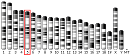
Mitochondrial fission 1 protein (FIS1) is a protein that in humans is encoded by the FIS1 gene on chromosome 7.[5][6][7] This protein is a component of a mitochondrial complex, the ARCosome, that promotes mitochondrial fission.[7][8] Its role in mitochondrial fission thus implicates it in the regulation of mitochondrial morphology, the cell cycle, and apoptosis.[7][8][9][10] By extension, the protein is involved in associated diseases, including neurodegenerative diseases and cancers.[11][12]
The protein encoded by this gene is a 16 kDa integral protein situated in the outer mitochondrial membrane (OMM).[9] It is composed of a transmembrane domain at the C-terminal and a cytosolic domain at the N-terminal.[9][13][14] The transmembrane domain anchors FIS1 in the OMM, though it has been observed to target different cellular compartments, such as the peroxisome, depending on its hydrophobicity, charge, and length.[14][15] Meanwhile, the cytosolic domain contains a bundle of six helices, four of which contain two tandem tetratricopeptide repeat (TPR)-like motifs. These motifs form a concave surface by their combined superhelical structure and potentially bind another FIS1 protein to form a dimer, or other proteins.[9][13] Moreover, the N-terminal arm can dock at, and thus obstruct, the TPR motifs, allowing the protein to exist in a dynamic equilibrium between "open" and "closed" states.[13]
FIS1 is indirectly involved in mitochondrial fission via binding dynamin-related protein 1 (DRP1).[12][15] By extension, FIS1 helps regulate the size and distribution of mitochondria in response to local demand for ATP or calcium ions.[13] In addition, mitochondrial fission may lead to release of cytochrome C, which eventually leads to cell death.[9]In a separate apoptotic signalling pathway, FIS1 interacts with BCAP31 to form a complex, the ARCosome. The ARCosome promotes cell death by bridging the mitochondria and the endoplasmic reticulum (ER), allowing FIS1 to transmit a proapoptotic signal from the mitochondria to the ER and activate procaspase-8. The ARCosome then forms a platform with procaspase-8 to increase calcium load in the mitochondria, resulting in apoptosis.[8][12]Additionally, FIS1 is involved in other modes of shaping mitochondrial morphology. For example, it interacts with TBC1D15 to regulate mitochondrial morphology, particularly with regard to lysosome and endosome fusion.[14] FIS1 also prevents mitochondria elongation, which would otherwise lead to cell cycle delay or arrest, and ultimately, senescence. Moreover, mitochondrial dysfunction results in elevated reactive oxygen species (ROS) levels, which cause DNA damage and induce transcriptional repression, as well as induce mitophagy.[9][10]
Clinical Significance
[edit]
As a fission factor, FIS1 is associated with neurodegenerative diseases.[11][12] Stress, such as NO, can trigger aberrant mitochondrial fission and fusion, resulting in mitophagy.[9][11] For example, increased mitochondrial fragmentation and FIS1 levels were observed in Alzheimer's disease (AD) patients. Thus, FIS1 could serve as a biomarker for early detection of AD.[11] FIS1 is also implicated in a variety of cancers, including acute myeloid leukemia, breast cancer, and prostate cancer.[12]
FIS1 has been shown to interact with:
- ^ a b c GRCh38: Ensembl release 89: ENSG00000214253 – Ensembl, May 2017
- ^ a b c GRCm38: Ensembl release 89: ENSMUSG00000019054 – Ensembl, May 2017
- ^ "Human PubMed Reference:". National Center for Biotechnology Information, U.S. National Library of Medicine.
- ^ "Mouse PubMed Reference:". National Center for Biotechnology Information, U.S. National Library of Medicine.
- ^ Stojanovski D, Koutsopoulos OS, Okamoto K, Ryan MT (Mar 2004). "Levels of human Fis1 at the mitochondrial outer membrane regulate mitochondrial morphology". Journal of Cell Science. 117 (Pt 7): 1201–10. doi:10.1242/jcs.01058. PMID 14996942.
- ^ Kong D, Xu L, Yu Y, Zhu W, Andrews DW, Yoon Y, Kuo TH (Apr 2005). "Regulation of Ca2+-induced permeability transition by Bcl-2 is antagonized by Drpl and hFis1". Molecular and Cellular Biochemistry. 272 (1–2): 187–99. doi:10.1007/s11010-005-7323-3. PMID 16010987. S2CID 21452703.
- ^ a b c "Entrez Gene: FIS1 fission 1 (mitochondrial outer membrane) homolog (S. cerevisiae)".
- ^ a b c d e Iwasawa R, Mahul-Mellier AL, Datler C, Pazarentzos E, Grimm S (Feb 2011). "Fis1 and Bap31 bridge the mitochondria-ER interface to establish a platform for apoptosis induction". The EMBO Journal. 30 (3): 556–68. doi:10.1038/emboj.2010.346. PMC 3034017. PMID 21183955.
- ^ a b c d e f g Gomes LC, Scorrano L (2008). "High levels of Fis1, a pro-fission mitochondrial protein, trigger autophagy". Biochimica et Biophysica Acta (BBA) - Bioenergetics. 1777 (7–8): 860–6. doi:10.1016/j.bbabio.2008.05.442. PMID 18515060.
- ^ a b Lee S, Park YY, Kim SH, Nguyen OT, Yoo YS, Chan GK, Sun X, Cho H (Feb 2014). "Human mitochondrial Fis1 links to cell cycle regulators at G2/M transition". Cellular and Molecular Life Sciences. 71 (4): 711–25. doi:10.1007/s00018-013-1428-8. PMC 11113609. PMID 23907611. S2CID 11694077.
- ^ a b c d Wang S, Song J, Tan M, Albers KM, Jia J (Jul 2012). "Mitochondrial fission proteins in peripheral blood lymphocytes are potential biomarkers for Alzheimer's disease". European Journal of Neurology. 19 (7): 1015–22. doi:10.1111/j.1468-1331.2012.03670.x. PMID 22340708. S2CID 21950507.
- ^ a b c d e Tian Y, Huang Z, Wang Z, Yin C, Zhou L, Zhang L, Huang K, Zhou H, Jiang X, Li J, Liao L, Yang M, Meng F (2014). "Identification of novel molecular markers for prognosis estimation of acute myeloid leukemia: over-expression of PDCD7, FIS1 and Ang2 may indicate poor prognosis in pretreatment patients with acute myeloid leukemia". PLOS ONE. 9 (1): e84150. Bibcode:2014PLoSO...984150T. doi:10.1371/journal.pone.0084150. PMC 3885535. PMID 24416201.
- ^ a b c d Lees JP, Manlandro CM, Picton LK, Tan AZ, Casares S, Flanagan JM, Fleming KG, Hill RB (Oct 2012). "A designed point mutant in Fis1 disrupts dimerization and mitochondrial fission". Journal of Molecular Biology. 423 (2): 143–58. doi:10.1016/j.jmb.2012.06.042. PMC 3456991. PMID 22789569.
- ^ a b c d Onoue K, Jofuku A, Ban-Ishihara R, Ishihara T, Maeda M, Koshiba T, Itoh T, Fukuda M, Otera H, Oka T, Takano H, Mizushima N, Mihara K, Ishihara N (Jan 2013). "Fis1 acts as a mitochondrial recruitment factor for TBC1D15 that is involved in regulation of mitochondrial morphology". Journal of Cell Science. 126 (Pt 1): 176–85. doi:10.1242/jcs.111211. PMID 23077178.
- ^ a b c Palmer CS, Elgass KD, Parton RG, Osellame LD, Stojanovski D, Ryan MT (Sep 2013). "Adaptor proteins MiD49 and MiD51 can act independently of Mff and Fis1 in Drp1 recruitment and are specific for mitochondrial fission". The Journal of Biological Chemistry. 288 (38): 27584–93. doi:10.1074/jbc.M113.479873. PMC 3779755. PMID 23921378.
- ^ Yoon Y, Krueger EW, Oswald BJ, McNiven MA (Aug 2003). "The mitochondrial protein hFis1 regulates mitochondrial fission in mammalian cells through an interaction with the dynamin-like protein DLP1". Molecular and Cellular Biology. 23 (15): 5409–20. doi:10.1128/MCB.23.15.5409-5420.2003. PMC 165727. PMID 12861026.
- Lai CH, Chou CY, Ch'ang LY, Liu CS, Lin W (May 2000). "Identification of novel human genes evolutionarily conserved in Caenorhabditis elegans by comparative proteomics". Genome Research. 10 (5): 703–13. doi:10.1101/gr.10.5.703. PMC 310876. PMID 10810093.
- James DI, Parone PA, Mattenberger Y, Martinou JC (Sep 2003). "hFis1, a novel component of the mammalian mitochondrial fission machinery". The Journal of Biological Chemistry. 278 (38): 36373–9. doi:10.1074/jbc.M303758200. PMID 12783892.
- Yoon Y, Krueger EW, Oswald BJ, McNiven MA (Aug 2003). "The mitochondrial protein hFis1 regulates mitochondrial fission in mammalian cells through an interaction with the dynamin-like protein DLP1". Molecular and Cellular Biology. 23 (15): 5409–20. doi:10.1128/MCB.23.15.5409-5420.2003. PMC 165727. PMID 12861026.
- Kikuchi M, Hatano N, Yokota S, Shimozawa N, Imanaka T, Taniguchi H (Jan 2004). "Proteomic analysis of rat liver peroxisome: presence of peroxisome-specific isozyme of Lon protease". The Journal of Biological Chemistry. 279 (1): 421–8. doi:10.1074/jbc.M305623200. PMID 14561759.
- Suzuki M, Jeong SY, Karbowski M, Youle RJ, Tjandra N (Nov 2003). "The solution structure of human mitochondria fission protein Fis1 reveals a novel TPR-like helix bundle". Journal of Molecular Biology. 334 (3): 445–58. doi:10.1016/j.jmb.2003.09.064. PMID 14623186.
- Dohm JA, Lee SJ, Hardwick JM, Hill RB, Gittis AG (Jan 2004). "Cytosolic domain of the human mitochondrial fission protein fis1 adopts a TPR fold". Proteins. 54 (1): 153–6. doi:10.1002/prot.10524. PMC 3047745. PMID 14705031.
- Frieden M, James D, Castelbou C, Danckaert A, Martinou JC, Demaurex N (May 2004). "Ca(2+) homeostasis during mitochondrial fragmentation and perinuclear clustering induced by hFis1". The Journal of Biological Chemistry. 279 (21): 22704–14. doi:10.1074/jbc.M312366200. PMID 15024001.
- Lee YJ, Jeong SY, Karbowski M, Smith CL, Youle RJ (Nov 2004). "Roles of the mammalian mitochondrial fission and fusion mediators Fis1, Drp1, and Opa1 in apoptosis". Molecular Biology of the Cell. 15 (11): 5001–11. doi:10.1091/mbc.E04-04-0294. PMC 524759. PMID 15356267.
- Koch A, Yoon Y, Bonekamp NA, McNiven MA, Schrader M (Nov 2005). "A role for Fis1 in both mitochondrial and peroxisomal fission in mammalian cells". Molecular Biology of the Cell. 16 (11): 5077–86. doi:10.1091/mbc.E05-02-0159. PMC 1266408. PMID 16107562.
- Yu T, Fox RJ, Burwell LS, Yoon Y (Sep 2005). "Regulation of mitochondrial fission and apoptosis by the mitochondrial outer membrane protein hFis1". Journal of Cell Science. 118 (Pt 18): 4141–51. doi:10.1242/jcs.02537. PMID 16118244. S2CID 21271384.
- Yoon YS, Yoon DS, Lim IK, Yoon SH, Chung HY, Rojo M, Malka F, Jou MJ, Martinou JC, Yoon G (Nov 2006). "Formation of elongated giant mitochondria in DFO-induced cellular senescence: involvement of enhanced fusion process through modulation of Fis1". Journal of Cellular Physiology. 209 (2): 468–80. doi:10.1002/jcp.20753. PMID 16883569. S2CID 22179820.
- Alirol E, James D, Huber D, Marchetto A, Vergani L, Martinou JC, Scorrano L (Nov 2006). "The mitochondrial fission protein hFis1 requires the endoplasmic reticulum gateway to induce apoptosis". Molecular Biology of the Cell. 17 (11): 4593–605. doi:10.1091/mbc.E06-05-0377. PMC 1635393. PMID 16914522.
- Kobayashi S, Tanaka A, Fujiki Y (May 2007). "Fis1, DLP1, and Pex11p coordinately regulate peroxisome morphogenesis". Experimental Cell Research. 313 (8): 1675–86. doi:10.1016/j.yexcr.2007.02.028. PMID 17408615.
- Lee S, Jeong SY, Lim WC, Kim S, Park YY, Sun X, Youle RJ, Cho H (Aug 2007). "Mitochondrial fission and fusion mediators, hFis1 and OPA1, modulate cellular senescence". The Journal of Biological Chemistry. 282 (31): 22977–83. doi:10.1074/jbc.M700679200. PMID 17545159.


