Protease-activated receptor-3 (PAR3) regulates PAR1 signaling by receptor dimerization (original) (raw)
Abstract
Thrombin activates endothelial cell signaling by cleaving the protease-activated receptor-1 (PAR1). However, the function of the apparently nonsignaling receptor PAR3 also expressed in endothelial cells is unknown. We demonstrate here the crucial role of PAR3 in potentiating the responsiveness of PAR1 to thrombin. We tested the hypothesis that PAR1/PAR3 heterodimerization and its effect in modifying G protein selectivity was responsible for PAR3 regulation of PAR1 sensitivity. Using bioluminescent resonance energy transfer-2, we showed that PAR1 had comparable dimerization affinity for PAR3 as for itself. We observed increased Gα13 coupling between the PAR1/3 heterodimer compared with the PAR1/1 homodimer. Moreover, knockdown of PAR3 moderated the PAR1-activated increase in endothelial permeability. These results demonstrate a role of PAR3 in allosterically regulating PAR1 signaling governing increased endothelial permeability. Because PAR3 is a critical determinant of PAR1 function, targeting of PAR3 may mitigate the effects of PAR1 in activating endothelial responses such as vascular inflammation.
Keywords: signal transduction, thrombin, endothelium, functional selectivity, G protein
Thrombin generation during thrombosis contributes to the pathophysiology of multiple thrombosis-related diseases such as acute lung injury resulting increased endothelial permeability (1), vascular inflammation (1, 2), and edema formation (for recent reviews, see refs. 3 and 4). These complications are the result of thrombin activation of protease-activated receptor-1 (PAR1) (5). Cleavage of the extracellular N terminus of PAR1 initiates signaling (6) by the concomitant activation of heterotrimeric GTP-binding proteins Gαq and Gα12/13 (7–9). The mechanisms by which PAR1 selectively activates G protein signaling remain unknown. Because the nonsignaling receptor PAR3 is also expressed in endothelial cells (10), we surmised that PAR1 and PAR3 are capable of interacting in such manner as to regulate PAR1 signaling. We found that PAR3 directly dimerizes with PAR1 to induce a specific PAR1/Gα13-binding conformation that favors Gα13 activation. We propose a model of PAR1 activation involving the interaction with PAR3, which alters the selectivity of PAR1 for Gα13 coupling and promotes endothelial barrier dysfunction. PAR3 functions as an essential allosteric modulator of PAR1 signaling through PAR1/3 dimerization and favors a distinct Gα13-activated downstream pathway.
Results
PAR3 Knockdown in Endothelial Cells Shifts the Potency of Thrombin Activation of PAR1.
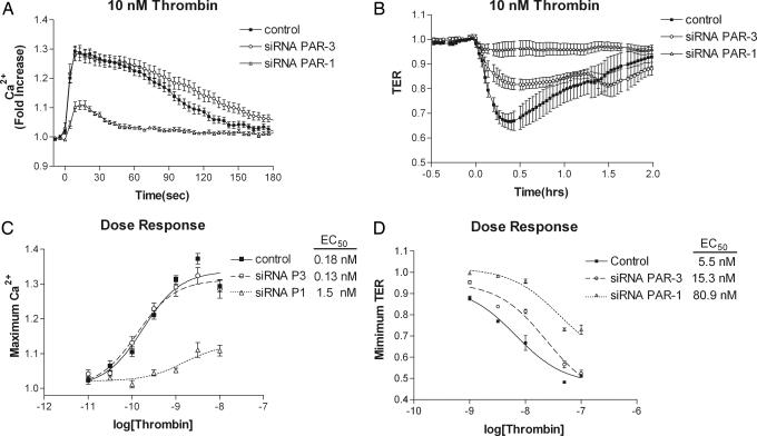
Endothelial cells express PAR1, -2, and –3, but not PAR4 (10), and of these only PAR1 and PAR3 are thrombin-sensitive (11). To address whether PAR3 alters thrombin signaling in endothelial cells, siRNA knockdown was used to reduce thrombin receptor expression in human pulmonary artery endothelial cells (HPAECs). PAR1 and PAR3 suppression was determined by semiquantitative RT-PCR. siRNA against PAR3 did not affect PAR1 expression, whereas it reduced PAR3 expression 8-fold. Conversely, siRNA against PAR1 only modestly affected PAR3 expression, whereas it suppressed PAR1 expression 9-fold [supporting information (SI) Fig. 6]. We next determined the effects of altered receptor expression by using two functional outputs: (i) intracellular calcium mobilization ([Ca2+]i) and (ii) endothelial barrier dysfunction. Thrombin-induced (0.1–10.0 nM) increase in [Ca2+]i was assessed in knockdown HPAECs. PAR1 knockdown showed a marked reduction in maximal peak height, whereas PAR3 knockdown did not (Fig. 1A). The lack of effect of PAR3 siRNA on [Ca2+]i was used to control for nonspecific effects of siRNA treatment. Dose–response curves (Fig. 1C) of maximum peak height yielded the following EC50 values and corresponding 95% confidence intervals: control = 0.18 nM (0.04–0.8), siRNA PAR3 = 0.13 nM (0.06–0.4), and siRNA PAR1 = 1.5 nM (0.2–11.1). PAR3 knockdown showed no statistically significant difference in [Ca2+]i, whereas PAR1 knockdown altered both thrombin potency and efficacy, thus indicating the efficacy of PAR1 siRNA treatment.
Fig. 1.
Differential effects of PAR1 and PAR3 siRNA knockdown in endothelial cells on thrombin-induced mobilization of intracellular calcium and thrombin-induced increase of endothelial monolayer permeability. Cultured HPAECs were electroporated with either buffer alone (control) or pools of siRNA specific for PAR3 or PAR1. (A) Resultant intracellular calcium mobilization traces for each knockdown and control group at a representative thrombin concentration of 10 nM. (B) Changes in endothelial monolayer permeability (TER) determined at a representative thrombin concentration of 10 nM. (C) Dose–response curves of the maximal-induced calcium response for each group. (D) Dose–response curves of the minimal-induced TER for each group. Each point represents the average of at least three separate experiments performed in duplicate. Error bars represent ± SEM.
We next addressed the effects of PAR1 or PAR3 knockdown on endothelial barrier function. Thrombin-induced (1–100 nM) transendothelial electrical resistance (TER) was assayed in knockdown HPAECs. PAR1 knockdown showed virtually no responsiveness at 10 nM thrombin compared with controls, whereas PAR3 knockdown reduced the response by 50% (Fig. 1B). Dose-response curves (Fig. 1D) of the minimal-induced TER yielded the following EC50 with 95% confidence intervals: control = 5.5 nM (4.1–7.5), siRNA PAR3 = 15.3 nM (8.0–29.4), and siRNA PAR1 = 80.9 nM (38.8–169). PAR3 knockdown showed a modest difference in thrombin-induced endothelial barrier dysfunction, whereas PAR1 knockdown markedly altered both thrombin potency and efficacy.
PAR1 and PAR3 Form Constitutive Heterodimers.
To address the mechanism of how down-regulation of PAR3 affects PAR1 signaling, we investigated interactions between PAR1 and PAR3 by using bioluminescent resonance energy transfer-2 (BRET2) (12–15). Using BRET2 PAR dimerization was assessed by transient expression of pairs of C-terminally tagged fusion proteins, one receptor tagged with Renilla luciferase (Rluc) and the other with modified GFP-2 (GFP2), followed by the addition of the cell permeable luciferase substrate and subsequent measurement of emission ratio (510/410 nm). Specific receptor/receptor interactions are characterized by a hyperbolic increase in BRET2 signal as the ratio of receptor-GFP2 to receptor-Rluc expression increases (16). Nonspecific interactions, conversely, increase in direct proportion to the acceptor:donor ratio and give a linear increase in BRET2 signal.
We cotransfected human embryonic kidney (HEK)293T cells with a fixed amount of PAR1-Rluc cDNA (0.5 μg) and increasing amounts (0.5, 1.0, 1.5, 2.0, and 2.5 μg) of GABABR2-GFP2 (as nonspecific control) or PAR1-GFP2 cDNA. The BRET2 signal and the GFP2/Rluc ratio were quantitatively determined as described in Materials and Methods.
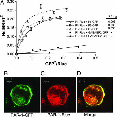
The nonspecific signal from the PAR1-Rluc/GABABR2-GFP2 interaction exhibited a small but linear increase, whereas PAR1-Rluc/PAR1-GFP2 exhibited a substantial and hyperbolic increase in signal (Fig. 2A). Similar results were observed for the PAR1/3 heterodimer and PAR3/3 homodimer. Thus, the dimerization of PARs is highly specific and not the result per se of overexpression or saturation of the signal. To ensure the BRET2 signal was originating from properly localized receptors, plasma membrane localization of PAR1-GFP2 was determined by using standard fluorescent confocal microscopy (Fig. 2B). The corresponding PAR1-Rluc was visualized in the same cells by immunohistochemistry by using a primary antibody against Rluc and a fluorescent-conjugated secondary antibody (Fig. 2C). We observed that corresponding PAR3 constructs showed a similar subcellular localization (data not shown).
Fig. 2.
Specificity and localization of PAR1/PAR3 homo- and heterodimers. A constant amount PAR1- or PAR3-Rluc cDNA was transiently transfected into HEK293T cells along with increasing amounts of PAR1-GFP2, PAR3-GFP2 or GABABR2-GFP2 cDNA. (A) Total Rluc, GFP, and BRET2 signals were determined as described in Materials and Methods. Each point represents the average of at least three separate experiments performed in duplicate. Error bars represent ± SEM. (B) A representative image of PAR1-GFP2 plasma membrane localization by fluorescent microscopy (green). (C) Rluc constructs were visualized in the same cells by immunohistochemistry using a primary antibody against Rluc and a fluorescent-conjugated secondary antibody (red). (D) Colocalization (merge) at the plasma membrane (yellow).
In addition to assessing PAR interactions, BRET2 was used to determine the relative binding affinities between the different partners (16). The rate at which the BRET2 signal is saturated as the GFP2/Rluc ratio is increased depends on the relative binding affinities of the two molecules (16). The value of the GFP2/Rluc ratio at 50% maximum NetBRET2, or BRET50 is an estimate of the relative affinities of the receptors for each other and follows the _K_d value; i.e., a lower BRET50 would indicate greater affinity. The following BRET50 values with 95% confidence intervals were obtained from data in Fig. 2A. PAR1/1 = 0.093 (0.05–0.13), PAR3/3 = 0.036 (0.001–0.071), and PAR1/3 = 0.036 (0.032–0.039).
Effects of PAR1 Activation After Dimerization with PAR3.
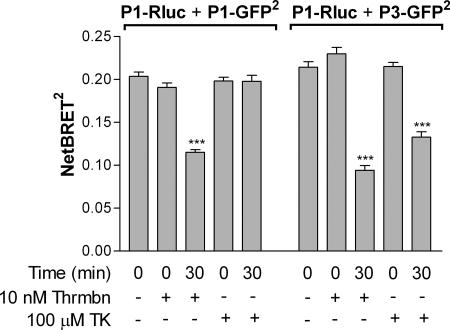
We next determined the effects of activation of PAR1 on its dimerization state with PAR3. We used thrombin as well as the PAR1-specific agonist peptide TFLLRNPNDK (TK). PAR1-Rluc was transiently coexpressed in HEK293T cells with either PAR1-GFP2 or PAR3-GFP2 at the optimal GFP2/Rluc ratio determined (as shown in Fig. 2). Cells were treated with 10 nM thrombin or 100 μM TK, and the BRET2 signal was immediately (time = 0) measured or measured after 30 min. The BRET2 signal was not initially affected by receptor activation induced by thrombin or TK for either PAR1/1 or PAR1/3. However, after 30 min, activation by thrombin produced 50% decrease in signal for both the PAR1/1 homodimer and PAR1/3 heterodimer. Importantly, 30-min activation with TK caused a decrease in signal only for the PAR1/3 heterodimer; PAR1/1 homodimer was unaffected (Fig. 3).
Fig. 3.
Effects of agonist-induced receptor activation on BRET2 signal resulting from receptor dimerization. PAR1-Rluc was transiently coexpressed in HEK293T cells with either PAR1-GFP2 or PAR3-GFP2, as indicated. Cells were stimulated with either 10 nM thrombin or 100 μM TK, after which BRET2 signal was determined immediately (time, 0 min), then again after 30 min. We report here differences between PAR1/1 and PAR1/3 dimer pairs in response to agonist peptide at 30 min, although we observed the same trend within 5 min after activation (data not shown). Each bar represents the average of at least three separate experiments performed in duplicate. Error bars represent ± SEM. ∗∗∗, P < 0.0001.
PAR Dimer Constitution Regulates G Protein Selectivity.
We next addressed the effects of receptor dimerization on Gαq and Gα13 G protein selectivity by using receptors tagged with Rluc and G protein tagged with GFP2. Specific receptor dimer expression was accomplished by cotransfection of 0.5 μg of PAR1-Rluc plasmid DNA into HEK293T cells with either 1.0 μg of untagged PAR1 or untagged PAR3 plasmid DNA. Coexpression of the untagged form of either PAR1 or PAR3 with labeled PAR1 would ensure that the BRET2 signal would originate only from the PAR1 subunit of the complex.
GFP2-labeled G proteins were introduced by two methods of cotransfecting 1.0 μg each of either (i) nontagged Gα-subunit, Gβ1 tagged on the N terminus with GFP2 (GFP2-Gβ1) and nontagged Gγ2 or (ii) internally tagged GFP2-Gα-subunit, nontagged Gβ1, and nontagged Gγ2.
When labeled on the N terminus, GFP2-Gβ1 is fully capable of G protein-coupled receptor (GPCR) coupling and signaling (13). In the case of the α subunit of Gαq and Gα13, the GFP2 cannot be attached at either terminus, because it interferes with GPCR coupling. Instead, the GFP moiety was incorporated internally. Following a precedent for Gαq in which insertion between amino acids E119 and N120 yielded functional protein (17), for Gα13, GFP2 was inserted between amino acids P130 and M131. All of the fluorescent G protein constructs were expressed in HEK293T cells and found to localize to the plasma membrane (data not shown).
Because the ability of GFP2-Gβ1 to couple to receptors is highly dependent on the specific (Gαq or Gα13) untagged Gα-subunit coexpressed (13), in this set of experiments, the BRET2 signal in the absence of expression of the corresponding nontagged Gα-subunit represents nonspecific binding. When GFP2-labeled Gα-subunits were used, nonspecific binding was determined by substitution of the GFP2-Gα with GABABR2-GFP2. The BRET2 signal from the receptor dimer/heterotrimeric G protein interaction was compared under basal and thrombin-induced activation conditions.
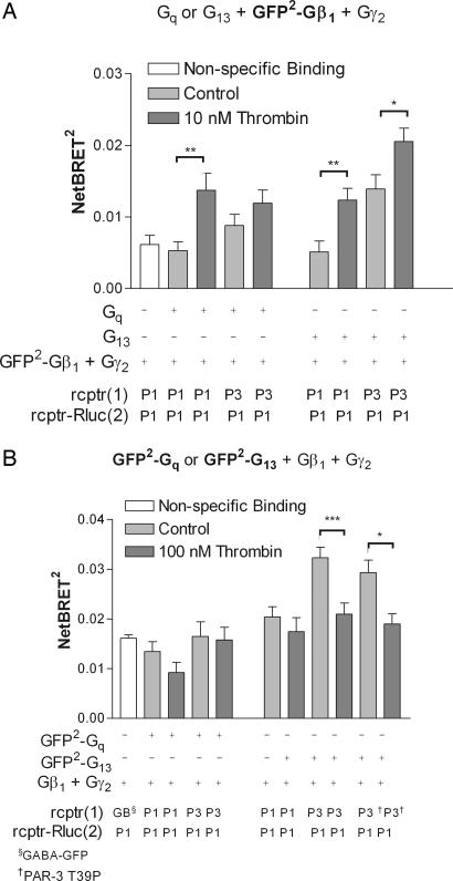
Interactions with Gαq were first probed by using GFP2-β1. Fig. 4A Left shows a significant agonist-dependent increase in BRET2 signal for the PAR1/1 homodimer interaction with GαqGFP2-β1γ2. The same trend was observed for the PAR1/3 heterodimer.
Fig. 4.
PAR1 and PAR3 dimer composition is a determinant of agonist-induced changes in G protein coupling. PAR1-Rluc was transiently coexpressed in HEK293T cells with either nontagged PAR1 or nontagged PAR3, as indicated, to create specific receptor dimers. GFP2-labeled G proteins were introduced by expressing each subunit of the heterotrimer with either the Gβ-subunit (A) or the Gα-subunit (B) tagged with GFP2. BRET2 signal due to nonspecific binding (white bars) was determined in the absence of expression of the corresponding nontagged Gα-subunit (A) or by substitution of the corresponding GFP2-Gα with GABABR2-GFP2 (B). BRET2 signal between receptor dimers and heterotrimeric G proteins under basal conditions (gray bars) was then compared with that under thrombin-induced activation (black bars). In B, a nontagged T39P, was also used where indicated. Each bar represents the average of at least three separate experiments performed in duplicate. Error bars represent ± SEM. ∗ and ∗∗, P < 0.05 and 0.01, respectively.
Interactions with Gα13 were then determined by using the same GFP2-β1 system. When Gα13GFP2-β1γ2 was used (Fig. 4A Right), the PAR1/1 homodimer showed an agonist-induced increase similar to that observed for Gαq. Strikingly, the PAR1/3 heterodimer showed a Gα13 interaction in nonstimulated cells (to the level seen in stimulated PAR1/1 homodimer), which also showed a further agonist-induced increase.
The Gαq counterpart experiments using the GFP2-Gαqβ1γ2 G protein (Fig. 4B Left) yielded no significant agonist-induced changes for either the PAR1/1 or PAR1/3 dimer. However, the Gα13 counterpart experiments using the GFP2-Gα13β1γ2 G protein (Fig. 4B Right) showed an agonist-induced change in BRET2 signal only in the PAR1/3 heterodimer and not in the PAR1/1. The trend was opposite that observed where the GFP2 was placed on the Gβ1 subunit; i.e., here, an elevated BRET2 signal was observed in nonstimulated cells, which decreased to nonspecific binding levels upon thrombin addition. Thus, PAR1/1 homodimer is capable of interacting with both Gαq and Gα13, whereas PAR1 interacting with PAR3 alters its Gα13 but not Gαq binding conformation.
We next determined whether the cleavage of PAR3 in the heterodimer contributed to the thrombin-induced-changes in Gα13 binding conformation. Fig. 4B shows substitution of a thrombin-insensitive PAR3 mutant (T39P) (18) for the wild type had no effect. Thus, cleavage of PAR1 and not of PAR3 in the PAR1/3 heterodimer is sufficient to induce signal transduction.
Discussion
Our study focuses on the crucial role of PAR3 in regulating PAR1 function. We show that PAR1/3 heterodimers are formed constitutively and the activated dimeric complex induces distinct signaling involving the selective coupling to Gα13 as compared with the PAR1/1 homodimer. Importantly, PAR1 has the ability to form heterodimers with PAR3, indicating that this heterodimer is readily formed under physiological conditions. Our findings are based on a differential BRET2 signal using different combinations of PAR1 and PAR3 as well as receptor dimers together with different combinations of G protein subtypes. In addition, we used PAR knockdown by siRNA in endothelial cells to demonstrate that PAR3 regulates PAR1-mediated increase in endothelial permeability such that the PAR1/3 heterodimer favors signaling through Gα13 to mediate the permeability response.
The role of PAR3 is of interest, because the function of this apparently nonsignaling receptor remains obscure (18–20). The ability of PAR3 to generate intracellular signal also remains in doubt, because it lacks the cytoplasmic tail domain shown in other PARs to couple with G-proteins (18). In mouse platelets, PAR3 was shown to function as a cofactor of PAR4 by presenting thrombin to low-affinity PAR4, thereby resulting in efficient receptor cleavage (21). We show here that PAR3 directly interacts with PAR1 in endothelial cells to alter the PAR1/Gα13 binding conformation and thus induce activation of downstream signaling pathways distinct from activation of the PAR1/1 homodimer.
Several models of PAR trans- and coactivation have been proposed (11, 21–25). Studies showed that platelet-specific PAR1/4 heterodimerization was responsible for thrombin-induced platelet aggregation (26). Our results using BRET2 demonstrated that PAR1 and PAR3 form highly specific homo- and heterodimer pairs in endothelial cells. The affinity of PAR1/3 interaction determined by BRET50 was comparable to that of PAR1/1 homodimer. Thus, it is likely that a significant portion of PAR1 is complexed with PAR3 under physiological conditions in vascular endothelial cells.
A limitation in interpreting BRET data is that BRET relies on photon emission of Rluc as a catalytic byproduct of substrate hydrolysis. The weak BRET signal (compared with FRET) is difficult to detect microscopically. To circumvent this concern, we showed using confocal microscopy and immunohistochemistry that the receptor constructs used were indeed localized appropriately on the plasma membrane.
The ability of PAR1 and PAR3 to form homo- as well as heterodimers is likely due to their high sequence homology suggesting they share a conserved over-lapping dimerization interface. We demonstrated that dimerization pairing of PAR1 altered signaling; however, importantly, there was no evidence that PAR3 directly participated in either receptor dimer cleavage and activation or G protein activation.
The PAR1/3 heterodimer also showed differences in receptor desensitization compared with the PAR1/1 homodimer. TK stimulation had no effect on the PAR1/1 BRET signal but within 15–30 min caused a reduction in PAR1/3 signal. We infer from this finding that the differential effects of TK were due to differences in desensitization between the PAR1/1 and PAR1/3 dimer pairs. As activation of receptors by TK showed desensitization only in the presence of PAR3, this further supports our contention that the PAR1/3 heterodimer has a unique signaling property distinct from PAR1/1 homodimer.
We addressed the effects of PAR1/3 heterodimerization on PAR1 G protein specificity. We observed that PAR1/1 homodimers interacted with both Gαq and Gα13 G proteins in an agonist-dependent manner. This finding is consistent with known interactions of PAR1 with G proteins (27, 28). However, we observed Gαq interactions using BRET2 only when the GFP2 label was placed on the Gβ1 subunit. Given the geometric constraints of the assay, the GFP2 expressed within the Gα-subunit may not have been within the 100-Å limitation for efficient energy transfer from the receptor. There were similar Gαq interactions for the PAR1/3 heterodimer as the PAR1/1 homodimer, which we interpret to mean that dimerization with PAR3 has little effect on PAR1-mediated Gαq activation. However, the presence of PAR3 in the receptor pair markedly increased the interaction of PAR1 with Gα13. Probing for Gα13 interaction using the fluorescent Gβ1 revealed that Gα13 bound with the PAR1/3 dimer in the basal state but not with PAR1/1 dimer. Gα13 coupling with PAR1/3 was further enhanced after stimulation with PAR1 agonist. In a related experiment using fluorescent Gα13, we also showed that only the PAR1/3 heterodimer resulted in agonist-induced changes in BRET2 signal and unexpectedly, this change was opposite in direction (i.e., elevated BRET2 signal in nonstimulated cells which decreased upon thrombin exposure). The opposite trend in BRET2 depended on which subunit of the G protein was labeled. Thus, our results suggest upon agonist activation, conformational changes in the G protein enabled rotation of the N terminus of Gβ-subunit toward the C terminus of the receptor concomitant with a movement of the Gα-subunit away from the C terminus of the receptor.
Our data demonstrate precoupling of the inactive PAR1/3 heterodimer with Gα13, which remains associated with the receptor heterodimer even after agonist activation of PAR1. This finding is consistent with studies of other GPCRs demonstrating unconventional receptor/G protein associations and kinetics (13). However, our findings contradict observations made in other systems that did not find precoupling of G-proteins with the receptor (29). We think that this discrepancy is likely due to different properties inherent in the G protein systems studied. Interestingly, precoupling to Gα13 was only observed for the PAR1/3 heterodimer, indicating that PAR1 achieves the unique Gα13 binding conformation only when PAR1 dimerized with PAR3.
The initial peak rise in thrombin-induced in [Ca2+]i is attributed to release from intracellular Ca2+ stores as the result of activation of Gαq (30). Knockdown of PAR3 in endothelial cells using siRNA had no effect on the peak Ca2+ rise. This lack of effect suggests that PAR3 did not alter PAR1-Gαq binding. PAR1 knockdown altered both thrombin potency of the Ca2+ response, suggesting that thrombin signaling of the rise [Ca2+]i was carried out primarily via the PAR1 subunit of the PAR1/3 heterodimer and formation of the heterodimer with PAR3 did not affect the PAR1-induced Gαq-mediated signaling.
The increase in endothelial permeability leading to vascular inflammation is the result of the integration of a network of signaling pathway dependent on Gαq as well as Gα13 signaling (7–9). Changes in the activation of one or the other or both G-proteins (Gαq and Gα13) may affect the permeability response to thrombin. We observed that PAR1 knockdown markedly altered both thrombin's potency and efficacy, whereas PAR3 knockdown showed only a modest decrease in potency. These results demonstrate that thrombin signaling in endothelial cells is transmitted primarily by PAR1 cleavage but PAR1 interaction with PAR3 results in maximal activation of the PAR1-induced increase in endothelial permeability. The basis of this finding is that PAR3 dimerization with PAR1 changes the selectivity of PAR1 to Gα13, and thereby enhances Gα13-mediated signaling. Thus, PAR3 is a novel therapeutic target that may be useful in preventing vascular inflammation secondary increased endothelial permeability induced by thrombin.
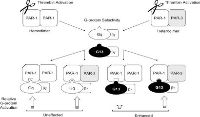
Our data using a thrombin-insensitive mutant of PAR3 showed that cleavage of PAR1, and not of PAR3, in the PAR1/3 heterodimer complex is sufficient to induce signal transduction. These data support a pentameric complex formed from two GPCRs and one G protein, a model postulated for rhodopsin, the archetypal GPCR (31), and proposed for other GPCRs (32). In the case of rhodopsin, one receptor molecule serves as the photon (ligand) acceptor whereas the second serves as a platform to support G protein binding. Our data support the model for PARs in which one receptor (PAR1) is activated, and serves as the thrombin substrate, whereas the second receptor (PAR3) serves as an allosteric modulator to mediate G protein selectivity (Fig. 5).
Fig. 5.
PAR3 modulates PAR1 signaling by receptor dimerization. PARs are constitutively expressed as homo- and/or heterodimeric receptors. Protease induced activation of the PAR1 subunit of either the PAR1/1 homodimer or the PAR1/3 heterodimer initiates multiple intracellular signaling cascades by selective activation of discrete G-proteins. PAR1 heterodimerization with PAR3 alters the PAR1/Gα13 binding conformation, enhancing Gα13 signaling. Heterodimerization likely does not affect PAR1/Gαq selectivity.
In summary, we addressed regulation of endothelial cell signaling by PAR1/3 interaction and the crucial role of this interaction in determining G protein-coupling selectivity. We show an interaction of PAR3 with PAR1 that alters the PAR1/Gα13 binding conformation. PAR1/1 homodimer is capable of interaction with Gαq and Gα13, whereas PAR1/3 heterodimer favors the Gα13 binding conformation. PAR3 therefore functions as an important allosteric modulator of PAR1 signaling.
Materials and Methods
Reagents.
All cell culture reagents were purchased from Invitrogen (Carlsbad, CA), except endothelial growth medium-2 (EBM-2), which was obtained from Clonetics (San Diego, CA). α-Thrombin was purchased from Enzyme Research (South Bend, IN). TK was purchased from GL Biochem (Shanghai, China) Mouse anti-Rluc monoclonal antibody was purchased from Chemicon International (Temecula, CA). Alexa Fluor 594 rabbit anti-mouse IgG (H+L) was obtained from Molecular Probes (Eugene, OR). Human cDNA clones for PAR1, Gαq, Gα13, Gβ1, and Gγ2 in pcDNA3.1(+) were purchased from University of Missouri cDNA Resource Center, University of Missouri (Rolla, MO). The BRET2 donor/acceptor fusion protein vectors pRluc-N and pGFP2-N were obtained from PerkinElmer (Boston, MA). All DNA primers, LipofectAMINE, TRIzol, and SuperScript III RT reagents were obtained from Invitrogen. siRNA was purchased from Dharmacon (Lafayette, CO). SYBR green PCR Master mix was purchased from Applied Biosystems (Branchburg, NJ).
Cell Culture.
HPAECs were purchased from Cambrex (East Rutherford, NJ). Cells were maintained in EBM-2 medium supplemented with 10% FBS in 5% CO2 at 37°C. All of the studies used cells between passages 5–8. HEK293T were maintained in DMEM supplemented with 10% FBS and penicillin/streptomycin (5,000 units/ml; 5,000 μg/ml) in 5% CO2 at 37°C.
cDNA Constructs.
PAR1 and PAR3 were subcloned by PCR amplification into both the pRluc-N1 and pGFP2-N1 vectors (PerkinElmer). GFP2 was amplified by PCR then inserted into Gαq in pcDNA3.1(+) between amino acid E119 and N120. The final protein sequence reads: M1… E119-EFGG-GFP2-GGEL-N120… stop. GFP2 in Gα13 in pcDNA3.1(+) final protein sequence reads: M1… P130-EFGG-GFP2-GGEL-M131… stop. GFP2 was amplified by PCR and subcloned upstream of Gβ1 in pcDNA3.1(+). A thrombin-insensitive mutant of PAR3 pcDNA3.1(+) was generated by mutating threonine 39 to a proline (18) by a PCR-based protocol. All constructs were verified by sequence analysis.
Transfections.
HEK293T were grown in six-well plates to ≈70% confluence. Between 0.5 and 2.5 μg of each purified plasmid DNAs were incubated with 10 μl of LipofectAMINE (Invitrogen) according to manufacturer's protocol. Cells were allowed to recover 48 h before experimentation.
Rluc Immunostaining and Confocal Microscopy.
Subcellular localization of Rluc fusion proteins in HEK293T cells was determined by immunohistochemistry as described (33) using mouse anti-Rluc Ab (1:500) coupled with Alexa Fluor 594 rabbit anti-mouse IgG. Images were acquired with the Zeiss (Oberkochen, Germany) LSM 510 confocal microscope using a 63 × 1.2 N.A. water immersion objective.
BRET2 Assay.
BRET2 assay was performed following the manufacturer's protocols (PerkinElmer). Briefly, HEK293T cells were grown in six-well plates and transiently transfected with fusion constructs of RlucGFP2. After 48 h, cells were washed two times in PBS and detached by gentle pipetting. Media were removed by decanting after centrifugation at 1,000 × g for 5 min, after which cells were resuspended in BRET buffer (PBS containing 1 mM MgCl2, 1 mM CaCl2, and 5 mM glucose) to a final density of ≈2.5 × 10⁶ cells/ml. Cells were then stimulated with agonist as indicated just before addition of BRET2 substrate DeepBlueC (PerkinElmer). BRET2 signal was measured by using the Victor2 (PerkinElmer) in luminescence mode by using bandpass filters for 410 and 515 nm. Values are reported as BRET2 (absolute ratio of emission at 515 to emission at 410) or as NetBRET2 (BRET2unknown − BRET2background), where BRET2background is taken to be the BRET2 signal obtained in the absence of an expressed GFP2 acceptor protein. The absolute GFP2 expression was measured by using the Victor2 in fluorescence mode with excitation at 405 nm and emission at 515 nm. The absolute Rluc expression in the same cells was measured by using the Victor2 in luminescence mode using no filters after addition of the non-BRET2 substrate MightyLight (Novagen, San Diego, CA), according to the manufacturer's protocols.
siRNA Knockdown.
HPAEC grown to confluence were harvested by detachment with 0.05% trypsin/0.5 mM EDTA followed by centrifugation, 1,000 × g for 10 min at room temperature. Each reaction consisted of 1 × 10⁶ cells resuspended in 100 μl of basic nucleofector solution (Amaxa, Cologne, Germany) containing a pool of three unique siRNA pairs for either PAR1 or PAR3 (0.25 nmol RNA each). Cells were then electroporated by using the Nucleofector Device according to manufacturer's protocols (Amaxa).
Semiquantitative RT-PCR.
Expression of PAR1 and PAR3 was monitored in HPAEC by real-time semiquantitative RT-PCR by using PCR SYBR green PCR Master mix (Applied Biosystems) was on a PRISM 7000 Sequence Detection System (Applied Biosystems) with GAPDH as the internal control as described (34). Briefly, after siRNA treatment described above, HPAEC were grown to confluence in six-well plates and total RNA was harvested, reverse-transcribed using oligo-dTs with SuperScript III RT, and real-time semiquantitative analysis was performed. Data analysis and calculations were done following the 2-ΔΔCT method (35). Each sample was assayed in triplicate.
Calcium Mobilization.
Calcium mobilization was determined as before (36). Briefly, after siRNA treatment, HPAEC were grown to confluence and serum deprived in EBM-2 containing 0.1% serum 3 h. Cells were then loaded with the FLIPR Calcium Plus kit dye and fluorescence measured on the FlexStation (Molecular Devices, Sunnyvale, CA).
TER.
In vitro endothelial barrier dysfunction was monitored by electric cell-substrate impedance sensor (37). TER was determine as described (36). Briefly, after siRNA treatment, HPAEC were seeded at 2 × 10⁵ cells/well and allowed to recover for 48 h. The monolayers were serum deprived in EBM-2 containing 0.1% serum 3 h before agonist addition.
Supplementary Material
Supporting Figure
Acknowledgments
We acknowledge the contributions of the following collaborators: Dr. Wadie F. Bahou (State University of New York, Stony Brook, NY) for the kind gift of the PAR3 construct; Dr. Michel Bouvier (Université de Montréal, Montréal, Canada) for assistance and the kind gift of the GABABR2-GFP2 construct; Dr. Richard D. Minshall (University of Illinois, Chicago) for assistance in confocal microscopy; Dr. Tatyana Voyno-Yasenetskaya for direction in designing the GFP2-Gα13 construct; and Dr. Shahid Siddiqui for design of the siRNA oligos.
Abbreviations
BRET2
bioluminescent resonance energy transfer-2
EBM-2
endothelial growth medium-2
GFP2
modified GFP
GPCR
G-protein coupled receptor
HPAEC
human pulmonary artery endothelial cells
PAR
protease-activated receptor
Rluc
Renilla luciferase
T39P
thrombin-insensitive mutant of PAR3
TER
transendothelial electrical resistance
TK
PAR1-specific agonist peptide TFLLRNPNDK.
Footnotes
The authors declare no conflict of interest.
References
- 1.Malik AB, Fenton JW., 2nd Semin Thromb Hemost. 1992;18:193–199. doi: 10.1055/s-2007-1002425. [DOI] [PubMed] [Google Scholar]
- 2.Rabiet MJ, Plantier JL, Dejana E. Br Med Bull. 1994;50:936–945. doi: 10.1093/oxfordjournals.bmb.a072935. [DOI] [PubMed] [Google Scholar]
- 3.Cepkova M, Matthay MA. J Intensive Care Med. 2006;21:119–143. doi: 10.1177/0885066606287045. [DOI] [PMC free article] [PubMed] [Google Scholar]
- 4.Bastarache JA, Ware LB, Bernard GR. Semin Respir Crit Care Med. 2006;27:365–376. doi: 10.1055/s-2006-948290. [DOI] [PubMed] [Google Scholar]
- 5.Jenkins RG, Su X, Su G, Scotton CJ, Camerer E, Laurent GJ, Davis GE, Chambers RC, Matthay MA, Sheppard D. J Clin Invest. 2006;116:1606–1614. doi: 10.1172/JCI27183. [DOI] [PMC free article] [PubMed] [Google Scholar]
- 6.Vu TK, Hung DT, Wheaton VI, Coughlin SR. Cell. 1991;64:1057–1068. doi: 10.1016/0092-8674(91)90261-v. [DOI] [PubMed] [Google Scholar]
- 7.Dudek SM, Garcia JG. J Appl Physiol. 2001;91:1487–1500. doi: 10.1152/jappl.2001.91.4.1487. [DOI] [PubMed] [Google Scholar]
- 8.Hart MJ, Jiang X, Kozasa T, Roscoe W, Singer WD, Gilman AG, Sternweis PC, Bollag G, Sternweis PM, Sharma S, et al. Science. 1998;280:2112–2114. doi: 10.1126/science.280.5372.2112. [DOI] [PubMed] [Google Scholar]
- 9.Tiruppathi C, Minshall RD, Paria BC, Vogel SM, Malik AB. Vascul Pharmacol. 2002;39:173–185. doi: 10.1016/s1537-1891(03)00007-7. [DOI] [PubMed] [Google Scholar]
- 10.Hirano K, Kanaide H. J Atheroscler Thromb. 2003;10:211–225. doi: 10.5551/jat.10.211. [DOI] [PubMed] [Google Scholar]
- 11.O'Brien PJ, Prevost N, Molino M, Hollinger MK, Woolkalis MJ, Woulfe DS, Brass LF. J Biol Chem. 2000;275:13502–13509. doi: 10.1074/jbc.275.18.13502. [DOI] [PubMed] [Google Scholar]
- 12.Pfleger KD, Eidne KA. Biochem J. 2005;385:625–637. doi: 10.1042/BJ20041361. [DOI] [PMC free article] [PubMed] [Google Scholar]
- 13.Gales C, Rebois RV, Hogue M, Trieu P, Breit A, Hebert TE, Bouvier M. Nat Methods. 2005;2:177–184. doi: 10.1038/nmeth743. [DOI] [PubMed] [Google Scholar]
- 14.Milligan G. Eur J Pharmacol Sci. 2004;21:397–405. doi: 10.1016/j.ejps.2003.11.010. [DOI] [PubMed] [Google Scholar]
- 15.Pfleger KD, Eidne KA. Pituitary. 2003;6:141–151. doi: 10.1023/b:pitu.0000011175.41760.5d. [DOI] [PubMed] [Google Scholar]
- 16.Mercier JF, Salahpour A, Angers S, Breit A, Bouvier M. J Biol Chem. 2002;277:44925–44931. doi: 10.1074/jbc.M205767200. [DOI] [PubMed] [Google Scholar]
- 17.Hughes TE, Zhang H, Logothetis DE, Berlot CH. J Biol Chem. 2001;276:4227–4235. doi: 10.1074/jbc.M007608200. [DOI] [PubMed] [Google Scholar]
- 18.Ishihara H, Connolly AJ, Zeng D, Kahn ML, Zheng YW, Timmons C, Tram T, Coughlin SR. Nature. 1997;386:502–506. doi: 10.1038/386502a0. [DOI] [PubMed] [Google Scholar]
- 19.Hansen KK, Saifeddine M, Hollenberg MD. Immunology. 2004;112:183–190. doi: 10.1111/j.1365-2567.2004.01870.x. [DOI] [PMC free article] [PubMed] [Google Scholar]
- 20.Kaufmann R, Schulze B, Krause G, Mayr LM, Settmacher U, Henklein P. Regul Pept. 2005;125:61–66. doi: 10.1016/j.regpep.2004.07.032. [DOI] [PubMed] [Google Scholar]
- 21.Nakanishi-Matsui M, Zheng YW, Sulciner DJ, Weiss EJ, Ludeman MJ, Coughlin SR. Nature. 2000;404:609–613. doi: 10.1038/35007085. [DOI] [PubMed] [Google Scholar]
- 22.Chen J, Ishii M, Wang L, Ishii K, Coughlin SR. J Biol Chem. 1994;269:16041–16045. [PubMed] [Google Scholar]
- 23.Shi X, Gangadharan B, Brass LF, Ruf W, Mueller BM. Mol Cancer Res. 2004;2:395–402. [PubMed] [Google Scholar]
- 24.Vesey DA, Cheung CW, Kruger WA, Poronnik P, Gobe G, Johnson DW. Kidney Int. 2005;67:1315–1329. doi: 10.1111/j.1523-1755.2005.00209.x. [DOI] [PubMed] [Google Scholar]
- 25.Damiano BP, Cheung W-M, Santulli RJ, Fung-Leung W-P, Ngo K, Ye RD, Darrow AL, Derian CK, de Garavilla L, Andrade-Gordon P. J Pharmacol Exp Ther. 1999;288:671–678. [PubMed] [Google Scholar]
- 26.Leger AJ, Jacques SL, Badar J, Kaneider NC, Derian CK, Andrade-Gordon P, Covic L, Kuliopulos A. Circulation. 2006;113:1244–1254. doi: 10.1161/CIRCULATIONAHA.105.587758. [DOI] [PubMed] [Google Scholar]
- 27.Hung DT, Wong YH, Vu TK, Coughlin SR. J Biol Chem. 1992;267:20831–20834. [PubMed] [Google Scholar]
- 28.Barr AJ, Brass LF, Manning DR. J Biol Chem. 1997;272:2223–2229. doi: 10.1074/jbc.272.4.2223. [DOI] [PubMed] [Google Scholar]
- 29.Hein P, Frank M, Hoffmann C, Lohse MJ, Bunemann M. EMBO J. 2005;24:4106–4114. doi: 10.1038/sj.emboj.7600870. [DOI] [PMC free article] [PubMed] [Google Scholar]
- 30.Lum H, Aschner JL, Phillips PG, Fletcher PW, Malik AB. Am J Physiol. 1992;263:L219–L225. doi: 10.1152/ajplung.1992.263.2.L219. [DOI] [PubMed] [Google Scholar]
- 31.Jastrzebska B, Fotiadis D, Jang G-F, Stenkamp RE, Engel A, Palczewski K. J Biol Chem. 2006;281:11917–11922. doi: 10.1074/jbc.M600422200. [DOI] [PMC free article] [PubMed] [Google Scholar]
- 32.Baneres JL, Parello J. J Mol Biol. 2003;329:815–829. doi: 10.1016/s0022-2836(03)00439-x. [DOI] [PubMed] [Google Scholar]
- 33.Sandoval R, Malik AB, Naqvi T, Mehta D, Tiruppathi C. Am J Physiol. 2001;280:L239–L247. doi: 10.1152/ajplung.2001.280.2.L239. [DOI] [PubMed] [Google Scholar]
- 34.McLaughlin JN, Mazzoni MR, Cleator JH, Earls L, Perdigoto AL, Brooks JD, Muldowney JA, III, Vaughan DE, Hamm HE. J Biol Chem. 2005;280:22172–22180. doi: 10.1074/jbc.M500721200. [DOI] [PubMed] [Google Scholar]
- 35.Livak KJ, Schmittgen TD. Methods. 2001;25:402–408. doi: 10.1006/meth.2001.1262. [DOI] [PubMed] [Google Scholar]
- 36.McLaughlin JN, Shen L, Holinstat M, Brooks JD, Dibenedetto E, Hamm HE. J Biol Chem. 2005;280:25048–25059. doi: 10.1074/jbc.M414090200. [DOI] [PubMed] [Google Scholar]
- 37.Tiruppathi C, Malik AB, Del Vecchio PJ, Keese CR, Giaever I. Proc Natl Acad Sci USA. 1992;89:7919–7923. doi: 10.1073/pnas.89.17.7919. [DOI] [PMC free article] [PubMed] [Google Scholar]
Associated Data
This section collects any data citations, data availability statements, or supplementary materials included in this article.
Supplementary Materials
Supporting Figure
