Stat1 Phosphorylation Determines Ras Oncogenicity by Regulating p27Kip1 (original) (raw)
Abstract
Inactivation of p27Kip1 is implicated in tumorigenesis and has both prognostic and treatment-predictive values for many types of human cancer. The transcription factor Stat1 is essential for innate immunity and tumor immunosurveillance through its ability to act downstream of interferons. Herein, we demonstrate that Stat1 functions as a suppressor of Ras transformation independently of an interferon response. Inhibition of Ras transformation and tumorigenesis requires the phosphorylation of Stat1 at tyrosine 701 but is independent of Stat1 phosphorylation at serine 727. Stat1 induces p27Kip1 expression in Ras transformed cells at the transcriptional level through mechanisms that depend on Stat1 phosphorylation at tyrosine 701 and activation of Stat3. The tumor suppressor properties of Stat1 in Ras transformation are reversed by the inactivation of p27Kip1. Our work reveals a novel functional link between Stat1 and p27Kip1, which act in coordination to suppress the oncogenic properties of activated Ras. It also supports the notion that evaluation of Stat1 phosphorylation in human tumors may prove a reliable prognostic factor for patient outcome and a predictor of treatment response to anticancer therapies aimed at activating Stat1 and its downstream effectors.
Introduction
The signal transducers and activators of transcription (Stats) are a family of cytoplasmic proteins that function as signal messengers and transcription factors involved in cellular responses induced by cytokines and growth factors [1], [2]. Stat1, the prototype of the family, is essential for innate immunity [2] and plays an important role in immune surveillance of tumors [3]. Specifically, Stat1 knockout (Stat1−/−) mice are highly susceptible to virus infection [4], [5] and more prone to the formation of tumors in response to carcinogens than normal mice [6]. Stat1 is also an important mediator of the anti-proliferative and pro-apoptotic functions of interferon-gamma (IFN-γ) and tumor necrosis factor-β (TNF-β) through its ability to upregulate caspase 1 and the cyclin dependent kinase (Cdk) inhibitor p21Cip1 [7]–[10]. At the molecular level, cytokines and growth factors induce Stat1 phosphorylation at tyrosine (Y) 701, which is essential for its homo-dimerization or hetero-dimerization with other Stats and binding to DNA [1], [2]. Tyrosine phosphorylation of Stat1 is mediated by cytokine receptor associated Janus tyrosine kinases (Jaks) as well as by receptor tyrosine kinases (RTKs) [2]. Phosphorylation of Stat1 at serine (S) 727 is mediated by various pathways and is required for the full induction of Stat1-dependent gene transactivation [11].
The Cdkn1b gene encodes for a 27 kDa protein (p27), which belongs to the Cip/Kip family of cyclin-dependent kinase inhibitors (CKIs) [12]. p27Kip1 acts in G0 and early G1 to inhibit cyclin-Cdk holoenzymes, particularly cyclin E-Cdk2, and impair cell cycle progression [12]. p27Kip1 levels decrease in response to mitogenic signaling thus permitting cell cycle progression and cell proliferation [12]. The human Cdkn1b gene is present on chromosome 12p13 and loss of one allele has been observed in a number of human malignancies [13]. Consistent with a tumor suppressor function, mice lacking one or both copies of the Cdkn1b gene have increased susceptibility to carcinogen-induced tumorigenesis [14]. p27Kip1 does not follow Knudson's classic “two-hit hypothesis” of tumor suppression because homozygous loss or silencing of the Cdkn1b locus in human tumors is extremely rare [13]. The Cdkn1b gene is rarely mutated in human cancers but decreased concentrations of p27Kip1 are implicated in human tumorigenesis [13]. There is an inverse correlation between p27Kip1 levels and prognosis in a variety of human cancers, including those of breast, colon, and prostate origin [13]. Expression of the Cdkn1b gene is regulated at the transcriptional, translational and post-translational levels. Transcription is controlled by several factors including Sp1 [15], Phox2a [16], members of the forkhead box (Fox) group of transcription factors [17] and Stat3 [18], [19]. Early findings provided evidence for a cell-cycle dependent regulation of Cdkn1b mRNA translation [20]. Subsequent studies found that translation of Cdkn1b mRNA is controlled by sequences within the 5′ untranslated region (5′ UTR) [21], [22] through a cap-independent mechanism [23] and the utilization of an internal ribosomal entry site (IRES) [24]. The post-translational control of p27Kip1 is fairly complex and involves phosphorylation, changes in subcellular distribution as well as proteasomal degradation [12].
The anti-tumor properties of Stat1 have mainly been linked to its function downstream of IFNs[3]. This prompted us to examine whether Stat1 plays a role in oncogenic signaling in the absence of an IFN effect. Herein, we present a novel functional link between Stat1, p27Kip1 and oncogenic Ras. We demonstrate that Stat1 subverts the inactivation of p27Kip1 in Ras transformed cells by positively regulating the transcription of the Cdkn1b gene. We further demonstrate that induction of p27Kip1 expression by Stat1 is essential for the suppression of Ras-mediated oncogenesis in vitro and in vivo via mechanisms that are affected by the phosphorylation of Stat1 at tyrosine 701 and serine 727.
Results
Stat1 counteracts the downregulation of p27Kip1 by activated Ras
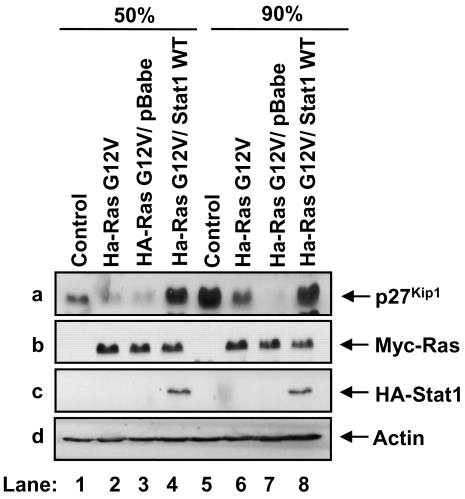
To examine the role of Stat1 in Ras transformation, we used primary mouse embryonic fibroblasts (MEFs) from Stat1 and p53 double-knock out animals (p53−/−Stat1−/− MEFs). We chose p53 deficient MEFs because p53 inactivation facilitates transformation by expression of cytoplasmic oncoproteins including activated Ras [25]. When primary p53−/− Stat1−/− MEFs were transfected with a Myc-tagged form of Ha-RasG12V, we noted that activated Ras decreased p27Kip1 levels as previously described [26] (Fig. 1, panel a, compare lane 1 with 2, and lane 5 with 6). The expression of p27Kip1 was affected by the density of the cells since p27Kip1 levels were proportional to the confluency of control cells (panel a, compare lane 1 with 5) and downregulation of p27Kip1 by activated Ras was more obvious in confluent than in sub-confluent cell cultures (Fig. 1, panel a, compare lanes 1 and 2 with 5 and 6). When the MEFs transfected with activated Ras were reconstituted with an HA-tagged form of human wild type (WT) Stat1 by retrovirus infection, we observed that Stat1 restored p27Kip1 protein levels in both sub-confluent and confluent cell cultures (panel a, lanes 4 and 8). Contrary to this, p27Kip1 levels remained low in MEFs infected with empty retroviruses (panel a, lanes 3 and 7). The expression of activated Ras (lane b) and reconstituted Stat1 (lane c) was not affected by the density of the cells as verified by immunoblotting. These data provided evidence that Stat1 subverts the donwregulation of p27Kip1 by activated Ras.
Figure 1. Stat1 prevents the decrease of p27Kip1 by activated Ras.
Primary p53−/−Stat1−/− MEFs (lanes 1 and 5) were transfected with Myc-Ha-Ras G12V (lanes 2 and 6) and reconstituted with HA-Stat1 WT by retrovirus infection (lanes 4 and 8). As control, Myc-Ha-Ras G12V expressing MEFs infected with empty retroviruses were used (lanes 3 and 7). Polyclonal populations were harvested at 50% (lanes 1–4) or 90% confluence (lanes 4–8) and cell extracts (50 µg of protein) were subjected to immunoblotting with anti-p27Kip1 monoclonal antibody (mAb) (panel a), anti-Myc mAb (panel b), anti-Stat1α mAb (panel c ) and anti-actin mAb (panel d).
p27Kip1 expression is controlled by phosphorylated Stat1 at the transcriptional level
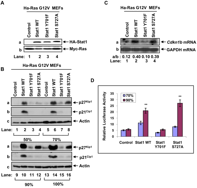
Previous findings showed that Stat1 is phosphorylated at Y701 and S727 in Ras transformed cells [27], [28]. To verify these observations, we used NIH3T3 cells transformed with Ha-RasG12V by retrovirus infection. We noted that activated Ras decreased Stat1 Y701 phosphorylation and increased Stat1 S727 phosphorylation compared to control cells in a manner that was dependent upon cell density (Fig. S1). To address the role of Stat1 phosphorylation in p27Kip1 expression, the Ras-transfected p53−/−Stat1−/− MEFs were reconstituted with HA-Stat1 forms bearing either the Y701F or S727A mutation by retrovirus infection. Immunoblot analysis of the reconstituted MEFs verified that expression of the Stat1 mutants was equal to that of Stat1 WT (Fig. 2A, panel a). When the MEFs were maintained at different levels of confluency, we noted the induction of p27Kip1 levels in cells expressing either HA-Stat1 WT or HA-Stat1S727A but not in cells expressing Stat1Y701F or devoid of Stat1 (Fig. 2B, panel a). The induction of p27Kip1 was proportional to the increased density of the cells and p27Kip1 levels were higher in cells reconstituted with Stat1WT than Stat1S727A (Fig. 2B, panel A). In parallel, we looked at the levels of p21Cip1 based on previous findings that Stat1 regulates p21Cip1 at the transcriptional level [9] and that p21Cip1 levels are upregulated by activated Ras [29]. We found that unlike p27Kip1, p21Cip1 levels were induced by Stat1 WT only (Fig. 2B, panel b) indicating that p21Cip1 expression in Ras transfected cells requires Stat1 phosphorylation at both Y701 and S727.
Figure 2. Induction of p27Kip1 by Stat1 in Ras-transformed cells depends on site-specific Stat1 phosphorylation.
(A) Whole cell extracts (50 µg of protein) from MEFs expressing Myc-Ha-RasG12V and reconstituted with the indicated HA-Stat1 proteins were subjected to immunoblotting with anti-Stat1α mAb (panel a) or anti-Myc mAb (panel b). (B) MEFs were harvested at 50% (lanes 1–4), 70% (lanes 5–8), 90% (lanes 9–12), or 100% confluence (lanes 13–16). Whole cell extracts (50 µg of protein) were subjected to immunoblotting with an anti-p27Kip1 mAb (panel a), anti-p21Cip1 rabbit polyclonal Ab (panel b) or anti-actin mAb (panel c). (C) Total RNA (15 µg) from MEFs harvested at 90% confluence were subjected to Northern blot analysis using [α-32P] dCTP-labeled Cdkn1b cDNA (panel a) and [α-32P] dCTP-labelled glyceraldehyde-3-phosphate dehydrogenase (GAPDH, panel b) as probes. The radioactive bands were detected by autoradiography and quantified by densitometry using the NIH Image 1.54 software. (D) Sub-confluent MEFs were transfected with the pGL2 vector containing the firefly luciferase reporter gene under the control of the full-length 1609-bp mouse Cdkn1b promoter. Forty eight or 72 hours after transfection, cells at 70% or 90% confluence were harvested and the luciferase activity was determined. The activity of Renilla luciferase expressed from a pGL2 vector lacking the Cdkn1b promoter was used as an internal transfection control. Results are expressed ±SD for 3 experiments performed in triplicate. **P<0.01.
To address the mechanism of regulation of p27Kip1 expression, we first looked at a possible transcriptional effect of Stat1. Northern blot analysis showed an increase in Cdkn1b mRNA levels in Ras-transfected MEFs reconstituted with either Stat1 WT or Stat1 S727A compared to control MEFs or MEFs reconstituted with Stat1Y701F (Fig. 2C, panel a). To further substantiate this finding, we assessed the transcriptional activation of the mouse 1.6-Kb Cdkn1b promoter by Stat1 in luciferase reporter assays [30]. We found that transcription of the Cdkn1b promoter was more highly induced in Ras-transfected MEFs expressing either Stat1 WT or Stat1S727A than in control MEFs or MEFs expressing Stat1Y701F (Fig. 2D). Consistent with the p27Kip1 protein levels (Fig. 2B), Stat1-dependent transcription from the mouse Cdkn1b promoter was proportional to the increased density of the MEFs (Fig. 2D). These data demonstrated a transcriptional role for Stat1 in Cdkn1b gene expression.
Transcriptional induction of the Cdkn1b gene by Stat1 requires Stat3
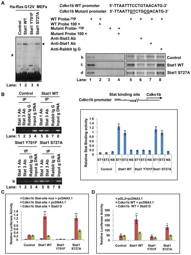
The mouse Cdkn1b promoter contains a Stat-binding site located at a position 1585 bp upstream of the transcription initiation site [18], [31]. To assess the role of the Stat-binding site in Cdkn1b transcription by Stat1, we performed electrophoretic mobility shift assays (EMSAs) using extracts from MEFs containing activated Ras and reconstituted with the various forms of Stat1, and a probe encompassing the Stat-binding site of the Cdkn1b promoter [18]. To increase the detection of DNA-binding, EMSAs were performed with protein extracts from 90% confluent cells in which Cdkn1b promoter activity was maximal (Fig. 2D). We detected the formation of a high molecular weight protein/DNA complex, whose intensity was enhanced in MEFs reconstituted with either HA-Stat1 WT or HA-Stat1 S727A (Fig. 3A, left panel, a). The formation of the complex was abolished when EMSAs were performed in the presence of a 100 fold excess of non-radioactive oligonucleotide (Fig. 3A, right panels b, c and d, lane 2) or when an oligonucleotide with mutations in the Stat-binding site was used as a probe (right panels b, c and d, lanes 3 and 4). To identify the proteins that form the complex, we performed the assay in the presence of antibodies against Stat1 or Stat3. We observed that formation of the complex in MEFs devoid of Stat1 was decreased by 50% after incubation with an antibody against Stat3 (right panel b, compare line 5 with 6) indicating the presence of Stat3 in the complex. Contrary to this, incubation with an antibody against Stat1 or rabbit IgG antibody, which served as a negative control, did not affect binding strength (Fig. 3A, right panel b, lanes 6 and 7). When the EMSAs were performed with protein extracts from MEFs reconstituted with either Stat1 WT (panel c) or Stat1S727A (panel d), we noted that the formation of the complex was decreased after incubation with antibodies against Stat3 (lane 6) or Stat1 (lane 7) but not after incubation with the rabbit IgG antibody (lane 8). The reduction but not elimination of complex formation after incubation with antibodies against Stat1 or Stat3 is consistent with a previous study showing that antibodies against Stat1 or Stat3 did not abolish binding of the Stat1/Stat3 complex to the Stat-site of the Cdkn1b promoter in mouse 32D lymphoid cells stimulated with G-CSF [18]. Nevertheless, the possibility remains that the Stat-site is also occupied by a third protein, which forms a complex with DNA and migrates with the same mobility as the Stat1/Stat3 complex in the polyacrylamide gels.
Figure 3. Transcriptional induction of Cdkn1b gene by Stat1 requires Stat3.
(A) (Left panel) Protein extracts from MEFs harvested at 90% confluence (panel a, lanes 1–4) were subjected to EMSA using a [32P]-labelled double-stranded oligonucleotide containing the Stat-binding site within the mouse Cdkn1b promoter. (Right panels) The same protein extracts from control MEFs (panel b) and MEFs reconstituted with either Stat1 WT (panel c) or Stat1S727A (panel d) were subjected to EMSAs with various specificity controls as indicated. (B) Detection of Stat1 and Stat3 binding to the Cdkn1b promoter by ChIP assays. Detection of Cdkn1b promoter DNA after immunoprecipitation with anti-Stat1, anti-Stat3 or rabbit IgG antibodies was performed by PCR. Primers were designed to amplify a 201 bp fragment containing the Stat-binding site of the promoter as indicated. Input gDNA refers to PCR amplification of the 201 bp fragment from genomic DNA purified from each type of MEFs. Quantification of Stat1 and Stat3 binding from 3 independent experiments is shown (graph in blue). (C and D) MEFs were transfected with either the pGL3 vector containing the firefly luciferase gene under the control of three tandem repeats of Stat-binding sites of the Cdkn1b promoter (C) or the pGL2 vector containing the firefly luciferase reporter gene under the control of the full-length 1609-bp mouse Cdkn1b promoter (D). As control, the same pGL3 vector with mutations in the Stat binding sites was used (C). Transfections included the Stat3-D cDNA expressed from the pcDNA3 vector. Firefly luciferase activity was measured 48 hours in confluent (C) or sub-confluent cells (D). The firefly luciferase levels were normalized to Renilla luciferase driven from the minimal promoter in the pGL3 vector utilized as an internal control. Results are expressed ±SD for 3 experiments performed in triplicate. ** P<0.01.
To confirm the DNA-binding data, we employed chromatin immunoprecipitation (ChIP) assays to assess binding of Stat1 and Stat3 to the Cdkn1b promoter in vivo (Fig. 3B). We detected the specific binding of Stat1 and Stat3 to the promoter in MEFs reconstituted with either Stat1 WT (panel a, lanes 5 and 6) or Stat1S727A (panel b, lanes 5 and 6). On the other hand, the intensity of Stat1 or Stat3 binding was equal to the intensity of binding detected after immunoprecipitation with irrelevant IgG in control MEFs (panel a, lane 2) as well as in MEFs reconstituted with Stat1Y701F (panel b, lanes 1 and 2). These data suggest a weak binding of Stat3 to the promoter DNA in the absence of Stat1 or in the presence of non tyrosine phosphorylated Stat1. Collectively, the above data showed that both Stat1 and Stat3 are bound to the Stat-binding site in the Cdkn1b promoter in a manner that is dependent on Y701 phosphorylation of Stat1.
We further examined the effect of Stat1 and Stat3 on the transcriptional activation of the Cdkn1b promoter. To this end, we employed a vector containing a luciferase reporter gene under the control of three tandem repeats of the Stat-binding site from the Cdkn1b promoter [18]. As control, the same vector containing three tandem repeats of a mutant form of the Stat-binding site was used [18]. We found that luciferase expression from the wild type Stat-binding site was significantly induced in Ras-transfected MEFs that were reconstituted with either Stat1 WT or Stat1 S727A compared to control MEFs or MEFs expressing the Stat1Y701F (Fig. 3C). Interestingly, co-expression of Stat3-D, a Stat3 mutant defective in transactivation activity that exerts a dominant negative effect [32], impaired luciferase expression in MEFs reconstituted with either Stat1 WT or Stat1S727A (Fig. 3C). The role of Stat3 was further verified in transient transfections of the Ras-transfected MEFs with a luciferase reporter gene under the control of the 1.6-Kb mouse Cdkn1b promoter (Fig. 3D). We found that the induction of expression of the reporter gene by Stat1 WT or Stat1S727A was blocked by the co-expression of Stat3-D mutant (Fig. 3D). Collectively, these data suggested that Stat1-dependent Cdkn1b gene expression requires the activity of Stat3.
p27Kip1 contributes to the inhibition of cell cycle progression by Stat1
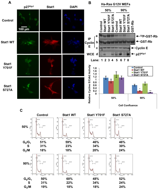
To better understand the biological significance of our findings, we looked at the localization of p27Kip1 in Ras-transfected MEFs. Nuclear localization of p27Kip1 is required for inhibition of cell cycle progression, which is counteracted in Ras transformed cells by the increased nucleocytoplasmic export and proteasomal degradation of p27Kip1 [12]. We found that p27Kip1 was both cytoplasmic and nuclear in MEFs lacking Stat1 (control cells) as well as in MEFs reconstituted with Stat1Y701F (Fig. 4A). However, p27Kip1 was predominantly nuclear in MEFs expressing either Stat1 WT or Stat1S727A compared to control MEFs or MEFs containing Stat1Y701F (Fig. 4A). On the other hand, Stat1 and its phosphorylation mutants were both nuclear and cytoplasmic in the reconstituted Ras-transfected MEFs (Fig. 4A). To determine whether increased p27Kip1 expression (Fig. 2B) had an effect on cell cycle progression, we measured the activity of the Cyclin E-Cdk2 complex, which is predominantly targeted by p27Kip1 [12]. To this end, Cyclin E-Cdk2 was purified by immunoprecipitation and subjected to in vitro kinase assays using GST-retinoblastoma (Rb) as substrate [33]. We found that Cyclin E-Cdk2 activity was reduced in Ras-transfected MEFs containing either Stat1 WT or Stat1 S727A (Fig. 4B, panel a). Although Cyclin E-Cdk2 activity declined with the increased density of all MEFs, the ability of either Stat1 WT or Stat1S727A to further inhibit CyclinE-Cdk2 activity was still evident in cells maintained at high confluence (Fig. 4B, see quantification in right panel). When we examined cell cycle progression by flow cytometry, we observed a blockade at G0/G1 phase in MEFs reconstituted with Stat1 WT when cells were maintained at either low or high confluence (Fig. 4C). Inhibition of cell cycle progression was also observed in confluent MEFs expressing Stat1S727A although to a lesser extent than in Stat1 WT MEFs (Fig. 4C). This difference between Stat1 WT and Stat1S727A can be explained by the ability of Stat1WT to upregulate both p21Cip1 and p27Kip1 and inhibit multiple Cyclin-Cdk complexes (Fig. 2B). These findings provided evidence that p27Kip1 expression contributes to the inhibition of cell cycle progression by Stat1 in a manner that is dependent on Stat1 phosphorylation and cell density.
Figure 4. p27Kip1 contributes to the inhibition of cell cycle progression by Stat1.
(A) MEFs maintained at 70% confluence were subjected to immunostaining with an anti-p27Kip1 mAb and a goat anti-mouse IgG conjugated to Alexa Fluro 488 (green). The nucleus was visualized by 4,6-diamidino-2-phenylindole (DAPI) staining. (B) Protein extracts (500 µg) from MEFs that reached 50% (lanes 1–4) or 90% confluence (lanes 5–8) were subjected to immunoprecipitation with an anti-Cyclin E antibody followed by in vitro kinase assays using GST-Rb (1 µg) and 1 µCi of [γ-32P] ATP (panel a). GST-Rb protein levels were visualized by Commassie blue staining (panel b). The levels of Cyclin E (panel c) and p27Kip1 (panel d) in the kinase assays were detected by immunoblotting. CyclinE-Cdk2 activity was assessed by normalizing GST-Rb phosphorylation levels to GST-Rb protein levels. The graph shows results expressed as ±SD from 3 independent experiments (*P<0.05; **P<0.01). (C) Cells were harvested at 50% (upper panel) or 90% confluence (lower panel), stained with propidium iodide and analyzed for DNA content by flow cytometry. The data shown represent one out of three reproducible experiments.
Phosphorylated Stat1 inhibits Ras-mediated transformation
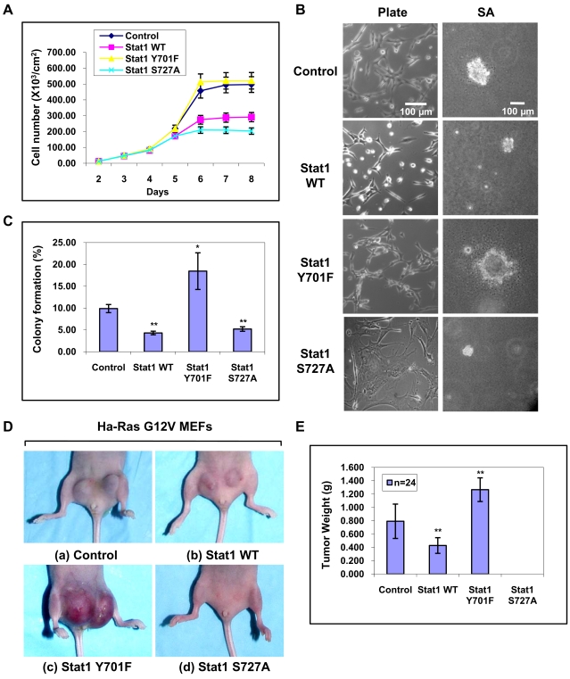
Next we examined the transforming potential of the Ras-transfected MEFs expressing various forms of Stat1. First, we observed that all MEFs propagated at similar rates when they were maintained at sub-confluent levels. However, in cells at high density, we observed a significant (50%) inhibition in the proliferation of MEFs reconstituted with either Stat1 WT or Stat1 S727A (Fig. 5A). Morphologically, Stat1 WT-expressing MEFs exhibited altered adhesive/spreading properties compared to control MEFs lacking Stat1 or MEFs reconstituted with each of the Stat1 phosphorylation mutants (Fig. 5A, left panels). When we looked at the growth of these cells in soft agar, we observed significant differences (Fig. 5B, right panels). Specifically, Ras-transfected MEFs reconstituted with either Stat1 WT or Stat1 S727A formed fewer colonies than control MEFs or MEFs reconstituted with Stat1Y701F (Fig. 5B, right panels). Furthermore, Ras-transfected MEFs with either Stat1 WT or Stat1 S727A yielded colonies that were smaller in size by 80% than the colonies derived from the control MEFs. On the other hand, MEFs reconstituted with Stat1 Y701F produced both the highest number of colonies and the largest colonies (Fig. 5C). Decreased colony formation by Stat1 WT or Stat1 S727A was not due to induction of Stat1-dependent apoptosis [34] as verified by annexin V staining and FACS analysis (Fig. S2). The differences in soft agar growth prompted us to examine the tumorigenic potential of the Ras-transfected MEFs. Tumor growth was assessed by the subcutaneous injection of the cells in athymic nude mice (Balb/c nu/nu). We found that control MEFs produced larger tumors than Stat1WT MEFs, which yielded ∼50% smaller tumors (Figs. 5D and E). On the other hand, MEFs reconstituted with Stat1Y701F yielded the largest tumors of all (Figs. 5D and E). Interestingly, MEFs expressing Stat1S727A did not produce tumors within the 3 week observation period (Figs. 5D and E) but yielded detectable tumors (∼2 mm) approximately 2.5 months after injection (data not shown). Histochemical analysis indicated that all tumors were high grade soft tissue sarcomas (data not shown). These data demonstrated that Stat1 functions as a suppressor of Ras-mediated oncogenesis in a manner that is dependent on site-specific Stat1 phosphorylation.
Figure 5. Stat1 inhibits Ras-mediated transformation.
(A) Growth rates of the MEFs were determined by counting the number of cells for the indicated periods of time. The results represent ±SD from two reproducible experiments performed in duplicate. (B) Morphological characteristics of MEFs grown on tissue culture dishes (left panel) or in soft agar (SA) (right panel). Bar, 100 µm. (C) Ras-transfected MEFs reconstituted with various Stat1 were plated in soft agar and let grow for 2 weeks. Colony formation of MEFs in soft agar was evaluated for clones larger than 100 µm. Data shown are ±SD from three independent experiments. *P<0.05; ** P<0.01. (D) MEFs were injected into 12 female athymic nude mice (Balb/c nu/nu). Each mouse received two subcutaneous injections (1×10⁶ cells for each of the 2 sites of injection) in the abdominal area proximal to the rear limbs (n = 2×12 = 24 injections). Mice were observed for tumor formation for ∼3 weeks until the largest tumor size became 2 cm in size at which point animals were sacrificed and tumors were excised and weighed. (E) Statistical analysis of tumor formation at 3 weeks post-injection. The average tumor weight (g) and ±SD are shown. **P<0.01.
p27Kip1 contributes to the inhibition of Ras-mediated transformation by Stat1
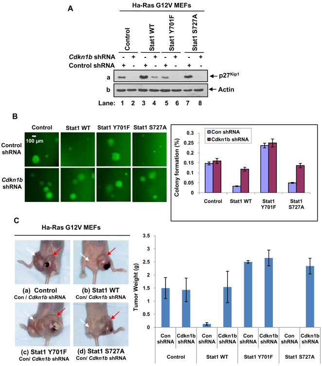
Previous findings established an essential role of p27Kip1 in the inhibition of Ras-mediated tumorigenesis [35]. To determine whether inhibition of Ras transformation by Stat1 involves p27Kip1, we assessed the transforming activity of the MEFs when endogenous p27Kip1 levels were decreased by shRNA. To this end, knockdown of p27Kip1 was achieved by infection of Ras transfected MEFs with retroviruses bearing Cdkn1b shRNA and the green fluorescence protein (GFP) as a marker [36]. As control, retroviruses bearing GFP and a shRNA against the luciferase reporter gene were used [36]. Decrease of p27Kip1 in the shRNA-treated MEFs was verified by immunoblotting (Fig. 6A). When the cells were plated in soft agar, we observed that anchorage-independent growth was restored in MEFs reconstituted with either Stat1 WT or Stat1S727A in which p27Kip1 was targeted by shRNA as indicated by the growth of the GFP-positive (green) colonies (Fig. 6B). On the other hand, decreased p27Kip1 levels did not further increase the ability of control MEFs lacking Stat1 or MEFs reconstituted with Stat1Y701F to form colonies in soft agar (Fig. 6B). The role of p27Kip1 in the inhibition of Ras-mediated tumorigenesis by Stat1 was further evaluated in nude mice. That is, tumor growth after subcutaneous injection of the MEFs in nude mice was significantly enhanced for MEFs reconstituted with either Stat1 WT or Stat1S727A and treated with shRNA against p27Kip1 (Fig. 6C). Contrary to this, downregulation of p27Kip1 in control MEFs or MEFs reconstituted with Stat1Y701 did not further enhance tumor growth (Fig. 6C). These finding demonstrated a major role of p27Kip1 in the inhibition of Ras-mediated oncogenesis by Stat1.
Figure 6. p27Kip1 mediates the inhibition of Ras-mediated transformation by Stat1.
(A) MEFs were infected with retroviruses bearing a shRNA against luciferase reporter gene (control shRNA) or shRNA against mouse Cdkn1b mRNA. Protein extracts (50 µg) were subjected to immunoblot analysis for p27Kip1 (panel a) and actin (panel b). (B) Cells expressing control shRNA or Cdkn1b shRNA were plated in soft agar and let grow for 2 weeks. Colonies expressing GFP (green) were evaluated for their ability to grow larger than 100 µm in size. Data shown are ±SD from three independent experiments. Colony formation (%) represents the number of cells forming colonies larger than 100 µm out of hundred plated cells. *P<0.05; **P<0.01. (C) MEFs treated with control shRNA or Cdkn1b shRNA were injected into 3 female athymic nude mice (Balb/c nu/nu). Each mouse received two subcutaneous injections (1×10⁶ cells per site of injection) in the abdomen proximal to the rear limbs. One injection contained MEFs treated with control shRNA (left side, white arrow) and the other injection contained MEFs treated with Cdkn1b shRNA (right side, red arrow). Mice were observed for tumor formation for ∼3 weeks until the largest tumor size became 2 cm in size at which point animals were sacrificed and tumors were excised and weighed. Statistical analysis of tumor formation at 3 weeks post-injection is shown in the graph. The average tumor weight (g) and ±SD are indicated. **P<0.01.
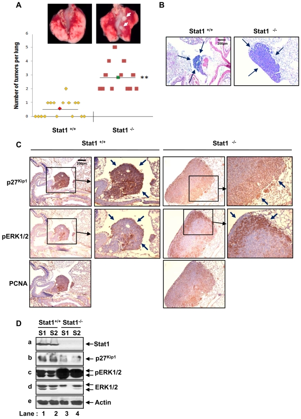
To further substantiate the importance of p27Kip1 and Stat1 in the suppression of Ras transformation, we examined the susceptibility of Stat1+/+ and Stat1−/− mice to urethane-induced tumorigenesis. Specifically, urethane treatment results primarily in the development of lung tumors that carry an activating mutation at codon 61 of K-Ras [37], [38]. Loss of p27Kip1 was shown to significantly increase the incidence and growth of lung tumors of mice treated urethane [39]. When Stat1+/+ and Stat1−/− mice were treated with a single intraperitoneal injection of urethane, we observed the development of tumors in both animal groups 28 weeks after treatment (Fig. 7A). However, only 50% of the animals in the Stat1+/+ group (8 out of 16) developed small tumors (<0.7 mm) as opposed to the Stat1−/− group in which all animals (n = 12) developed large tumors (>2 mm). Histological analysis indicated that the lung tumors were a mixture of bronchioalveolar adenomas and papillary adenomas (Fig. 7B). Immunohistochemical analysis further showed a high amount of p27Kip1 in lung tumors from Stat1+/+ mice compared to lung tumors from Stat1−/− mice (Fig. 7C). Consistent with tumor growth, we detected a higher amount of phosphorylated Erk1/2 in Stat1−/− than in Stat1+/+ lung tumors, which indicated the induction of the Ras-MAPK pathway by activated K-Ras (Fig. 7C). Interestingly, Erk1/2 phosphorylation levels were inversely proportional to p27Kip1 levels in the lung tumors as detected by immunohistochemistry (Fig. 7C) and immunoblotting (Fig. 7D). These data further indicated that both p27Kip1 and Stat1 function together to suppress Ras-mediated oncogenesis in vivo.
Figure 7. Stat1 inhibits lung tumor formation by activated K-Ras.
(A) Lungs were dissected and tumors (indicated by arrows) were counted by visual inspection 28 weeks after urethane treatment. Tumor multiplicity was increased in Stat1−/− mice (n = 12) compared to Stat1+/+ mice (n = 15). ** P<0.01. (B) Eosin and hematoxylin staining of lungs tissues from Stat1+/+ and Stat1−/− mice. The location of tumors in the stained lung tissue is indicated by arrows. Tumors were classified as bronchioalveolar adenomas and papillary adenomas. (C) Lung tissue from urethane-treated Stat1+/+ or Stat1−/− mice was subjected to immunohistochemical analysis for p27Kip1(top panels), phospho-(p)Erk1/2 (middle panels) and proliferating cell nuclear antigen (PCNA) (bottom panels). Staining of the areas in rectangles is shown in higher magnification in the right panels. The location of tumors is indicated by arrows. (D) Protein extracts (50 µg) from Stat1+/+(T1, T2) and Stat1−/−(T3, T4) lung tumors were separated by SDS-PAGE and subjected to immunoblot analysis for Stat1, p27Kip1, phospho(p)-Erk1/2, total Erk1/2 and actin.
Discussion
Our findings uncover an important function of Stat1 in the regulation of p27Kip1 with implications in Ras-mediated tumorigenesis. The ability of Stat1 to act upstream of p27Kip1 is a property of Ras transformation because Stat1 did not exhibit similar effects on Cdkn1b gene transcription or expression and localization of p27Kip1 in immortalized MEFs (Fig. S3). Moreover, induction of Cdkn1b gene transcription by Stat1 in Ras-transformed MEFs is independent of p53 (Fig. S4). The transcriptional effect of Stat1 on the Cdkn1b promoter depends on Y701 phosphorylation but is independent of S727 phosphorylation. Given the essential role of S727 phosphorylation in the transactivation properties of Stat1 in response to IFNs [40], [41], the dispensable role of S727 phosphorylation in the induction of the Cdkn1b gene indicated that transactivation of the Cdkn1b promoter was mediated by a protein other than Stat1. Consistent with this notion, our data demonstrated that the transcriptional induction of the Cdkn1b gene by Stat1 occurs in cooperation with Stat3. Stat3 was previously shown to activate the Cdkn1b gene during myeloid cell differentiation in response to IL-6 and G-CSF [18], [42]. The ability of Stat1 to cooperate with Stat3 in the transcriptional activation of the Cdkn1b gene suggests that Stat1 is capable of re-programming the biological function of Stat3 by converting it from a positive to a negative regulator of cell proliferation. At first glance, the ability of Stat3 to induce the expression of p27Kip1 in Ras transformed cells was not in line with its well characterized role as a positive regulator of cell proliferation and tumorigenesis [43], [44]. However, recent findings support the notion that Stat3 also possesses the capacity to impair cell proliferation and oncogenesis in a manner that depends on the signalling pathway and the genetic background of the target cells [45]. Although transcriptional control of the Cdkn1b gene by Stat1 plays a major role in regulating p27Kip1 levels in Ras transformed cells, the possibility that Stat1 can also regulate Cdkn1b gene expression at the post-transcriptional level can not be ruled out. This notion is supported by the observation that p27Kip1 is more highly expressed in Ras transformed MEFs reconstituted with Stat1 WT than with Stat1S727A (Fig. 2B) although both Stat1 proteins induce Cdkn1b gene transcription at comparable levels (Figs. 2C and D). Possible post-transcriptional regulation of Cdkn1b gene expression may occur at the level of mRNA translation and/or protein stability. At the translational level, Stat1 was previously shown to signal to the cellular translational machinery via physical and functional interactions with the eIF2α kinase PKR [46], [47]. At the post-translational level, the potential effects of Stat1 may be exerted through its ability to inhibit the transcription of c-myc [48], which induces the expression of proteins, including Cyclin D1, that sequester and inhibit p27Kip1 [13]. This is consistent with previous findings showing that Stat1 impairs c-myc and induces p27Kip1 expression in human monocytic U-937 cells in response to all-trans retinoid acid [49].
The anti-proliferative effects of Stat1 in response to IFNs are partly mediated by its ability to inhibit cell cycle progression [9], [48]. Our findings show that Stat1 is required for the upregulation of both p21Cip1 and p27Kip1 in Ras transformed cells in the absence of IFN treatment. It has been well documented that mitogens increase p21Cip1 levels through the activation of Ras and Raf-MAPK signalling [29] which results in increased transcription of the Cdkn1a gene [50]. Consistent with these findings, we found that induction of p21Cip1 in Ras transformed cells is dependent on Stat1 and is mediated at the transcriptional level (data not shown). However, unlike p27Kip1, the induction of p21Cip1 levels in Ras transformed cells depends on both tyrosine and serine phosphorylation of Stat1. Although Stat1 upregulates p21Cip1 and p27Kip1 levels in Ras transformed cells through separate mechanisms, both Cdk inhibitors appear to be involved in G0/G1 arrest (Fig. 4). It is of interest that the cell cycle inhibitory effects of Stat1 were increased in confluent cell cultures indicating a role of intercellular adhesion signalling in this process. Consistent with this observation, it was shown that Stat1 becomes activated by the focal adhesion kinase (FAK) with important implications in the regulation of cell adhesion and migration [51]. Given that p27Kip1 plays an important role in cell motility independent of its cell cycle regulatory functions [52], regulation of p27Kip1 levels by Stat1 may also have profound roles in cell migration and tumor metastasis [53].
Several findings support the anti-tumor function of Stat1 [43], [44]. Specifically, Stat1−/−mice are more prone to chemical induced carcinogenesis than Stat1+/+ mice, and Stat1−/− mice bred onto p53−/− background develop spontaneous tumors more rapidly than the p53−/− mice [6]. The high incidence of tumor formation in Stat1−/− animals is partly explained by impaired tumor immunosurveillance caused by defects in IFN-γ-signalling and natural killer cell activity [3]. Previous work established that the sensitivity of tumors to IFN-γ is required for the development of an anti-tumor response in immunocompetent hosts [6]. Because nude mice are not completely immunodeficient [54], the observed differences in tumor growth of the Ras transformed MEFs might have been attributed to their responsiveness to IFN-γ. However, we found that the responsiveness of the Ras transformed MEFs to IFN-γ did not correlate with their growth properties in nude mice. That is, although IFN-γ-mediated gene transactivation was impaired in Ras transformed cells expressing Stat1S727A (Fig. S5), these cells were barely tumorigenic in nude mice (Fig. 5). These observations argued against a role of tumor immunosurveillance in regulation of tumor growth in nude mice in our system. Also, growth of Ras-transformed cells in soft agar correlated with their growth in nude mice (Fig. 5) further supporting a direct role of Stat1 in suppression of Ras-mediated tumorigenesis. Our approach with shRNA clearly demonstrated that the anti-tumor activity of Stat1 is dependent on p27Kip1 (Fig. 6). It is of interest that tumor growth of Ras-transformed MEFs in nude mice is more highly suppressed by Stat1S727A than Stat1 WT (Fig. 5). Inasmuch as both Stat1 [53] and p27Kip1 [55] are involved in suppression of angiogenesis and Stat1 phosphorylation is affected by tumor hypoxia [56], tumor microenvironment may have more pronounced effects on the inhibition of tumor growth of Ras-transformed cells containing Stat1S727A than cells containing Stat1 WT. The effects of Stat1 are not confined to Ras-transformed MEFs only since activation of the K-Ras pathway in lung tissue by urethane results in a higher tumor incidence in Stat1−/− than in Stat1+/+ mice (Fig. 7). Although the increased tumor formation in urethane treated Stat1−/− mice could partly involve defects in tumor immunosurveillance [54], the higher incidence of lung tumor formation in Stat1−/− compared to Stat1+/+ mice was proportional to Ras-MAPK activation and inversely proportional to p27Kip1. Given that urethane-treated Cdkn1b −/− mice were more prone to lung tumorigenesis than Cdkn1b +/+ mice [57], together these data suggest that Stat1 and p27Kip1 act in the same pathway to inhibit Ras-mediated oncogenesis. It is of interest that ERK1/2 phosphorylation was diminished in lung tumors containing Stat1 compared to tumors devoid of Stat1 (Fig. 7C and D). This result indicated that Stat1 may have an inhibitory effect on Ras-MAPK signalling. Consistent with this notion, we noted that reconstitution of Ras-transfected Stat1−/−p53−/− MEFs with Stat1 WT resulted in a significant inhibition of ERK1/2 phosphorylation compared to control MEFs or MEFs reconstituted with each of the Stat1 phosphorylation mutants (Fig. S6). The molecular basis of this inhibition is not immediately clear and represents the focus of future experiments. Given that activation of the Ras-MAPK pathway results in the degradation of p27Kip1 [13], inhibition of the Ras-MAPK pathway by Stat1 may also reveal its ability to regulate p27Kip1 at the post-translational level.
There has been an established link between Stat1 phosphorylation and human cancer [44]. Specifically, Stat1 is constitutively phosphorylated at Y701 in many blood tumors including multiple myeloma, erythroleukemia and acute myelogenous leukemia (AML) [58]. In the case of solid tumors, Y701 phosphorylation of Stat1 has been detected in breast as well as in head and neck cancers [44]. Moreover, S727 phosphorylation of Stat1 is induced in chronic lymphocytic leukemia (CLL) [58], in Wilms' tumors [59] as well as in tumor cells deficient in tuberous sclerosis complex (TSC) 1 and 2 [60]. Given that phosphorylation is essential for Stat1 activation, detection of phosphorylated Stat1 in human tumors appears to be inconsistent with its anti-proliferative and tumor suppressor activities. However, recent findings showed that the anti-tumor function of Stat1 is determined by the type of the tumor and the oncogenic signalling within it. That is, Stat1 was shown to act as a promoter of leukemogenesis induced by v-abl and TEL-Jak2 oncogenes [61]. Our findings suggest a different regulation of the anti-tumor activity of Stat1 by site-specific phosphorylation. That is, activated Ras has the capacity to decrease Y701 phosphorylation and increase S727 phosphorylation of Stat1 (Fig. S1). These differences in Stat1 phosphorylation may significantly contribute to Ras-mediated tumorigenesis based on the ability of Stat1S727A or Stat1Y701F to compromise or promote the transforming activity of Ras in MEFs respectively (Figs. 5, 6). As such, it is reasonable to speculate that differences in the equilibrium between serine and tyrosine phosphorylation of Stat1 could determine the outcome of an oncogenic insult and the efficacy of chemotherapies aimed at inducing Stat1 phosphorylation [62]–[65]. Although Stat1 phosphorylation mutants have not been identified in human cancers, our findings indicate that Stat1 phosphorylation in tumors may interfere with the normal function of Stat1 and that the occurrence and frequency of site-specific phosphorylated Stat1 in human cancers could be of significant diagnostic and prognostic value. Consistent with this notion, tyrosine phosphorylation of Stat1 was shown to be a marker in the prognostic evaluation of breast tumors [66] as well as of head and neck tumors [67].
Materials and Methods
Animals and treatments
BALB/c Stat1−/− mice [4] and wild type BALB/c mice from Harlan labs were maintained as previously described [4]. Athymic mice (Balb/c nu/nu), female and 4–6 weeks old, were provided by Charles River. Urethane treatment was carried out using a previously described protocol [57]. Mice were sacrificed after 28 weeks, dissected and examined for lung tumors. Lungs were fixed in formalin, embedded in paraffin, and slides (4 µm thick) were subjected to immuno-histochemical analysis. Tumors in athymic mice were monitored daily for ∼3 weeks to ensure that the conditions and good welfare of the animals were not compromised. The mice were sacrificed when the tumor size reached 2 cm at which point they became cumbersome or necrotic. The animal experiments were performed in accordance with approved protocols and regulations by the Animal Welfare Committee of McGill University (protocol #3271).
Cell culture procedures
Mouse embryonic fibroblasts (MEFs) and NIH3T3 cells (ATCC CRL-1658) were maintained in Dulbecco's modified Eagle's medium (DMEM) (Gibco) supplemented with 10% calf serum and antibiotics. Infection with pBabe-expressing retroviruses was described elsewhere [47]. The luciferase assays were performed with the Dual-Luciferase Reporter Assay System (Promega) using Renilla luciferase as an internal control. Soft agar growth assays were performed as described [68].
Plasmids and antibodies
Myc-tagged Ha-RasG12V cDNA was subcloned into the EcoRV site of pcDNA3.1/Hygro (Invitrogen). The pBabe vector containing wild type (WT) HA-Stat1 was described previously [47]. HA-Stat1S727A was produced by the QuickChange site-directed mutagenesis (Stratagene) using the primers 5′- CAACCTGCTCCCCATGGCACCTGAGGAGTTTGACGAGG-3′ and 5′-CCTCGTCA AACTCCTCAGGTGCCATGGGGAGCAGGTTG-3′ on wild type template vector. HA-Stat1S727A and HA-Stat1Y701F cDNA [69] were subcloned into the SnaB I site of the pBabe vector. Cdkn1b shRNA and luciferase shRNA in a pSIREN vector were reported elsewhere [36]. The pGL2 vector containing the luciferase reporter gene under the control of full length mouse Cdkn1b promoter (−1609 to + 178 bp) was described [30]. The PGL3 vectors containing the luciferase gene under the control of wild type or mutant Stat-binding site of the Cdkn1b promoter was described [18]. The Stat3-D cDNA in pcDNA expression vector was previously described [32].
Anti-Stat1α p91(C-111), anti-Myc (9E10), anti-Stat3(C-20), anti-Stat1(M-23) and anti-p21(C-19) antibodies were purchased from Santa Cruz Biotechnology; anti-pY701-Stat1, anti-pS727-Stat1 and phosphor-p44/p42 MAPK (Thr202/Tyr204) antibodies from Cell Signalling; anti-actin (C4) from Biosource International; anti-p27Kip1 antibody from BD Transduction Laboratories; anti-Cyclin E rabbit serum was provided by Dr. A. Besson. The horseradish peroxidase (HRP)-conjugated anti-mouse IgG antibody and HRP anti-rabbit IgG antibody were from Amersham Pharmacia Biotech. The Alexa Fluor 488 conjugated goat anti-mouse IgG and Alexa Fluor 546 conjugated goat anti-rabbit IgG antibodies were from Molecular Probes.
Immunoblottings, immunoprecipitations, immunofluorescence and flow cytometry
Immunoblottings and immunoprecipitations were performed as described [70] whereas cell cycle analysis was based on a established protocol [71]. Immunofluorescence analysis was performed as reported [72].
Northern blotting and electrophoretic mobility shift assays (EMSAs)
Northern blotting using 15 µg of total RNA was performed as described [70]. EMSAs were performed based on a previously established protocol [46] using an oligonucleotide encompassing the Stat-binding site of the mouse Cdkn1b promoter in wild type form (5′-TTAATTTCCTGTAACATG-3′) or in its mutant form (5′- TTAATTGTCTGCGACATG-3′; mutations are underlined) as reported [18].
Chromatin immunoprecipitation (ChIP) assays
ChIP assays were carried out based on a protocol described elsewhere [73]. Polymerase chain reaction (PCR) was performed using the Cdkn1b forward primer 5′-GTGGCTAAGAAAACAAGTCAAT-3′ and reverse primer 5′-TAGCCAGGCCTGTCGTATCTCA-3′. The conditions were: 94°C for 5 min, 30 cycles at 94°C for 1 min, 55°C for 1 min, 72°C for 30 sec and a final elongation at 72°C for 10 min.
Cyclin E –Cdk2 kinase assay
Immunoprecipitation of Cyclin E-Cdk2 and in vitro kinase assays using GST-Rb were performed as previously described [33].
Supporting Information
Figure S1
Control of Stat1 phosphorylation by activated Ras. NIH3T3 cells were infected with pBabe retroviruses lacking (control; Con) or bearing activated Ha-RasG12V. After selection in 2 µg/ml puromycin for 2 weeks, polyclonal populations were maintained at different levels of confluency (50–100%). Protein extracts (50 µg) were subjected to immunoblot analysis for Stat1 phosphorylated at Y701 (panel a) or S727 (panel b), total Stat1 (panel c), ERK1/2 phosphorylated at Thr202/Tyr204 (panel d), total ERK1/2 (panel e) or actin (panel f). The doublet recognized by the Stat1 Y701 phosphospecific antibody most likely represents the two isoforms (α and β) of Stat1.
(6.40 MB TIF)
Figure S2
Evaluation of apoptosis in Ras-transformed Stat1−/−p53−/− MEFs reconstituted with the various forms of Stat1. Sub-confluent Ras-transformed Stat1−/−p53−/− MEFs lacking (Control) or reconstituted with either Stat1 WT or Stat1S727A were subjected to staining with Annexin V-propidium iodide (PI) staining according to the manufacturer's specifications (Biosource). Cells were then subjected to flow cytometry analysis by using FACScan (Becton Dickinson), and data were analyzed by using WinMDI version 2.8 software (The Scripps Institute). The data represent one out of two reproducible experiments.
(4.84 MB TIF)
Figure S3
Detection of p27Kip1 localization and Cdkn1b mRNA levels in immortalized MEFs containing Stat1 WT or Stat1 phosphorylation mutants. (A) Spontaneously immortalized isogenic Stat1−/− MEFs as well as Stat1−/− MEFs reconstituted with Stat1 WT were subjected to immunostaining for endogenous p27Kip1 and Stat1 as described in Fig. 4A. (B) Immortalized Stat1−/− MEFs reconstituted with either Stat1 WT or Stat1 phosphorylation mutants (i.e. Stat1Y701F, Stat1S727A) were maintained at 90% confluency and subjected to Northern blot analysis for detection of endogenous Cdkn1b (a) and GAPDH mRNA levels (b) as described in Fig. 2C. The levels of reconstituted Stat1 proteins were detected by immunoblot analysis (panel c). The data represent one out of two reproducible experiments.
(5.93 MB TIF)
Figure S4
Examination of the role of p53 in induction of Cdkn1b gene transcription by Stat1. Ras-transformed Stat1−/−p53−/− MEFs (Control) and Ras-trasnformed Stat1−/−p53−/− MEFs reconstituted with Stat1 WT (Stat1 WT) were transfected with pCL2 vector containing the firefly luciferase reporter gene under the control of the full length mouse Cdkn1b promoter (Cdkn1bWT) together with the pcDNA3.0 vector lacking (pcDNA3) or containing the mouse wild type p53 cDNA (p53). As control, pCL2 vector containing the firefly luciferase gene but lacking the Cdkn1b promoter was used. The firefly luciferase levels were normalized to Renilla luciferase driven from the minimal promoter in the pGL3 vector used as an internal control. Results are expressed ±SD for 3 experiments performed in triplicate.
(3.95 MB TIF)
Figure S5
Control of IFN-γ-mediated gene transactivation in Ras-trasnfected MEFs. MEFs were transiently transfected with a firefly luciferase reporter gene under the control of a promoter containing two IFN-γ-activated sites (GAS) from the IFP53 gene (pGL-2XIFP53 GAS luciferase). Thirty two hours post transfection cells were left untreated or treated with 500 IU/ml of mouse IFN-γ (Biosource) for 12 hours. Cells were harvested and assayed for firefly luciferase activity and normalized to an internal control consisting of a renilla luciferase reporter. Results are expressed ±SD for 3 experiments performed in triplicate.
(3.37 MB TIF)
Figure S6
Detection of ERK1/2 phosphorylation in Ras-transfromed MEFs. Protein extracts (50 µg) from confluent cells were subjected to immunoblotting for ERK1/2 phosphorylated at Thr202/Tyr204 (panel a) as well as for total ERK1/2 (panel b). The ratio of phosphorylated to non-phosphorylated ERK1/2 for each lane is indicated. The data represent one out of two reproducible experiments.
(3.77 MB TIF)
Acknowledgments
We thank A. Besson for p27Kip1−/− MEFs and anti-Cyclin E polyclonal antibody; Y. Gotoh for p27Kip1shRNA construct; T. Hirano for HA-Stat1 Y701F cDNA and Stat3-D cDNA; N. Lamarche-Vane for Myc-tagged RasG12V cDNA; Dr. J. Chen for pGL2 vector containing the mouse Cdkn1b promoter; I.P.Touw for pGL3 vector containing either the WT or mutant form of Stat-binding site of the mouse Cdkn1b promoter; G. Ferbeyre for pBabe-RasG12V retrovirus; T.A. Bismar and N. Benlimame for immunohistochemical analysis. T. Taniguchi, S. Richard and G. Ferbeyre for helpful comments.
Footnotes
Competing Interests: The authors have declared that no competing interests exist.
Funding: The work was funded with a grant from the Canadian Breast Cancer Research Alliance (CBCRA) awarded through the Canadian Institutes of Health Research (CIHR) to AEK. JFR is the recipient of a U.S. Army Pre-doctoral Traineeship Award. The funders had no role in study design, data collection and analysis, decision to publish, or preparation of the manuscript.
References
- 1.Schindler C, Levy DE, Decker T. JAK-STAT signaling: from interferons to cytokines. J Biol Chem. 2007;282:20059–20063. doi: 10.1074/jbc.R700016200. [DOI] [PubMed] [Google Scholar]
- 2.Levy DE, Darnell JE., Jr Stats: transcriptional control and biological impact. Nat Rev Mol Cell Biol. 2002;3:651–662. doi: 10.1038/nrm909. [DOI] [PubMed] [Google Scholar]
- 3.Dunn GP, Koebel CM, Schreiber RD. Interferons, immunity and cancer immunoediting. Nat Rev Immunol. 2006;6:836–848. doi: 10.1038/nri1961. [DOI] [PubMed] [Google Scholar]
- 4.Durbin JE, Hackenmiller R, Simon MC, Levy DE. Targeted disruption of the mouse Stat1 gene results in compromised innate immunity to viral disease. Cell. 1996;84:443–450. doi: 10.1016/s0092-8674(00)81289-1. [DOI] [PubMed] [Google Scholar]
- 5.Meraz MA, White JM, Sheehan KC, Bach EA, Rodig SJ, et al. Targeted disruption of the Stat1 gene in mice reveals unexpected physiologic specificity in the JAK-STAT signaling pathway. Cell. 1996;84:431–442. doi: 10.1016/s0092-8674(00)81288-x. [DOI] [PubMed] [Google Scholar]
- 6.Kaplan DH, Shankaran V, Dighe AS, Stockert E, Aguet M, et al. Demonstration of an interferon gamma-dependent tumor surveillance system in immunocompetent mice. Proc Natl Acad Sci U S A. 1998;95:7556–7561. doi: 10.1073/pnas.95.13.7556. [DOI] [PMC free article] [PubMed] [Google Scholar]
- 7.Bromberg JF, Horvath CM, Wen Z, Schreiber RD, Darnell JE., Jr Transcriptionally active Stat1 is required for the antiproliferative effects of both interferon alpha and interferon gamma. Proc Natl Acad Sci U S A. 1996;93:7673–7678. doi: 10.1073/pnas.93.15.7673. [DOI] [PMC free article] [PubMed] [Google Scholar]
- 8.Chin YE, Kitagawa M, Kuida K, Flavell RA, Fu XY. Activation of the STAT signaling pathway can cause expression of caspase 1 and apoptosis. Mol Cell Biol. 1997;17:5328–5337. doi: 10.1128/mcb.17.9.5328. [DOI] [PMC free article] [PubMed] [Google Scholar]
- 9.Chin YE, Kitagawa M, Su WC, You ZH, Iwamoto Y, et al. Cell growth arrest and induction of cyclin-dependent kinase inhibitor p21 WAF1/CIP1 mediated by STAT1. Science. 1996;272:719–722. doi: 10.1126/science.272.5262.719. [DOI] [PubMed] [Google Scholar]
- 10.Kumar A, Commane M, Flickinger TW, Horvath CM, Stark GR. Defective TNF-alpha-induced apoptosis in STAT1-null cells due to low constitutive levels of caspases. Science. 1997;278:1630–1632. doi: 10.1126/science.278.5343.1630. [DOI] [PubMed] [Google Scholar]
- 11.Decker T, Kovarik P. Serine phosphorylation of STATs. Oncogene. 2000;19:2628–2637. doi: 10.1038/sj.onc.1203481. [DOI] [PubMed] [Google Scholar]
- 12.Besson A, Dowdy SF, Roberts JM. CDK inhibitors: cell cycle regulators and beyond. Dev Cell. 2008;14:159–169. doi: 10.1016/j.devcel.2008.01.013. [DOI] [PubMed] [Google Scholar]
- 13.Chu IM, Hengst L, Slingerland JM. The Cdk inhibitor p27 in human cancer: prognostic potential and relevance to anticancer therapy. Nat Rev Cancer. 2008;8:253–267. doi: 10.1038/nrc2347. [DOI] [PubMed] [Google Scholar]
- 14.Fero ML, Randel E, Gurley KE, Roberts JM, Kemp CJ. The murine gene p27Kip1 is haplo-insufficient for tumour suppression. Nature. 1998;396:177–180. doi: 10.1038/24179. [DOI] [PMC free article] [PubMed] [Google Scholar]
- 15.Wei Q, Miskimins WK, Miskimins R. The Sp1 family of transcription factors is involved in p27(Kip1)-mediated activation of myelin basic protein gene expression. Mol Cell Biol. 2003;23:4035–4045. doi: 10.1128/MCB.23.12.4035-4045.2003. [DOI] [PMC free article] [PubMed] [Google Scholar]
- 16.Paris M, Wang WH, Shin MH, Franklin DS, Andrisani OM. Homeodomain transcription factor Phox2a, via cyclic AMP-mediated activation, induces p27Kip1 transcription, coordinating neural progenitor cell cycle exit and differentiation. Mol Cell Biol. 2006;26:8826–8839. doi: 10.1128/MCB.00575-06. [DOI] [PMC free article] [PubMed] [Google Scholar]
- 17.Myatt SS, Lam EW. The emerging roles of forkhead box (Fox) proteins in cancer. Nat Rev Cancer. 2007;7:847–859. doi: 10.1038/nrc2223. [DOI] [PubMed] [Google Scholar]
- 18.de Koning JP, Soede-Bobok AA, Ward AC, Schelen AM, Antonissen C, et al. STAT3-mediated differentiation and survival and of myeloid cells in response to granulocyte colony-stimulating factor: role for the cyclin-dependent kinase inhibitor p27(Kip1). Oncogene. 2000;19:3290–3298. doi: 10.1038/sj.onc.1203627. [DOI] [PubMed] [Google Scholar]
- 19.Kortylewski M, Heinrich PC, Mackiewicz A, Schniertshauer U, Klingmuller U, et al. Interleukin-6 and oncostatin M-induced growth inhibition of human A375 melanoma cells is STAT-dependent and involves upregulation of the cyclin-dependent kinase inhibitor p27/Kip1. Oncogene. 1999;18:3742–3753. doi: 10.1038/sj.onc.1202708. [DOI] [PubMed] [Google Scholar]
- 20.Hengst L, Reed SI. Translational control of p27Kip1 accumulation during the cell cycle. Science. 1996;271:1861–1864. doi: 10.1126/science.271.5257.1861. [DOI] [PubMed] [Google Scholar]
- 21.Millard SS, Vidal A, Markus M, Koff A. A U-rich element in the 5′ untranslated region is necessary for the translation of p27 mRNA. Mol Cell Biol. 2000;20:5947–5959. doi: 10.1128/mcb.20.16.5947-5959.2000. [DOI] [PMC free article] [PubMed] [Google Scholar]
- 22.Gopfert U, Kullmann M, Hengst L. Cell cycle-dependent translation of p27 involves a responsive element in its 5′-UTR that overlaps with a uORF. Hum Mol Genet. 2003;12:1767–1779. doi: 10.1093/hmg/ddg177. [DOI] [PubMed] [Google Scholar]
- 23.Miskimins WK, Wang G, Hawkinson M, Miskimins R. Control of cyclin-dependent kinase inhibitor p27 expression by cap-independent translation. Mol Cell Biol. 2001;21:4960–4967. doi: 10.1128/MCB.21.15.4960-4967.2001. [DOI] [PMC free article] [PubMed] [Google Scholar]
- 24.Jiang H, Coleman J, Miskimins R, Srinivasan R, Miskimins WK. Cap-independent translation through the p27 5′-UTR. Nucleic Acids Res. 2007;35:4767–4778. doi: 10.1093/nar/gkm512. [DOI] [PMC free article] [PubMed] [Google Scholar]
- 25.Hanahan D, Weinberg RA. The hallmarks of cancer. Cell. 2000;100:57–70. doi: 10.1016/s0092-8674(00)81683-9. [DOI] [PubMed] [Google Scholar]
- 26.Kerkhoff E, Rapp UR. Cell cycle targets of Ras/Raf signalling. Oncogene. 1998;17:1457–1462. doi: 10.1038/sj.onc.1202185. [DOI] [PubMed] [Google Scholar]
- 27.Song JH, So EY, Lee CE. Increased serine phosphorylation and activation of STAT1 by oncogenic Ras transfection. Mol Cells. 2002;13:322–326. [PubMed] [Google Scholar]
- 28.Evdonin AL, Medvedeva ND, Pospelova TV, Pospelov VA. [Transcription factor p91/Stat1 is constitutively phosphorylated through tyrosine and is bound to MAP-kinase in rats embryonal fibroblasts, transformed by E1A and Ha-Ras oncogenes]. Mol Biol (Mosk) 1998;32:712–716. [PubMed] [Google Scholar]
- 29.Coleman ML, Marshall CJ, Olson MF. RAS and RHO GTPases in G1-phase cell-cycle regulation. Nat Rev Mol Cell Biol. 2004;5:355–366. doi: 10.1038/nrm1365. [DOI] [PubMed] [Google Scholar]
- 30.Wang C, Hou X, Mohapatra S, Ma Y, Cress WD, et al. Activation of p27Kip1 Expression by E2F1. A negative feedback mechanism. J Biol Chem. 2005;280:12339–12343. doi: 10.1074/jbc.C400536200. [DOI] [PubMed] [Google Scholar]
- 31.Kwon TK, Nagel JE, Buchholz MA, Nordin AA. Characterization of the murine cyclin-dependent kinase inhibitor gene p27Kip1. Gene. 1996;180:113–120. doi: 10.1016/s0378-1119(96)00416-7. [DOI] [PubMed] [Google Scholar]
- 32.Nakajima K, Yamanaka Y, Nakae K, Kojima H, Ichiba M, et al. A central role for Stat3 in IL-6-induced regulation of growth and differentiation in M1 leukemia cells. EMBO J. 1996;15:3651–3658. [PMC free article] [PubMed] [Google Scholar]
- 33.Besson A, Yong VW. Involvement of p21(Waf1/Cip1) in protein kinase C alpha-induced cell cycle progression. Mol Cell Biol. 2000;20:4580–4590. doi: 10.1128/mcb.20.13.4580-4590.2000. [DOI] [PMC free article] [PubMed] [Google Scholar]
- 34.Kim HS, Lee MS. STAT1 as a key modulator of cell death. Cell Signal. 2007;19:454–465. doi: 10.1016/j.cellsig.2006.09.003. [DOI] [PubMed] [Google Scholar]
- 35.Pruitt K, Der CJ. Ras and Rho regulation of the cell cycle and oncogenesis. Cancer Lett. 2001;171:1–10. doi: 10.1016/s0304-3835(01)00528-6. [DOI] [PubMed] [Google Scholar]
- 36.Itoh Y, Masuyama N, Nakayama K, Nakayama KI, Gotoh Y. The cyclin-dependent kinase inhibitors p57 and p27 regulate neuronal migration in the developing mouse neocortex. J Biol Chem. 2007;282:390–396. doi: 10.1074/jbc.M609944200. [DOI] [PubMed] [Google Scholar]
- 37.Meuwissen R, Berns A. Mouse models for human lung cancer. Genes Dev. 2005;19:643–664. doi: 10.1101/gad.1284505. [DOI] [PubMed] [Google Scholar]
- 38.Horio Y, Chen A, Rice P, Roth JA, Malkinson AM, et al. Ki-ras and p53 mutations are early and late events, respectively, in urethane-induced pulmonary carcinogenesis in A/J mice. Mol Carcinog. 1996;17:217–223. doi: 10.1002/(SICI)1098-2744(199612)17:4<217::AID-MC5>3.0.CO;2-A. [DOI] [PubMed] [Google Scholar]
- 39.Besson A, Gurian-West M, Chen X, Kelly-Spratt KS, Kemp CJ, et al. A pathway in quiescent cells that controls p27Kip1 stability, subcellular localization, and tumor suppression. Genes Dev. 2006;20:47–64. doi: 10.1101/gad.1384406. [DOI] [PMC free article] [PubMed] [Google Scholar]
- 40.Wen Z, Zhong Z, Darnell JE., Jr Maximal activation of transcription by Stat1 and Stat3 requires both tyrosine and serine phosphorylation. Cell. 1995;82:241–250. doi: 10.1016/0092-8674(95)90311-9. [DOI] [PubMed] [Google Scholar]
- 41.Varinou L, Ramsauer K, Karaghiosoff M, Kolbe T, Pfeffer K, et al. Phosphorylation of the Stat1 transactivation domain is required for full-fledged IFN-gamma-dependent innate immunity. Immunity. 2003;19:793–802. doi: 10.1016/s1074-7613(03)00322-4. [DOI] [PubMed] [Google Scholar]
- 42.Kortylewski M, Heinrich PC, Mackiewicz A, Schniertshauer U, Klingmuller U, et al. Interleukin-6 and oncostatin M-induced growth inhibition of human A375 melanoma cells is STAT-dependent and involves upregulation of the cyclin-dependent kinase inhibitor p27/Kip1. Oncogene. 1999;18:3742–3753. doi: 10.1038/sj.onc.1202708. [DOI] [PubMed] [Google Scholar]
- 43.Bromberg J. Stat proteins and oncogenesis. J Clin Invest. 2002;109:1139–1142. doi: 10.1172/JCI15617. [DOI] [PMC free article] [PubMed] [Google Scholar]
- 44.Yu H, Jove R. The stats of cancer–new molecular targets come of age. Nat Rev Cancer. 2004;4:97–105. doi: 10.1038/nrc1275. [DOI] [PubMed] [Google Scholar]
- 45.de la Inglesia N, Konopka G, Puram SV, Chan JA, Bachoo RM, et al. Identification of a PTEN-regulated STAT3 brain tumor suppressor pathway. Genes Dev. 2008;22:449–462. doi: 10.1101/gad.1606508. [DOI] [PMC free article] [PubMed] [Google Scholar]
- 46.Wong AH, Tam NW, Yang YL, Cuddihy AR, Li S, et al. Physical association between STAT1 and the interferon-inducible protein kinase PKR and implications for interferon and double-stranded RNA signaling pathways. EMBO J. 1997;16:1291–1304. doi: 10.1093/emboj/16.6.1291. [DOI] [PMC free article] [PubMed] [Google Scholar]
- 47.Wong AH, Durbin JE, Li S, Dever TE, Decker T, et al. Enhanced antiviral and antiproliferative properties of a STAT1 mutant unable to interact with the protein kinase PKR. J Biol Chem. 2001;276:13727–13737. doi: 10.1074/jbc.M011240200. [DOI] [PubMed] [Google Scholar]
- 48.Ramana CV, Grammatikakis N, Chernov M, Nguyen H, Goh KC, et al. Regulation of c-myc expression by IFN-gamma through Stat1-dependent and -independent pathways. EMBO J. 2000;19:263–272. doi: 10.1093/emboj/19.2.263. [DOI] [PMC free article] [PubMed] [Google Scholar]
- 49.Dimberg A, Karlberg I, Nilsson K, Oberg F. Ser727/Tyr701-phosphorylated Stat1 is required for the regulation of c-Myc, cyclins, and p27Kip1 associated with ATRA-induced G0/G1 arrest of U-937 cells. Blood. 2003;102:254–261. doi: 10.1182/blood-2002-10-3149. [DOI] [PubMed] [Google Scholar]
- 50.Liu Y, Martindale JL, Gorospe M, Holbrook NJ. Regulation of p21WAF1/CIP1 expression through mitogen-activated protein kinase signaling pathway. Cancer Res. 1996;56:31–35. [PubMed] [Google Scholar]
- 51.Xie B, Zhao J, Kitagawa M, Durbin J, Madri JA, et al. Focal adhesion kinase activates Stat1 in integrin-mediated cell migration and adhesion. J Biol Chem. 2001;276:19512–19523. doi: 10.1074/jbc.M009063200. [DOI] [PubMed] [Google Scholar]
- 52.Besson A, Gurian-West M, Schmidt A, Hall A, Roberts JM. p27Kip1 modulates cell migration through the regulation of RhoA activation. Genes Dev. 2004;18:862–876. doi: 10.1101/gad.1185504. [DOI] [PMC free article] [PubMed] [Google Scholar]
- 53.Huang S, Bucana CD, Van Arsdall M, Fidler IJ. Stat1 negatively regulates angiogenesis, tumorigenicity and metastasis of tumor cells. Oncogene. 2002;21:2504–2512. doi: 10.1038/sj.onc.1205341. [DOI] [PubMed] [Google Scholar]
- 54.Dunn GP, Bruce AT, Ikeda H, Old LJ, Schreiber RD. Cancer immunoediting: from immunosurveillance to tumor escape. Nat Immunol. 2002;3:991–998. doi: 10.1038/ni1102-991. [DOI] [PubMed] [Google Scholar]
- 55.Goukassian D, ez-Juan A, Asahara T, Schratzberger P, Silver M, et al. Overexpression of p27(Kip1) by doxycycline-regulated adenoviral vectors inhibits endothelial cell proliferation and migration and impairs angiogenesis. FASEB J. 2001;15:1877–1885. doi: 10.1096/fj.01-0065com. [DOI] [PubMed] [Google Scholar]
- 56.Terui K, Haga S, Enosawa S, Ohnuma N, Ozaki M. Hypoxia/re-oxygenation-induced, redox-dependent activation of STAT1 (signal transducer and activator of transcription 1) confers resistance to apoptotic cell death via hsp70 induction. Biochem J. 2004;380:203–209. doi: 10.1042/BJ20031891. [DOI] [PMC free article] [PubMed] [Google Scholar]
- 57.Besson A, Gurian-West M, Chen X, Kelly-Spratt KS, Kemp CJ, et al. A pathway in quiescent cells that controls p27Kip1 stability, subcellular localization, and tumor suppression. Genes Dev. 2006;20:47–64. doi: 10.1101/gad.1384406. [DOI] [PMC free article] [PubMed] [Google Scholar]
- 58.Lin TS, Mahajan S, Frank DA. STAT signaling in the pathogenesis and treatment of leukemias. Oncogene. 2000;19:2496–2504. doi: 10.1038/sj.onc.1203486. [DOI] [PubMed] [Google Scholar]
- 59.Timofeeva OA, Plisov S, Evseev AA, Peng S, Jose-Kampfner M, et al. Serine-phosphorylated STAT1 is a prosurvival factor in Wilms' tumor pathogenesis. Oncogene. 2006 doi: 10.1038/sj.onc.1209742. [DOI] [PubMed] [Google Scholar]
- 60.El-Hashemite N, Zhang H, Walker V, Hoffmeister KM, Kwiatkowski DJ. Perturbed IFN-gamma-Jak-signal transducers and activators of transcription signaling in tuberous sclerosis mouse models: synergistic effects of rapamycin-IFN-gamma treatment. Cancer Res. 2004;64:3436–3443. doi: 10.1158/0008-5472.CAN-03-3609. [DOI] [PubMed] [Google Scholar]
- 61.Kovacic B, Stoiber D, Moriggl R, Weisz E, Ott RG, et al. STAT1 acts as a tumor promoter for leukemia development. Cancer Cell. 2006;10:77–87. doi: 10.1016/j.ccr.2006.05.025. [DOI] [PubMed] [Google Scholar]
- 62.Youlyouz-Marfak I, Gachard N, Le CC, Najjar I, Baran-Marszak F, et al. Identification of a novel p53-dependent activation pathway of STAT1 by antitumour genotoxic agents. Cell Death Differ. 2008;15:376–385. doi: 10.1038/sj.cdd.4402270. [DOI] [PubMed] [Google Scholar]
- 63.Thomas M, Finnegan CE, Rogers KM, Purcell JW, Trimble A, et al. STAT1: a modulator of chemotherapy-induced apoptosis. Cancer Res. 2004;64:8357–8364. doi: 10.1158/0008-5472.CAN-04-1864. [DOI] [PubMed] [Google Scholar]
- 64.Fryknas M, Dhar S, Oberg F, Rickardson L, Rydaker M, et al. STAT1 signaling is associated with acquired crossresistance to doxorubicin and radiation in myeloma cell lines. Int J Cancer. 2007;120:189–195. doi: 10.1002/ijc.22291. [DOI] [PubMed] [Google Scholar]
- 65.Townsend PA, Cragg MS, Davidson SM, McCormick J, Barry S, et al. STAT-1 facilitates the ATM activated checkpoint pathway following DNA damage. J Cell Sci. 2005;118:1629–1639. doi: 10.1242/jcs.01728. [DOI] [PubMed] [Google Scholar]
- 66.Widschwendter A, Tonko-Geymayer S, Welte T, Daxenbichler G, Marth C, et al. Prognostic significance of signal transducer and activator of transcription 1 activation in breast cancer. Clin Cancer Res. 2002;8:3065–3074. [PubMed] [Google Scholar]
- 67.Laimer K, Spizzo G, Obrist P, Gastl G, Brunhuber T, et al. STAT1 activation in squamous cell cancer of the oral cavity: a potential predictive marker of response to adjuvant chemotherapy. Cancer. 2007;110:326–333. doi: 10.1002/cncr.22813. [DOI] [PubMed] [Google Scholar]
- 68.Koromilas AE, Roy S, Barber GN, Katze MG, Sonenberg N. Malignant transformation by a mutant of the IFN-inducible dsRNA-dependent protein kinase. Science. 1992;257:1685–1689. doi: 10.1126/science.1382315. [DOI] [PubMed] [Google Scholar]
- 69.Sekimoto T, Nakajima K, Tachibana T, Hirano T, Yoneda Y. Interferon-gamma-dependent nuclear import of Stat1 is mediated by the GTPase activity of Ran/TC4. J Biol Chem. 1996;271:31017–31020. doi: 10.1074/jbc.271.49.31017. [DOI] [PubMed] [Google Scholar]
- 70.Wang S, Raven JF, Baltzis D, Kazemi S, Brunet DV, et al. The catalytic activity of the eukaryotic initiation factor-2alpha kinase PKR is required to negatively regulate Stat1 and Stat3 via activation of the T-cell protein-tyrosine phosphatase. J Biol Chem. 2006;281:9439–9449. doi: 10.1074/jbc.M504977200. [DOI] [PubMed] [Google Scholar]
- 71.Kazemi S, Papadopoulou S, Li S, Su Q, Wang S, et al. Control of alpha subunit of eukaryotic translation initiation factor 2 (eIF2 alpha) phosphorylation by the human papillomavirus type 18 E6 oncoprotein: implications for eIF2 alpha-dependent gene expression and cell death. Mol Cell Biol. 2004;24:3415–3429. doi: 10.1128/MCB.24.8.3415-3429.2004. [DOI] [PMC free article] [PubMed] [Google Scholar]
- 72.Qu L, Huang S, Baltzis D, Rivas-Estilla AM, Pluquet O, et al. Endoplasmic reticulum stress induces p53 cytoplasmic localization and prevents p53-dependent apoptosis by a pathway involving glycogen synthase kinase-3beta. Genes Dev. 2004;18:261–277. doi: 10.1101/gad.1165804. [DOI] [PMC free article] [PubMed] [Google Scholar]
- 73.Hartman SE, Bertone P, Nath AK, Royce TE, Gerstein M, et al. Global changes in STAT target selection and transcription regulation upon interferon treatments. Genes Dev. 2005;19:2953–2968. doi: 10.1101/gad.1371305. [DOI] [PMC free article] [PubMed] [Google Scholar]
Associated Data
This section collects any data citations, data availability statements, or supplementary materials included in this article.
Supplementary Materials
Figure S1
Control of Stat1 phosphorylation by activated Ras. NIH3T3 cells were infected with pBabe retroviruses lacking (control; Con) or bearing activated Ha-RasG12V. After selection in 2 µg/ml puromycin for 2 weeks, polyclonal populations were maintained at different levels of confluency (50–100%). Protein extracts (50 µg) were subjected to immunoblot analysis for Stat1 phosphorylated at Y701 (panel a) or S727 (panel b), total Stat1 (panel c), ERK1/2 phosphorylated at Thr202/Tyr204 (panel d), total ERK1/2 (panel e) or actin (panel f). The doublet recognized by the Stat1 Y701 phosphospecific antibody most likely represents the two isoforms (α and β) of Stat1.
(6.40 MB TIF)
Figure S2
Evaluation of apoptosis in Ras-transformed Stat1−/−p53−/− MEFs reconstituted with the various forms of Stat1. Sub-confluent Ras-transformed Stat1−/−p53−/− MEFs lacking (Control) or reconstituted with either Stat1 WT or Stat1S727A were subjected to staining with Annexin V-propidium iodide (PI) staining according to the manufacturer's specifications (Biosource). Cells were then subjected to flow cytometry analysis by using FACScan (Becton Dickinson), and data were analyzed by using WinMDI version 2.8 software (The Scripps Institute). The data represent one out of two reproducible experiments.
(4.84 MB TIF)
Figure S3
Detection of p27Kip1 localization and Cdkn1b mRNA levels in immortalized MEFs containing Stat1 WT or Stat1 phosphorylation mutants. (A) Spontaneously immortalized isogenic Stat1−/− MEFs as well as Stat1−/− MEFs reconstituted with Stat1 WT were subjected to immunostaining for endogenous p27Kip1 and Stat1 as described in Fig. 4A. (B) Immortalized Stat1−/− MEFs reconstituted with either Stat1 WT or Stat1 phosphorylation mutants (i.e. Stat1Y701F, Stat1S727A) were maintained at 90% confluency and subjected to Northern blot analysis for detection of endogenous Cdkn1b (a) and GAPDH mRNA levels (b) as described in Fig. 2C. The levels of reconstituted Stat1 proteins were detected by immunoblot analysis (panel c). The data represent one out of two reproducible experiments.
(5.93 MB TIF)
Figure S4
Examination of the role of p53 in induction of Cdkn1b gene transcription by Stat1. Ras-transformed Stat1−/−p53−/− MEFs (Control) and Ras-trasnformed Stat1−/−p53−/− MEFs reconstituted with Stat1 WT (Stat1 WT) were transfected with pCL2 vector containing the firefly luciferase reporter gene under the control of the full length mouse Cdkn1b promoter (Cdkn1bWT) together with the pcDNA3.0 vector lacking (pcDNA3) or containing the mouse wild type p53 cDNA (p53). As control, pCL2 vector containing the firefly luciferase gene but lacking the Cdkn1b promoter was used. The firefly luciferase levels were normalized to Renilla luciferase driven from the minimal promoter in the pGL3 vector used as an internal control. Results are expressed ±SD for 3 experiments performed in triplicate.
(3.95 MB TIF)
Figure S5
Control of IFN-γ-mediated gene transactivation in Ras-trasnfected MEFs. MEFs were transiently transfected with a firefly luciferase reporter gene under the control of a promoter containing two IFN-γ-activated sites (GAS) from the IFP53 gene (pGL-2XIFP53 GAS luciferase). Thirty two hours post transfection cells were left untreated or treated with 500 IU/ml of mouse IFN-γ (Biosource) for 12 hours. Cells were harvested and assayed for firefly luciferase activity and normalized to an internal control consisting of a renilla luciferase reporter. Results are expressed ±SD for 3 experiments performed in triplicate.
(3.37 MB TIF)
Figure S6
Detection of ERK1/2 phosphorylation in Ras-transfromed MEFs. Protein extracts (50 µg) from confluent cells were subjected to immunoblotting for ERK1/2 phosphorylated at Thr202/Tyr204 (panel a) as well as for total ERK1/2 (panel b). The ratio of phosphorylated to non-phosphorylated ERK1/2 for each lane is indicated. The data represent one out of two reproducible experiments.
(3.77 MB TIF)
