Notch signaling controls multiple steps of pancreatic differentiation (original) (raw)
Abstract
Multiple cell types of the pancreas appear asynchronously during embryogenesis, which requires that pancreatic progenitor cell potential changes over time. Loss-of-function studies have shown that Notch signaling modulates the differentiation of these progenitors, but it remains unclear how and when the Notch pathway acts. We established a modular transgenic system to heritably activate mouse Notch1 in multiple types of progenitors and differentiated cells. We find that misexpression of activated Notch in _Pdx1_-expressing progenitor cells prevents differentiation of both exocrine and endocrine lineages. Progenitors remain trapped in an undifferentiated state even if Notch activation occurs long after the pancreas has been specified. Furthermore, endocrine differentiation is associated with escape from this activity, because _Ngn3_-expressing endocrine precursors are susceptible to Notch inhibition, whereas fully differentiated endocrine cells are resistant.
The adult pancreas is a confederation of different cell types, including exocrine acini that produce digestive enzymes, ducts that channel those enzymes into the gut, and islets of Langerhans that themselves include multiple classes of endocrine hormone-producing cells. Although all of these cells arise embryonically from a pair of _Pdx1_-expressing endodermal patches (1, 2), their development is not synchronous.
Pdx1 expression initiates at approximately embryonic day 8.5 (E8.5) (2, 3), and glucagon-producing α cells begin to appear as soon as 1 day later (4, 5). Endocrine cells, including the first mature insulin-producing β cells, are born in increasing numbers beginning at approximately E13.5, and exocrine cells appear shortly thereafter (4, 5). The Pdx1+ progenitor populations at E8.5, E13.5, and later are thus quite distinct, owing to either a changing inductive milieu or an intrinsic timing program. For example, temporally regulated lineage tracing has uncovered a major distinction among Pdx1+ cells in that E9.5–E11.5 Pdx1+ progenitors contain a population that will contribute to adult duct cells as well as exocrine and endocrine cells, whereas Pdx1+ progenitors in older embryos are restricted to acinar and islet fates (1).
Loss-of-function experiments have shown that the Notch signaling pathway promotes pancreatic progenitor cell self-renewal and/or exocrine lineage commitment (6, 7). Although pancreata lacking the Notch signaling components Delta1 or _RBPJ-_κ exhibit excess α cell production (6), these mutant mice die between E9.5 and E12.5, before effects on β cell and acinar differentiation can be ascertained. Moreover, although Hes1 mutants survive to late gestation stages, they do not exhibit major defects in β cell or exocrine cell differentiation (7), raising the possibility that Notch primarily regulates α cell numbers. Because all of these experiments ablate Notch signaling at the earliest steps of pancreas development, they do not address how Notch might affect pancreatic progenitors at stages relevant to the full range of exocrine and endocrine fates.
To compare the effects of Notch activation across multiple lineages and time points, we exploited a bigenic Cre-lox system that combines multiple Cre transgenic lines with a single floxed Notch transgene. We find that, throughout embryogenesis, Pdx1+ progenitors are sensitive to Notch signaling, which acts cell-autonomously to trap them in an undifferentiated state that then persists into adulthood. Although Neurogenin3 (Ngn3)-expressing endocrine precursors are thought to arise from cells that evade Notch signaling and therefore might be insensitive to the pathway, we find that activation of Notch specifically in Ngn3+ precursors blocks their differentiation as well. Once differentiation is complete, however, endocrine cells become resistant to this activity. These findings highlight the utility of a bigenic approach, which can be extended to the study of additional lineages, organs, and signaling pathways.
Materials and Methods
Targeting NotchIC to the Rosa26 Locus. We transferred a DNA fragment (generously provided by Sean Megason and Andy McMahon, Harvard University) that encoded (from 5′ to 3′) an intracellular fragment of mouse Notch1 (amino acids 1749–2293, lacking the C-terminal PEST domain), an internal ribosome entry sequence, and nuclear-localized enhanced GFP (EGFP) into a previously described Rosa26 targeting vector (8). The resulting construct was linearized and electroporated into AK7 embryonic stem cells (129/SvJaeSor, supplied by Phil Soriano, Fred Hutchison Cancer Research Center, Seattle). Three of 12 colonies initially screened by Southern blotting showed correct targeting; one of these three was used to generate chimeras.
Animals. Chimeric males were bred to C57BL/6 females, and the resulting line (designated RosaNotch) was maintained on a hybrid 129 × C57BL/6 background. RosaNotch mice are healthy and fertile as heterozygotes or homozygotes. PCR genotyping was performed as described (9). Pdx1-Cre, Pdx1-CreER, and Ngn3-Cre mice (1) were maintained on an ICR background. For conditional Cre recombination, tamoxifen (TM; Sigma T-5648) was dissolved in corn oil and introduced by i.p. injection at doses of 1.5–4 mg.
Tissue Analyses. Embryonic and adult tissue was fixed in 4% paraformaldehyde/PBS for 1–4 h at 4°C and processed for serial 7-μm wax (immunohistochemistry and in situ hybridization) or 10- to 12-μm frozen (immunofluorescence) sectioning by standard techniques. Ngn3 expression was detected by in situ hybridization with a DIG-labeled probe (provided by David Anderson, California Institute of Technology, Pasadena). Primary antibodies used for immunohistochemistry and immunofluorescence were as follows: guinea pig anti-rat C-peptide (1:2,500; Linco Research Immunoassay, St. Charles, MO), guinea pig anti-glucagon (1:2500; Linco Research Immunoassay), rabbit anti-human amylase (1:500; Sigma), and guinea pig anti-Pdx1 (1:1,000; a gift from Chris Wright, Vanderbilt University, Nashville, TN). Secondary antibodies (biotinylated or rhodamine red X-conjugated) were purchased from Jackson ImmunoResearch, and histochemical staining was performed with Vectastain reagents and diaminobenzidene substrate from Vector Laboratories. Confocal images were acquired on a Nikon TE2000U inverted microscope equipped with a Perkin–Elmer Ultraview Spinning Disk Confocal head and a Orca ER cooled charge-coupled device camera (Hamamatsu, Middlesex, NJ) and analyzed by using metamorph software (Universal Imaging, Media, PA). Brightfield images were collected by using a Nikon E800 upright microscope equipped with a Insight Camera (Diagnostic Instruments, Sterling Heights, MI) and analyzed with metamorph.
Statistical Analysis. For each embryo, positive cells were counted on 6–11 evenly spaced sections spanning the volume of the developing pancreas, and the average number of cells per section was calculated. For each genotype we present the mean of these averages ± SD; P values were determined by using a two-tailed t test.
Results
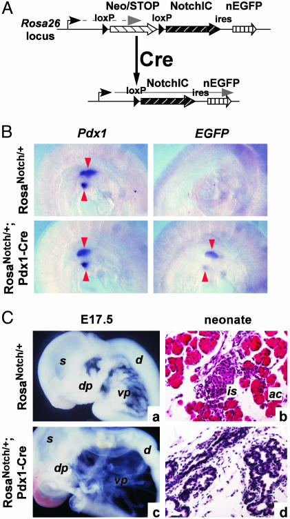
Directed Misexpression of Activated Notch1. The truncated cytoplasmic domains of Notch molecules exhibit constitutive signaling activity (10, 11). We targeted a cDNA encoding an intracellular fragment of mouse Notch1 (NotchIC; amino acids 1749–2293) to the ubiquitously expressed Rosa26 locus (9), followed by internal ribosome entry sequence–nuclear EGFP, and preceded by a STOP fragment flanked by loxP sites (Fig. 1_A_). In the absence of Cre recombinase, transcription of the modified locus (RosaNotch) is blocked by the STOP sequence; in the presence of Cre, STOP will be deleted, yielding heritable, constitutive coexpression of NotchIC and nuclear EGFP.
Fig. 1.
Cre-activated misexpression of activated Notch1. (A) Structure of the RosaNotch locus. Transcription (gray arrow) is initially blocked by the STOP cassette, which is deleted by Cre recombinase, permitting transcription of NotchIC and internal ribosome entry sequence–nuclear EGFP (nEGFP). (B) Whole-mount in situ hybridizations (purple staining) for Pdx1 and EGFP at E9.5, when transgene activation is first detected in the pancreatic buds (arrowheads) of bigenic RosaNotch;Pdx1-Cre embryos. (C) Gross phenotype of monogenic and bigenic pancreata. (a and c) At E17.5, bigenic pancreas is translucent and cystic. (b and d) Hematoxylin/eosin-stained sections of neonatal pancreata reveal tubular epithelia and absence of normal acinar and islet tissue in RosawNotch;Pdx1-Cre bigenics. ac, acini; d, duodenum; dp, dorsal pancreas; is, islet; s, stomach; vp, ventral pancreas.
When we crossed RosaNotch mice with mice expressing Cre under control of the Pdx1 promoter (Pdx1-Cre; ref. 1), we observed transgene activation from approximately E9.5 within the dorsal and ventral pancreatic buds (Fig. 1_B_). Although monogenic embryos (i.e., Pdx1-Cre or RosaNotch alone) were indistinguishable from wild type at all stages examined, pancreata of bigenic embryos exhibited a visually obvious phenotype at E14.5 and later. As shown in Fig. 1_Ca_, the wild-type pancreas at E17.5 is made up largely of nearly opaque acinar tissue, which at high magnification resolves into tight clusters of cells with microscopic lumens. In RosaNotch/+;Pdx1-Cre embryos, by contrast, the pancreatic epithelium is translucent and comprised of highly branched, tubular epithelium with few distinct acini (Fig. 1_Cc_). Whereas newborn pancreata exhibit obvious acinar and islet tissue in histological section (Fig. 1_Cb_), those of bigenic littermates are dominated by convoluted epithelium in a fibro-blastic stroma (Fig. 1_Cd_).
Notch Signaling Inhibits both Exocrine and Endocrine Development. The differentiation of NotchIC-expressing pancreata was assessed by immunohistochemistry. NotchIC expression in the developing Pdx1 lineage was associated with a significant decrease in the numbers of endocrine α and β cells, producing glucagon and insulin, respectively. At E15.5, α cell numbers were depressed 8-fold (53.0 ± 11.8 cells per monogenic RosaNotch/+ section, n = 3 embryos vs. 6.7 ± 4.1 per bigenic RosaNotch/+;Pdx1-Cre section, n = 5; P < 0.001), and β cell numbers were repressed nearly 100-fold (monogenic 103 ± 55 cells per section vs. bigenic 1.22 ± 1.23 cells per section; P < 0.005). A similar effect was observed with the rarer somatostatin- and pancreatic polypeptide-producing cells (data not shown).
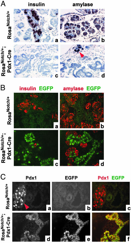
The epithelial tubules formed in the RosaNotch/+; Pdx1-Cre pancreata are negative for endocrine markers such as insulin (Fig. 2_A a_ and c) and exocrine markers such as amylase (Fig. 2 A b and d). We observe no apoptosis within the EGFP+ epithelium, as detected by staining for cleaved caspase-3 (data not shown). Confocal immunofluorescence indicates that most of the residual differentiated cells in the bigenic pancreata are EGFP-negative (Fig. 2_B_), indicating that they arose from progenitors that failed to activate RosaNotch or underwent transgene silencing. NotchIC thus appears to impose a formidable barrier to both exocrine and endocrine differentiation.
Fig. 2.
Early activation of RosaNotch inhibits endocrine and exocrine development. (A) Insulin and amylase expression in E15.5 monogenics (a and b) and bigenics (c and d) stained by diaminobenzidene (brown) with hematoxylin counterstain. Bigenic RosaNotch;Pdx1-Cre pancreata contain tubular epithelial cells devoid of differentiation markers. Arrowhead in d indicates a cluster of acinar cells in the bigenic pancreas. (B) Confocal immunofluorescence indicates that residual differentiated cells (red) in bigenics (c and d) are largely EGFP-negative (green), indicating that they do not express NotchIC. Green signal in monogenic sections (a and b) represents background autofluorescence. (C) Pdx1 (a and d) and EGFP (b and e) expression detected by confocal immunofluorescence in neonatal pancreata. In RosaNotch monogenics, Pdx1 is largely restricted to β cells (arrowhead in a indicates an islet), whereas bigenic pancreata maintain broad Pdx1 expression in EGFP+ cells (merged signal in c and f; overlap is yellow).
The NotchIC-expressing epithelium remains Pdx1+ both in embryos (data not shown), when Pdx1 expression is broad in the developing pancreas, and in newborns (Fig. 2_C_), when Pdx1 is normally restricted to differentiated β cells (ref. 3 and data not shown). Expression of Glut2, a marker of undifferentiated pancreatic epithelium (12), is similarly widespread in the NotchIC-expressing epithelium (data not shown). Rather than promote exocrine fates at the expense of endocrine, NotchIC appears to block both lineages, possibly by arresting progenitor cell differentiation.
Progenitor Cells Remain Sensitive to NotchIC During Later Pancreatic Development. The phenotype observed in RosaNotch/+; Pdx1-Cre bigenic embryos reflects transgene expression in E9.5 progenitor cells and their offspring. To determine whether these early Pdx1+ cells represent a unique population with respect to Notch responsiveness, we used a transgenic strain, Pdx1-CreER (1), in which the Pdx1 promoter drives expression of a fusion protein between Cre recombinase and a TM-dependent estrogen receptor (ER™). This protein is active only when bound to 4-hydroxytamioxifen, a metabolite of TM. i.p. TM injection rapidly activates Cre-ER™, allowing it to catalyze loxP recombination; the spatial extent of recombination is proportional to the dose of TM (13). We crossed RosaNotch and Pdx1-CreER mice, administered TM to pregnant females at different stages of gestation, and analyzed embryos several days later.
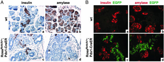
We find that progenitor cells remain susceptible to NotchIC-mediated inhibition even following the early stages of pancreas specification. Sequential injection of TM at E13.5 and E14.5, 1.5–2 mg each day, resulted in a phenotype at E17.5 similar to that of RosaNotch/+;Pdx1-Cre bigenics, characterized by tubular epithelium devoid of endocrine and exocrine markers (Fig. 3_A c_ and d). Sequential injection at E11.5 and E12.5 also recapitulated the RosaNotch/+;Pdx1-Cre phenotype at E14.5 (data not shown). TM had no effect on nontransgenic or monogenic animals (Fig. 3_A a_ and b and data not shown).
Fig. 3.
Late activation of RosaNotch inhibits endocrine and exocrine development. (A) Embryos were treated with sequential doses of TM at E13.5 and E14.5 and examined at E17.5. Expression of insulin and amylase (brown) is inhibited in bigenics (c and d) compared with wild-type littermates (a and b), concomitant with appearance of undifferentiated epithelial tubules. (B) Embryos were administered a single dose of TM at E11.5 and examined at E15.5 by immunofluorescence for insulin and amylase (red; EGFP is shown in green). EGFP expression in bigenics (c and d) is excluded from differentiated cells.
Widespread misexpression of NotchIC results in aberrant morphogenesis as well as impaired differentiation. To reduce the number of affected cells and hence minimize effects on morphogenesis that might indirectly influence differentiation, we administered a single TM dose (1.5 mg) to E11.5 embryos. When harvested at E15.5, no gross difference was seen between monogenic and bigenic guts. Nonetheless, immunofluorescence reveals the existence of a population of EGFP+ cells localized to core regions of the bigenic pancreata (Fig. 3_B c_ and d) lacking endocrine or exocrine marker expression. NotchIC signaling thus inhibits Pdx1+ progenitor cell differentiation at multiple stages of embryogenesis, cell-autonomously and independent of aberrant morphogenesis.
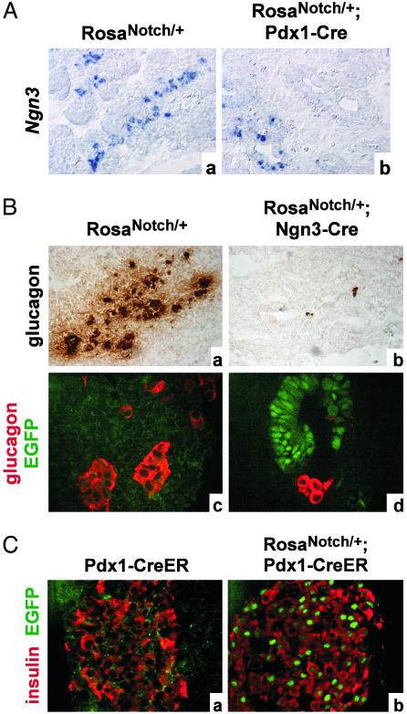
Effects of Activated Notch on Committed Endocrine Precursors and Differentiated Islet Cells. A subset of pancreatic progenitors normally activate Ngn3 expression and initiate endocrine differentiation (1, 14); loss of Notch signaling expands this population (6). Conversely, we find that NotchIC expression reduces Ngn3+ cell numbers by ≈4-fold at E15.5 (82.4 ± 24.5 cells per monogenic RosaNotch/+ section, n = 5 embryos vs. 19.8 ± 9.4 per bigenic RosaNotch/+;Pdx1-Cre section, n = 10; P < 0.0001) (Fig. 4_A_). Unlike differentiated cells, however, most residual Ngn3-expressing cells in bigenic embryos are EGFP+ (data not shown), suggesting that the NotchIC-expressing epithelium retains a limited capacity for lineage selection.
Fig. 4.
Stages of endocrine lineage commitment responsive to NotchIC inhibition. (A) Ngn3 expression in E15.5 monogenic RosaNotch/+ (a) and bigenic RosaNotch/+;Pdx1-Cre (b) pancreata. Ngn3+ cells are reduced in number in the bigenic and are absent from most epithelial tubules. (B) Glucagon expression in E13.5 monogenic RosaNotch;Ngn3-Cre and bigenic pancreata, stained in brightfield (brown, a and b) or by immunofluorescence (red, merged with EGFP in green, c and d). Bigenic embryos exhibit dramatic reduction of glucagon-positive cells, and remaining cells are EGFP-negative, indicating that they do not express NotchIC. (C) Widespread expression of NotchIC in adult β cells, by adult activation of Pdx1-CreER (b), does not affect insulin expression (red; EGFP is shown in green) or islet morphology.
As described above, further endocrine differentiation of these EGFP+, Notch-expressing progenitors appears severely attenuated (Fig. 2 A and B). To determine whether Notch signaling can directly inhibit differentiation of cells already committed to the endocrine lineage, we crossed RosaNotch mice with mice expressing Cre under control of the Ngn3 promoter (Ngn3-Cre; ref. 1) to activate NotchIC specifically in endocrine precursors.
For reasons that are unclear, presumably because of Cre activity outside of the pancreas, we were unable to recover viable RosaNotch/+;Ngn3-Cre embryos past E13.5. Because β cell differentiation is minimal at this stage, we were restricted to analyzing glucagon-producing α cells. As shown in Fig. 4_B_, the monogenic E13.5 pancreas contains abundant glucagon+ cells, whereas the bigenic RosaNotch/+;Ngn3-Cre pancreas is almost devoid of them. The remaining glucagon-positive cells in bigenic pancreata lack EGFP expression, likely indicating that they failed to activate RosaNotch in the course of differentiation (Fig. 4_Bd_). Although abundant EGFP+ cells are found in the RosaNotch/+;Ngn3-Cre pancreas, their development appears deranged: like normal descendants of Ngn3+ cells, they do not sustain Ngn3 expression, but they are also negative for Isl1, an early marker of endocrine differentiation (data not shown). NotchIC thus imposes at least two blockades to endocrine differentiation: It inhibits Pdx1+ progenitors from activating Ngn3 and initiating the cascade of endocrine lineage commitment (Fig. 4_A_), and it prevents Ngn3+ cells from executing their differentiation program (Fig. 4_B_).
Epithelial expression of Pdx1 progressively fades during development and becomes restricted to the insulin-producing β cells near birth (3). We therefore performed an experiment to determine the effect of expressing NotchIC in adult β cells by injecting adult RosaNotch;Pdx1-CreER bigenic animals with TM (four i.p. doses of 4 mg each over a 1-week period). To detect both acute and longer-term effects of NotchIC expression, we examined the bigenic pancreas 1 day after the injection series: at this time point, the islets should contain a heterogenous population of β cells that have expressed NotchIC for 1–8 days. Although most islets exhibited widespread activation of the transgene, as detected by EGFP expression, the β cells continued to express insulin at levels indistinguishable from their EGFP-negative neighbors (Fig. 4_C_); there was evidence of neither a dedifferentiated population arising in response to NotchIC nor cell death (data not shown). These cells also express Pdx1 and Glut2, markers of mature β cells, at levels indistinguishable from monogenics (data not shown). Identical results were obtained in islets examined 1 week after the injection series, i.e., in β cells expressing NotchIC for 8–15 days (data not shown). NotchIC expression apparently does not perturb mature endocrine cells, suggesting that its effects in the endocrine lineage are restricted to progenitors.
Maintenance of Undifferentiated Epithelial Cells Through Adulthood. Having shown that NotchIC prevents endocrine and exocrine differentiation, we wanted to establish the ultimate fate of the abnormal epithelium in bigenic animals. One possibility is that NotchIC collaborates with another signal, present specifically during embryogenesis, and that as epithelial cells age they progressively differentiate or are eliminated. To investigate this, we bred RosaNotch and Pdx1-Cre mice and let offspring grow to adulthood. As expected from the lack of endocrine and exocrine cells in developing bigenics, the majority of such animals die perinatally: in crosses of RosaNotch/Notch males with hemizygous Pdx1-Cre females, we recovered 11 Pdx1-Cre+ bigenics at weaning compared with 83 Cre-negative monogenics. Nonetheless, the observed mosaicism of Pdx1-Cre activity (Fig. 2_B_) permits the survival of subset of bigenics.
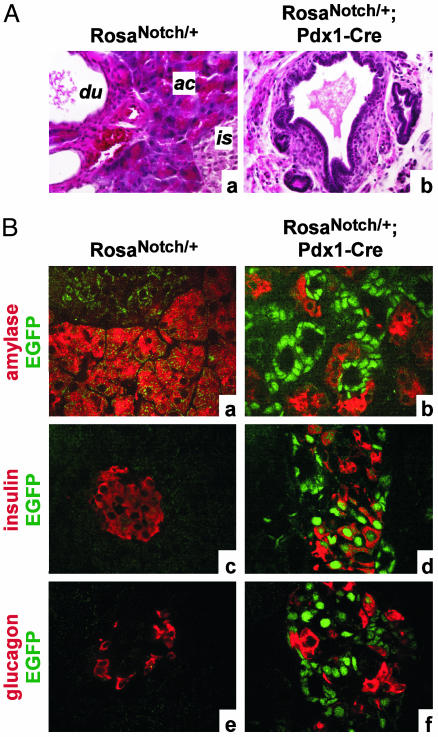
If the blockade to exocrine or endocrine differentiation in NotchIC+ cells required an embryonic environment, we would expect these cells to differentiate or die in adulthood. Instead, the pancreata of bigenic adults contain abundant epithelial cells histologically similar to those seen during development (Fig. 5_A_). As in embryos, there is a clear segregation of differentiated acinar tissue and EGFP+ tubular epithelium (Fig. 5_B a_ and b); however, β cells frequently express EGFP (Fig. 5_B c_ and d), whereas α cells in the same islet are mostly EGFP-negative (Fig. 5_B e_ and f). This presumably reflects Pdx1-Cre activation of RosaNotch in already differentiated β cells as shown in Fig. 4_C_. Thus, Notch-inhibited progenitors can survive and proliferate (data not shown) into adulthood, whereas differentiated β cells remain unaffected by the transgene.
Fig. 5.
Persistence of undifferentiated cells in adult RosaNotch;Pdx1-Cre bigenics. (A) Wild-type pancreas (a) comprises islet, acinar, and duct tissue, whereas the bigenic adult pancreata contains large regions of tubular epithelium (b) similar to that seen in embryos. (B) As in the embryo, EGFP+ epithelial cells are largely negative for amylase (a and b) and glucagon (e and f) expression, whereas insulin-positive cells frequently coexpress EGFP (c and d), suggesting RosaNotch activation subsequent toβ cell differentiation (differentiation markers are shown in red, and EGFP is shown in green). ac, acini; du, duct; is, islet.
Discussion
The pancreas is typical of many complex organs in that its multiple cell types arise over distinct developmental periods yet apparently derive from similar early progenitor cells (1). Although little is known about the mechanism that governs the changing potential of pancreatic progenitors, it presumably involves a combination of extrinsic cues and intrinsic programming. This implies that a progenitor cell present in the newly specified pancreas, shortly after Pdx1 expression initiates at E8.5, will be different from a lineal descendant of that cell at E11.5 or E13.5 in terms of both its environment and its responsiveness to that environment.
The differentiated cell types themselves reflect such differences: early endocrine cells formed prior to E13.5 differ in marker expression and genetic requirements from those comprising the mature islet (15, 16). In addition, temporal lineage tracing reveals that duct cell precursors, uniquely, arise before E11.5 (1). These factors confound traditional studies of gene and pathway function. In a knockout mouse, the gene function of interest will be absent at the onset of organogenesis, long before mature cell types appear. Any role that the gene might play during differentiation could therefore be obscured by an earlier role in organ specification or growth. Similar problems apply to misexpression experiments in the pancreas, which have typically relied on the Pdx1 promoter. This promoter will drive transgene expression in newly specified pancreatic epithelium, again several days before the appearance of mature endocrine and exocrine cells.
Our modular strategy offers certain advantages over prior approaches. For example, because the misexpressor (RosaNotch) is inactive before Cre recombination, a single founder line can be established, eliminating any concern about artifactual differences in expression levels or pattern from one line to the next. Integration of the transgene in a single, defined genomic locus ensures its productive activation by Cre. Most importantly, we can direct transgene expression in progenitor cells after the early events of organogenesis are complete, when mature cell type specification is actually underway.
Our results indicate that expression of activated Notch1 throughout the pancreas is sufficient to prevent both endocrine and exocrine development and appears to trap both early and late progenitors in an undifferentiated state. Despite the lack of pertinent data from loss-of-function experiments (6, 7), our results indicate that Notch signaling is sufficient to arrest progenitor cell differentiation at stages relevant to acinar and β cell formation. Furthermore, we find that cells undergoing endocrine differentiation lose responsiveness to Notch, because NotchIC activation in Ngn3+ endocrine precursors prevents their differentiation, whereas activation in mature β cells is without effect. In the wild-type pancreas, transient Notch signaling may delay the differentiation of Ngn3+ cells, which normally proliferate before differentiating (17). It will be interesting to observe whether specific removal of Notch function in the Ngn3 lineage leads to a loss of this expansion process.
Hald et al. (18) recently published transient transgenesis experiments in which the Pdx1 promoter was used to misexpress a similar form of activated Notch1. Our data regarding RosaNotch;Pdx1-Cre embryos are similar to theirs, although they report a more profound defect in foregut morphogenesis, leading to near agenesis of the pancreas. This phenotype may be the result of directly misexpressing NotchIC under control of the Pdx1 promoter, which likely results in earlier transgene expression than in our two-step Cremediated approach (approximately E8.5, compared with E9.5). Part of the phenotype described by Hald et al. (18) may thus pertain to a role for Notch in the earliest events of organ specification.
Our results further emphasize the potential clinical importance of Notch signaling in the pancreas. Because its activity inhibits β cell development without promoting exocrine fates, Notch stimulation might serve to expand multipotent pancreatic progenitors in vitro, to generate β cells for transplantation. On the other hand, it is important to note that NotchIC-expressing pancreatic cells are not hyperproliferative, and as development proceeds the cell mass of the bigenic organ appears to lag behind that of wild type, despite a near absence of apoptosis (data not shown). In light of the recent finding that Notch is activate in pancreatic tumorigenesis (19), our data suggest that its role is to prevent differentiation of cancer precursors, permitting other signals to drive their proliferation. If Notch could be inhibited, tumor cells might initiate a differentiation program that would render them refractory to growth-promoting signals.
Acknowledgments
We thank Jolanta Dubauskaite for blastocyst injections; Jennifer Waters Shuler (Nikon Imaging Center, Harvard Medical School) for assistance with microscopy; Sean Megason, Andy McMahon, Chris Wright, Phil Soriano, Elizabeth Robertson, David Anderson, andFrank Constantini for generous gifts of reagents and advice; and members of the Melton laboratory for feedback and patience. D.A.M. is a Howard Hughes Medical Institute Investigator. This work was supported by fellowships from the Helen Hay Whitney Foundation (to L.C.M.), the Howard Hughes Medical Institute, and the National Institutes of Health (to B.Z.S.).
Abbreviations: EGFP, enhanced GFP; TM, tamoxifen; E_n_, embryonic day n.
References
- 1.Gu, G., Dubauskaite, J. & Melton, D. A. (2002) Development (Cambridge, U.K.) 129**,** 2447–2457. [DOI] [PubMed] [Google Scholar]
- 2.Ohlsson, H., Karlsson, K. & Edlund, T. (1993) EMBO J. 12**,** 4251–4259. [DOI] [PMC free article] [PubMed] [Google Scholar]
- 3.Guz, Y., Montminy, M. R., Stein, R., Leonard, J., Gamer, L. W., Wright, C. V. & Teitelman, G. (1995) Development 121**,** 11–18. [DOI] [PubMed] [Google Scholar]
- 4.Pictet, R. & Rutter, W. J. (1972) in Handbook of Physiology, Section 7, eds. Steiner, D. & Freinkel, N. (Williams & Williams, Baltimore), Vol. 1, pp. 25–66. [Google Scholar]
- 5.Slack, J. M. (1995) Development (Cambridge, U.K.) 121**,** 1569–1580. [DOI] [PubMed] [Google Scholar]
- 6.Apelqvist, A., Li, H., Sommer, L., Beatus, P., Anderson, D. J., Honjo, T., Hrabe de Angelis, M., Lendahl, U. & Edlund, H. (1999) Nature 400**,** 877–881. [DOI] [PubMed] [Google Scholar]
- 7.Jensen, J., Pedersen, E. E., Galante, P., Hald, J., Heller, R. S., Ishibashi, M., Kageyama, R., Guillemot, F., Serup, P. & Madsen, O. D. (2000) Nat. Genet. 24**,** 36–44. [DOI] [PubMed] [Google Scholar]
- 8.Srinivas, S., Watanabe, T., Lin, C. S., William, C. M., Tanabe, Y., Jessell, T. M. & Costantini, F. (2001) BMC Dev. Biol. 1**,** 4. [DOI] [PMC free article] [PubMed] [Google Scholar]
- 9.Soriano, P. (1999) Nat. Genet. 21**,** 70–71. [DOI] [PubMed] [Google Scholar]
- 10.Kopan, R., Nye, J. S. & Weintraub, H. (1994) Development (Cambridge, U.K.) 120**,** 2385–2396. [DOI] [PubMed] [Google Scholar]
- 11.Mizutani, T., Taniguchi, Y., Aoki, T., Hashimoto, N. & Honjo, T. (2001) Proc. Natl. Acad. Sci. USA 98**,** 9026–9031. [DOI] [PMC free article] [PubMed] [Google Scholar]
- 12.Pang, K., Mukonoweshuro, C. & Wong, G. G. (1994) Proc. Natl. Acad. Sci. USA 91**,** 9559–9563. [DOI] [PMC free article] [PubMed] [Google Scholar]
- 13.Danielian, P. S., Muccino, D., Rowitch, D. H., Michael, S. K. & McMahon, A. P. (1998) Curr. Biol. 8**,** 1323–1326. [DOI] [PubMed] [Google Scholar]
- 14.Schwitzgebel, V. M., Scheel, D. W., Conners, J. R., Kalamaras, J., Lee, J. E., Anderson, D. J., Sussel, L., Johnson, J. D. & German, M. S. (2000) Development (Cambridge, U.K.) 127**,** 3533–3542. [DOI] [PubMed] [Google Scholar]
- 15.Sander, M., Sussel, L., Conners, J., Scheel, D., Kalamaras, J., Dela Cruz, F., Schwitzgebel, V., Hayes-Jordan, A. & German, M. (2000) Development (Cambridge, U.K.) 127**,** 5533–5540. [DOI] [PubMed] [Google Scholar]
- 16.Wilson, M. E., Kalamaras, J. A. & German, M. S. (2002) Mech. Dev. 115**,** 171–176. [DOI] [PubMed] [Google Scholar]
- 17.Jensen, J., Heller, R. S., Funder-Nielsen, T., Pedersen, E. E., Lindsell, C., Weinmaster, G., Madsen, O. D. & Serup, P. (2000) Diabetes 49**,** 163–176. [DOI] [PubMed] [Google Scholar]
- 18.Hald, J., Hjorth, J. P., German, M. S., Madsen, O. D., Serup, P. & Jensen, J. (2003) Dev. Biol. 260**,** 426–437. [DOI] [PubMed] [Google Scholar]
- 19.Miyamoto, Y., Maitra, A., Ghosh, B., Zechner, U., Argani, P., Iacobuzio-Donahue, C. A., Sriuranpong, V., Iso, T., Meszoely, I. M., Wolfe, M. S., et al. (2003) Cancer Cell 3**,** 565–576. [DOI] [PubMed] [Google Scholar]
